
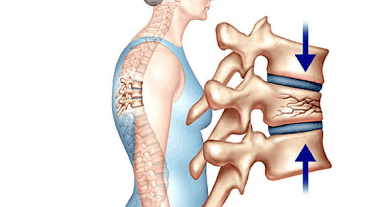
Spine compression fractures are the most common type of osteoporotic fracture, yet they remain poorly understood by patients and, in many cases, poorly recognised by clinicians. In the United States alone, approximately 1.5 million vertebral compression fractures occur each year — far more than hip or wrist fractures — but two-thirds are never clinically diagnosed because they produce no specific symptoms or their symptoms are attributed to ordinary back pain. This diagnostic gap matters enormously: a single vertebral compression fracture approximately quintuples the risk of a second vertebral fracture in the following year, and multiple compression fractures compound to produce height loss, progressive spinal deformity, chronic pain, respiratory restriction, and increased mortality. A useful rule of thumb: any adult over 50 with new or worsening back pain — particularly localised mid-back or lower-back pain that is worse with movement — should be assessed for VCF before assuming a muscular or degenerative cause. Understanding what spine compression fractures are, how they present, and what can be done about them — both at the time and to prevent subsequent fractures — is an important part of bone health literacy for adults over 50.
What Is a Vertebral Compression Fracture?
The vertebral column consists of 33 vertebrae stacked in a column from the skull to the pelvis. Each vertebra has a roughly cylindrical body of trabecular (spongy) bone — the structural element that bears compressive load. In a vertebral compression fracture (VCF), this vertebral body partially or completely collapses under a compressive force that weakened bone can no longer withstand.
VCFs are classified by their morphology:
- Wedge fractures: The most common type. The anterior (front) of the vertebral body collapses while the posterior (back) remains intact, creating a wedge shape. Wedge fractures, particularly at the thoracic (mid-back) level, drive the forward-curving spinal deformity (kyphosis) associated with osteoporosis and colloquially known as “dowager’s hump.” Each wedge fracture adds a few degrees of forward flexion to the spine; multiple fractures compound to produce significant kyphosis and height loss (typically 1–2 cm per vertebra fractured).
- Biconcave fractures: Both the upper and lower endplates of the vertebral body cave inward, creating a biconcave “fish vertebra” appearance on X-ray. Common in the lumbar spine.
- Crush fractures: Complete collapse of the vertebral body across all dimensions. Typically the most severe morphology, associated with the greatest pain and functional impairment.
The thoracic spine (particularly T6–T12) is the most common site for osteoporotic VCFs, followed by the thoracolumbar junction (T12–L1) and upper lumbar spine (L1–L4). These locations reflect the sites of greatest compressive loading and highest trabecular bone proportion — the same structural properties that make these areas most vulnerable to osteoporotic failure.
How Do Spinal Compression Fractures Happen?
In people with severe osteoporosis, a VCF can occur with trivial provocation — bending forward to pick up a bag, sneezing, coughing, or even simply rising from a chair. In less severe osteoporosis, a more significant force — a forward fall onto the hands, landing heavily from a step, carrying a heavy load — is typically required. The common thread is that the compressive force applied to the vertebral body exceeds the load-bearing capacity of the weakened trabecular bone.
Unlike hip and wrist fractures, which typically require a fall onto a specific anatomical structure, VCFs can occur in any posture that loads the anterior spine — which is essentially any forward-flexed position. Activities such as reaching forward while seated, bending at the waist to garden, or even strong muscle contractions during exertion can generate sufficient anterior compressive force in severely osteoporotic vertebrae. This explains why many VCFs occur during routine daily activities and why some patients cannot identify a specific precipitating event.
Symptoms — Including the Silent Majority
The most important clinical point about VCFs is that approximately two-thirds produce no acute symptoms or produce pain that is attributed to non-specific back pain rather than a fracture. This silent majority is the reason why many adults walk around with multiple vertebral compression fractures discovered only when a DEXA scan or spinal X-ray is done for another reason — or not discovered at all until a more severe event occurs.
When VCFs do produce symptoms, the typical presentation of an acute fracture is:
- Sudden onset of localised back pain at the level of the fractured vertebra, typically in the mid-back or lower back
- Pain that worsens with movement, weight-bearing, and sitting, and is partially relieved by lying flat
- Tenderness on percussion over the spinous process of the affected vertebra
- Pain that radiates around the chest wall or abdomen (if thoracic) or into the buttocks or thighs (if lumbar), occasionally misattributed to cardiac, pleural, or visceral causes
- Acute height loss of 1–2 cm in severe cases
Chronic consequences of multiple VCFs — the long-term effects that develop over months to years — include progressive kyphosis (forward spinal curvature), cumulative height loss (some patients lose 10–15 cm of height from multiple fractures), chronic back pain, and reduction in rib cage volume. This last consequence is clinically significant: thoracic kyphosis from multiple VCFs reduces the space available for lung expansion, contributing to reduced respiratory capacity, susceptibility to pneumonia, and in severe cases restrictive respiratory failure. Our guide to fracture risk: what adults should know covers the cascade risk from a first VCF.
Diagnosis — X-ray, MRI, and Bone Scan
Plain X-ray is the primary diagnostic tool for VCFs and can identify vertebral height loss, wedge deformity, and endplate irregularity. However, X-ray cannot distinguish between an acute (recent) and a chronic (old) fracture — which is clinically important for management decisions, since acute fractures are more likely to benefit from interventional procedures and require more urgent pain management.
MRI is the investigation of choice when X-ray findings are equivocal, when an acute fracture needs to be distinguished from a chronic one (bone marrow oedema on STIR sequences indicates acute fracture), when there is concern about spinal cord or nerve root compression, or when a pathological fracture (fracture from tumour or infection) needs to be excluded. MRI is particularly important when neurological symptoms are present — weakness, numbness, or bowel/bladder dysfunction associated with a vertebral fracture is an emergency requiring urgent neurosurgical review.
DEXA and FRAX should follow any VCF diagnosis — the fracture itself is strong evidence of bone fragility, and osteoporosis assessment is indicated regardless of prior BMD testing. DEXA at this point establishes a baseline for monitoring treatment response. It is also important to look for incidental vertebral fractures on any DEXA scan that includes a lateral spine view (vertebral fracture assessment, or VFA) — this can identify silent fractures in people having DEXA for other reasons and is an opportunity for earlier intervention. If you have a DEXA scan and your report does not mention a VFA, you can ask whether this assessment was performed or whether lateral spine imaging would be appropriate.
Treatment — Conservative Management
The majority of acute VCFs are managed conservatively, with the goals of pain control, early mobilisation, and prevention of subsequent fractures:
Analgesia: Paracetamol is the first-line option; NSAIDs (ibuprofen, naproxen) may be used if no contraindication but are avoided in older adults with renal impairment or GI risk. Short-term opioid analgesia is sometimes required for severe acute pain but is associated with constipation, falls risk, and cognitive effects in older adults — duration should be minimised. Calcitonin nasal spray has a modest evidence base for acute VCF pain reduction and may reduce opioid requirements in the short term.
Spinal bracing: A spinal orthosis (brace) may be prescribed for acute fractures causing significant pain with movement. Bracing reduces pain by limiting spinal movement but does not prevent fracture progression. There are two main types: soft thoracolumbar braces (for comfort) and more rigid orthopaedic braces (for fractures at the thoracolumbar junction where structural support is more relevant). Prolonged brace use beyond 6–8 weeks is discouraged as it promotes muscle deconditioning — the target is early mobilisation, not prolonged immobilisation.
Physiotherapy: Targeted physiotherapy — focusing on back extensor strengthening, postural correction, and safe movement patterns — is the cornerstone of long-term management after an acute VCF. Strengthening the spinal extensor muscles (erector spinae, multifidus) provides dynamic support to the fractured vertebral column, reduces pain chronicity, and helps prevent subsequent fractures by maintaining better spinal loading mechanics. The Sinaki protocol and other evidence-based back extensor programmes have demonstrated measurable reductions in subsequent vertebral fracture rates in postmenopausal women compared to controls who performed forward-flexion exercises. Forward-flexion exercises (such as standard sit-ups or toe-touches) are typically avoided in the acute phase as they increase anterior vertebral loading and can precipitate additional fractures in osteoporotic bone.
Pain management for long-term back pain: Chronic post-VCF back pain affects a significant proportion of patients, particularly those with multiple fractures. A pain management approach combining physiotherapy-directed strengthening, graded activity, pain medication review, and psychological support (acceptance and coping strategies) is more effective than prolonged analgesia alone. Input from a specialist pain service may be appropriate for those with severe chronic pain that limits function significantly.
Osteoporosis treatment — bisphosphonates, vitamin D, calcium, and in severe cases teriparatide — is the most important element of long-term fracture prevention after a VCF and is discussed in our guide to bone health after age 60.
Surgical Options — Vertebroplasty and Kyphoplasty
Two minimally invasive procedures — vertebroplasty and kyphoplasty — are available for acute vertebral compression fractures that are not responding adequately to conservative management:
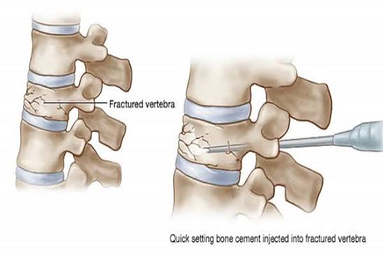
Vertebroplasty involves injecting bone cement (polymethylmethacrylate, PMMA) under imaging guidance directly into the collapsed vertebral body, stabilising the fracture. The procedure typically takes 30–60 minutes under local anaesthesia or light sedation.
Kyphoplasty involves first inflating a balloon inside the vertebral body to partially restore vertebral height and create a cavity, then filling the cavity with bone cement. It is associated with slightly lower cement leakage rates than vertebroplasty and may provide modest height restoration.
The evidence for both procedures is genuinely controversial. Two major randomised controlled trials comparing vertebroplasty to a sham procedure (INVEST, 2009; VERTOS-II, 2010) found no significant advantage for vertebroplasty over placebo in terms of pain reduction — challenging the clinical rationale for the procedure. A subsequent trial (VAPOUR, 2016) showed significant benefit in acute fractures (less than 6 weeks old), suggesting that patient selection — particularly early treatment of acute, highly painful fractures — may be key. Current NICE guidance limits vertebroplasty and kyphoplasty to specific circumstances where conservative management has failed and fractures are acute. The decision is made by a specialist spine team.
Long-Term Consequences and the Fracture Cascade
A first vertebral compression fracture should be treated as a clinical emergency for secondary fracture prevention. The 5-fold increase in second VCF risk in the year after a first fracture represents the rapid phase of the fracture cascade — before treatment has been initiated. Initiating bisphosphonate or denosumab treatment promptly after a first VCF reduces this cascade risk significantly. Our guide to osteoporosis in women after menopause covers pharmacological treatment choices in detail.
Multiple vertebral fractures accumulate to produce severe functional consequences: progressive kyphosis that causes chronic back pain, impairs gait (the forward posture shifts the centre of gravity, increasing falls risk), restricts lung expansion (increasing respiratory morbidity), and produces a characteristic change in appearance (height loss, protruding abdomen from thoracic compression, forward head posture) that causes significant psychological distress. Teriparatide — the anabolic bone-building agent used for severe osteoporosis — is specifically indicated for multiple vertebral fractures or rapidly progressing spinal disease where the urgency of building new bone exceeds what bisphosphonates can provide. Our guides to bone health after age 50 and calcium and bone health cover the nutritional foundations of bone protection.
Frequently Asked Questions
Can a spine compression fracture heal on its own?
Yes — vertebral compression fractures do heal without surgical intervention in the majority of cases. Over 6–12 weeks, the acute fracture stabilises, bone callus forms, and pain typically improves substantially. However, healing does not restore original vertebral height — the collapsed bone heals in its compressed position, meaning the height loss and any associated kyphosis are permanent changes. The underlying bone fragility that allowed the fracture also remains — or worsens — if osteoporosis is not treated. Conservative management (analgesia, early mobilisation, physiotherapy) manages the acute pain and function; osteoporosis treatment (bisphosphonates, vitamin D, calcium) is required to prevent subsequent fractures.
How long does pain from a compression fracture last?
Acute pain from an uncomplicated vertebral compression fracture typically peaks in the first 1–4 weeks and gradually improves over 6–12 weeks as the fracture heals and bone callus stabilises. Most patients are significantly more comfortable by 3 months, though some residual back pain at the fracture site persists longer, particularly with movement or prolonged sitting. Chronic back pain — persisting beyond 3 months — is more common after multiple VCFs, where the cumulative mechanical disruption of the spine is greater. Physical therapy-directed back extensor strengthening helps manage chronic post-fracture back pain by improving dynamic support of the fractured vertebral column and correcting posture.
Is bed rest recommended for a spinal compression fracture?
Short-term rest during the period of severe acute pain (typically the first few days to two weeks) is reasonable, but prolonged bed rest is harmful. Immobility accelerates muscle deconditioning, bone loss, and increases the risk of DVT, pressure sores, and respiratory complications. NICE guidance, the Royal Osteoporosis Society, and clinical physiotherapy guidelines all recommend early return to gentle activity and mobilisation as soon as pain levels allow — typically with a walking frame or stick in the acute phase. Maintaining some movement, even if limited, preserves muscle strength, helps prevent further bone loss, and reduces the risk of a subsequent fall from deconditioning. A physiotherapist can advise on safe movement and graduated activity resumption.
Should I see a doctor for back pain if I have osteoporosis?
Yes — any new or worsened back pain in a person with known osteoporosis or significant fracture risk factors (age over 65, prior fragility fracture, long-term glucocorticoid use) should prompt medical review to exclude a new vertebral fracture. This is particularly important if the pain is localised to a specific spinal level, worse with movement and weight-bearing, associated with height loss, or associated with any neurological symptoms (weakness, numbness, bladder or bowel changes). An X-ray is usually the first investigation. Many VCFs are discovered incidentally on imaging performed for other reasons — if a vertebral height change is reported on any spinal or chest imaging, follow-up with a GP or specialist to ensure osteoporosis is assessed and treated is appropriate.
What exercises are safe after a compression fracture?
Safe exercise after a vertebral compression fracture focuses on movements that do not increase anterior compressive loading on the spine. Back extensor strengthening exercises (such as prone lying with gentle arm and leg lifts), standing hip extension, and wall squats are typically recommended by physiotherapists in the recovery phase. Forward-flexion exercises (sit-ups, toe-touches, rowing) are generally avoided in the acute phase and approached with caution long-term in those with osteoporosis. Walking is encouraged from early in recovery as a low-impact weight-bearing activity. Balance exercises (standing on one leg, supported tandem walking) help reduce falls risk. All exercise after a VCF should be guided by a physiotherapist familiar with osteoporosis management, as the appropriate starting point and progression depends on the severity of the fracture, the number of fractures, and the individual’s pain and fitness levels.
What is the difference between vertebroplasty and kyphoplasty?
Both procedures involve injecting bone cement into a collapsed vertebral body to stabilise it. Vertebroplasty injects cement directly into the collapsed bone under imaging guidance. Kyphoplasty first inflates a balloon inside the vertebral body to create a cavity and partially restore vertebral height, then fills the cavity with cement. Kyphoplasty has a slightly lower rate of cement leakage (which can cause nerve irritation if it tracks into the spinal canal) and may offer modest height restoration. Both procedures are minimally invasive, typically performed under local anaesthesia, and take 30–60 minutes. The clinical evidence for both is genuinely debated — two randomised controlled trials found no benefit over sham procedure, while a later trial showed benefit in acute fractures under 6 weeks old. Current NICE guidance limits both procedures to cases where conservative management has failed in acute fractures. A spine surgeon or interventional radiologist specialist can advise on appropriateness.
How many compression fractures are considered severe osteoporosis?
Two or more vertebral fractures, or a single fracture with a T-score below −3.5, constitutes severe osteoporosis by clinical definition. Severe osteoporosis is an indication for teriparatide (Forsteo) — an anabolic (bone-building) agent that stimulates osteoblasts and produces larger BMD gains than bisphosphonates. Teriparatide is reserved for this indication because of its higher cost and the practicality of daily injection, but it is specifically indicated when the pace of bone loss from multiple VCFs is outrunning the protection that bisphosphonates provide. After an 18–24 month course of teriparatide, an antiresorptive agent (bisphosphonate or denosumab) is given to consolidate the BMD gains. If you have sustained two or more vertebral fractures, referral to a specialist osteoporosis service for review of treatment options is appropriate.
Summary
Vertebral compression fractures are the most common osteoporotic fracture, affecting approximately 1.5 million people per year in the US alone, yet two-thirds are never clinically identified — they either cause no symptoms or their symptoms are misattributed to non-specific back pain, leaving the underlying bone fragility unaddressed. They range from entirely silent to severely painful, and their long-term consequences — progressive kyphosis, height loss, chronic pain, and respiratory restriction — reflect the cumulative effect of multiple fractures on spinal structure and function. The fracture cascade risk (5-fold increase in second VCF after first) makes prompt secondary fracture prevention — bisphosphonates, denosumab, or teriparatide for severe disease — the most clinically important intervention after a first VCF. Conservative management (analgesia, physiotherapy, early mobilisation) addresses the acute fracture; osteoporosis treatment addresses the underlying disease that made the fracture possible. Both must be addressed together — treating only the pain without treating the osteoporosis leaves the patient exposed to the fracture cascade. Our guides to fracture risk: what adults should know and vitamin D and bone health cover the evidence-based prevention foundations.
Medical disclaimer: This article is for general educational purposes and does not constitute medical advice. Consult a qualified healthcare professional for personalised bone health assessment and management.
References:
NICE CG146. Osteoporosis: assessing the risk of fragility fracture. NICE. 2023.
Royal Osteoporosis Society. Vertebral fracture information. theros.org.uk. 2023.
Kallmes DF et al. A randomised trial of vertebroplasty for osteoporotic spinal fractures (INVEST). NEJM. 2009.
Clark W et al. Vertebroplasty for acute painful osteoporotic fractures (VAPOUR). The Lancet. 2016.
NHS. Back pain. nhs.uk. 2023.
I want to share my experience because I had no idea spine compression fractures could be silent. At 67 I had a DEXA scan arranged by my GP because I had an early menopause at 44. The results came back showing a T-score of −2.6 at the spine and −2.1 at the hip, but they also included something called a VFA — vertebral fracture assessment — which showed I had two old wedge fractures at T8 and T11. I have never had acute back pain severe enough to seek medical care. My GP explained that these were silent fractures — they probably occurred at some point in my 60s without my knowing, possibly attributed to back aches I had put down to posture or age. The news was quite shocking. I was immediately started on alendronate, calcium and vitamin D. My GP explained that these two existing fractures meant I was at particularly high risk of further fractures and that treatment was urgent. The article’s point about two-thirds of VCFs being silent and the importance of VFA on DEXA scans is exactly what happened to me — I would never have known about these fractures without the VFA, and I might have had further fractures without treatment.
Evelyn, your case is a textbook illustration of why VFA is such a clinically important addition to standard DEXA reporting — and why the discovery of two silent T8 and T11 fractures in an asymptomatic patient with a T-score of −2.6 changes the clinical picture entirely. Without VFA, you would have been assessed as an osteoporosis patient without fracture history, warranting treatment based on FRAX and T-score alone. With the fracture history revealed by VFA, you are in the severe osteoporosis category — two existing VCFs plus a T-score below −2.5 — where the urgency of treatment is higher and the absolute risk of further fracture is substantially greater. Your GP’s decision to start alendronate immediately is exactly correct; the fracture cascade risk in your first year post-discovery, now that the fractures are identified, needs to be addressed without delay. Thomas, your clinical outcome with vertebroplasty is consistent with the evidence profile for acute fractures treated early — the VAPOUR trial specifically enrolled patients with acute, severely painful fractures under 6 weeks old, exactly matching your presentation at 5 weeks. The pain reduction you describe is the expected outcome in this subgroup. The important next step is ensuring the bisphosphonate therapy continues long-term to prevent the fracture cascade from producing further VCFs — vertebroplasty stabilises the acute fracture but does not address the underlying bone fragility. Your T12 fracture already approximately quintuples your risk of another VCF in the coming year, and bisphosphonate treatment substantially reduces that risk.
I had a severe acute compression fracture at T12 three months ago — the pain was so severe I couldn’t stand upright or take a full breath. I was admitted to hospital and given strong analgesia but was struggling to mobilise. The spine team offered me vertebroplasty. I had researched it online and found conflicting information about whether it works — some sources citing the INVEST trial showing no benefit over a sham procedure, others saying it can be very effective. My spine surgeon explained the evidence carefully — that the INVEST and VERTOS-II trials were controversial partly because they included many older, chronic fractures where cement was unlikely to help, and that the VAPOUR trial showed clear benefit in acute, painful fractures under 6 weeks old — which was exactly my situation. I had vertebroplasty at 5 weeks post-fracture. The pain reduction was significant within 48 hours — I went from barely able to stand to walking with a stick. I started bisphosphonates after the procedure. The article’s balanced presentation of the vertebroplasty evidence — noting both the trials showing no benefit and the VAPOUR trial showing benefit in acute fractures — reflects the nuance I wish I had understood better when I was first trying to research it.