Blood circulation is one of the body’s most fundamental and continuous processes. Every organ, tissue, and cell depends on the circulatory system for oxygen, nutrients, and the removal of metabolic waste products. The heart beats approximately 100,000 times per day, driving blood through roughly 60,000 miles of blood vessels to maintain this constant delivery. Understanding how blood circulates through the body — what drives it, where it goes, what it delivers, and what happens when it fails — is the foundation for understanding cardiovascular health.
The Two Circuits — Pulmonary and Systemic
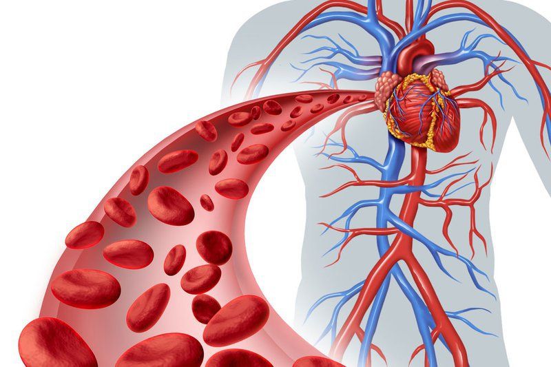
Blood does not follow a single circuit through the body. It follows two distinct circuits that operate simultaneously, in series, with the heart serving as the pump that drives both.
The pulmonary circuit carries blood from the right side of the heart to the lungs and back. Its purpose is gas exchange: delivering carbon dioxide (the waste product of cellular metabolism) to the lungs for exhalation and collecting freshly oxygenated blood to return to the heart.
The systemic circuit carries blood from the left side of the heart to every organ and tissue in the body, delivering oxygen and nutrients, collecting carbon dioxide and metabolic waste, and returning deoxygenated blood to the right side of the heart.
A single red blood cell completing one full circuit travels through the right heart, both lungs, the left heart, and the entire systemic circulation in approximately one minute at rest. At rest, approximately 64 percent of total blood volume resides in the veins — which is why veins are called “capacitance vessels.” When a person stands from lying flat, gravity shifts blood into the leg veins, reducing venous return to the heart — the physiological basis of orthostatic hypotension.
The Pulmonary Circuit — How Blood Gets Oxygenated
The pulmonary circuit begins when the right ventricle contracts and ejects deoxygenated blood into the pulmonary artery — the only artery in the body that carries deoxygenated blood. The pulmonary artery branches into the right and left pulmonary arteries, then progressively smaller arterioles, and finally into the dense capillary network surrounding the alveoli (tiny air sacs) of both lungs.
At the alveolar-capillary membrane — a barrier only about 0.5 micrometers thick — gas exchange occurs by simple diffusion: carbon dioxide (at higher concentration in blood than in alveolar air) diffuses from blood into the alveoli to be exhaled; oxygen (at higher concentration in alveolar air) diffuses from the alveoli into the blood, where it binds to hemoglobin. Each hemoglobin molecule carries four oxygen molecules; fully oxygenated arterial blood achieves a hemoglobin oxygen saturation of approximately 95 to 100 percent in healthy lungs.
Oxygenated blood flows through pulmonary venules and collects into the four pulmonary veins — two from each lung — draining into the left atrium and completing the pulmonary circuit.
A critical feature of the pulmonary circuit is its low pressure. Normal pulmonary artery systolic pressure is approximately 25 mmHg, compared to systemic arterial systolic pressure of 120 mmHg. This low pressure is essential: the thin-walled alveolar capillaries would force fluid into the alveolar space (pulmonary edema) at high pressure. Conditions that elevate pulmonary artery pressure — pulmonary arterial hypertension or left heart failure (which backs up pressure through the pulmonary circuit) — produce progressive right ventricular strain.
The Systemic Circuit — Delivering Oxygen to Every Organ
The systemic circuit begins when the left ventricle contracts and ejects oxygenated blood into the aorta — the body’s largest artery, approximately 2.5 cm in diameter at its root. From the aorta, blood is distributed through major branches to every organ and tissue:
- Coronary arteries: arising immediately above the aortic valve, supplying the heart muscle itself
- Carotid and subclavian arteries (via the brachiocephalic artery): supplying the head, neck, and upper limbs
- Celiac trunk, superior mesenteric, and inferior mesenteric arteries: supplying the abdominal organs (stomach, intestines, liver, spleen, pancreas)
- Renal arteries: supplying the kidneys, which receive approximately 20 percent of total cardiac output — a disproportionately large fraction reflecting their continuous metabolic demands for filtration
- Iliac and femoral arteries: supplying the pelvis and lower limbs
From major arteries, blood flows into progressively smaller arteries → arterioles → capillaries → venules → veins → superior and inferior vena cava → right atrium, completing the systemic circuit.
What Happens in the Capillaries — The Purpose of Circulation
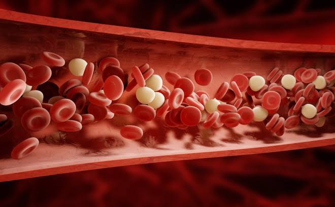
The capillaries are where the actual physiological work of circulation takes place — the reason the entire circulatory system exists. Capillary walls are only one cell thick (a single layer of endothelium), allowing oxygen, carbon dioxide, nutrients, and waste products to diffuse rapidly between blood and surrounding tissue fluid.
Oxygen delivery: in metabolically active tissues, the partial pressure of oxygen is lower than in arterial blood, driving diffusion of oxygen from blood into cells. The more active a tissue — working muscle, for example — the lower its oxygen partial pressure and the more oxygen it extracts from passing blood.
Carbon dioxide removal: cellular metabolism produces CO2, which diffuses from cells into the bloodstream. Approximately 70 percent is converted to bicarbonate, 23 percent is carried bound to hemoglobin, and 7 percent dissolves in plasma. CO2 is ultimately delivered to the lungs and exhaled.
Fluid balance: the balance of fluid between the capillary and surrounding interstitial space is governed by Starling forces — capillary hydrostatic pressure (pushing fluid out of the capillary) versus plasma oncotic pressure (pulling fluid back in via dissolved proteins). When hydrostatic pressure rises (as in heart failure or venous obstruction) or oncotic pressure falls (as in liver disease or severe malnutrition with low albumin), excess fluid accumulates in the interstitial space as edema.
Special Circulations — Brain, Heart, Kidneys, Liver
Coronary circulation: the heart muscle cannot absorb oxygen from blood within its chambers — it depends entirely on the coronary arteries. Coronary blood flow occurs primarily during diastole, because the contracting myocardium during systole compresses intramural coronary vessels. Local metabolites including adenosine regulate coronary arteriolar tone to match flow to myocardial oxygen demand. Atherosclerotic narrowing reduces this supply-demand flexibility, producing angina during exertion and heart attack when occlusion is complete.
Cerebral circulation: despite comprising only about 2 percent of body weight, the brain receives 15 to 20 percent of resting cardiac output and consumes approximately 20 percent of total body oxygen. Consciousness is lost within seconds and irreversible neuronal damage begins within minutes of complete cerebral blood flow cessation. The Circle of Willis — an anastomotic loop at the base of the brain formed by branches of the internal carotid and vertebral arteries — provides collateral supply that can partially compensate for obstruction of one feeding vessel. The blood-brain barrier (BBB) — formed by tight junctions between cerebral endothelial cells — strictly restricts the passage of most molecules from blood into brain tissue, protecting the neural environment but limiting drug delivery to the brain.
Renal circulation: the kidneys’ extraordinary blood flow (20 percent of cardiac output) supports their role as the body’s primary filtration organ. Blood enters the glomerulus under higher-than-normal hydrostatic pressure, forcing fluid and small molecules into the tubule for processing, while retaining proteins and blood cells. The glomerular filtration rate (GFR) — approximately 125 mL per minute in healthy adults — generates about 180 liters of filtrate per day, of which more than 99 percent is reabsorbed, producing approximately 1.5 liters of urine.
Portal circulation: blood from the intestines, stomach, pancreas, and spleen collects into the portal vein and flows to the liver before joining the systemic circulation. This arrangement allows the liver to process absorbed nutrients, perform first-pass metabolism of orally ingested drugs and toxins, and filter bacteria crossing the intestinal barrier. Portal hypertension — most commonly from cirrhosis — causes blood to bypass the liver through collateral veins, producing esophageal varices, splenomegaly, and ascites.
Lymphatic Circulation — The Return System
Alongside blood capillaries runs a parallel network of lymphatic capillaries that drain the interstitial space. Approximately 3 liters of interstitial fluid per day that is not reabsorbed by blood capillaries is collected by the lymphatic system, flowing through lymph vessels and lymph nodes (where immune surveillance occurs) until it drains via the thoracic duct into the subclavian vein, returning to the systemic circulation.
When lymphatic drainage is impaired — due to lymph node removal (as after cancer surgery), radiation damage, or parasitic infection — lymph accumulates in the affected limb, producing lymphedema: a chronic, protein-rich swelling prone to infection and difficult to treat.
How Blood Pressure Drives Circulation
Blood pressure — created by left ventricular contraction and maintained by arteriolar resistance — is the driving force of the entire circulatory system. Mean arterial pressure (MAP) — calculated as diastolic blood pressure plus one-third of the pulse pressure — is the most physiologically meaningful measure of organ perfusion pressure. A MAP of approximately 60 mmHg is considered the minimum adequate for perfusing the brain and kidneys.
Blood pressure is regulated at three time scales:
- Baroreceptors (seconds): pressure sensors in the carotid sinus and aortic arch detect blood pressure changes and reflexively adjust heart rate and vasoconstriction within seconds
- RAAS (minutes to hours): when blood pressure falls, the kidneys release renin, triggering production of angiotensin II (vasoconstrictor) and aldosterone (promotes sodium and water retention)
- Renal fluid balance (hours to days): the long-term determinant of blood pressure — the kidneys adjust sodium and water excretion to maintain the blood pressure that achieves sodium balance; this is why low-sodium diets reduce blood pressure over time
When Circulation Fails — Clinical Consequences
Peripheral artery disease: atherosclerosis in the leg arteries reduces flow to working muscles. The hallmark is claudication — reproducible pain or cramping in the calf or thigh that appears with walking and resolves within minutes of rest. Severe PAD can cause ischemic rest pain, non-healing ulcers, and limb-threatening gangrene.
Heart failure: when the heart cannot generate adequate cardiac output, tissues receive insufficient blood flow (hypoperfusion) and fluid backs up behind the failing ventricle (congestion). Left heart failure backs up pressure into the pulmonary veins, producing pulmonary edema and breathlessness. Right heart failure backs up pressure into the systemic veins, producing peripheral edema and ascites.
Orthostatic hypotension: when the baroreceptor reflex is impaired — from dehydration, autonomic neuropathy, medications, or aging — blood pressure drops transiently on standing, causing lightheadedness, vision changes, or syncope.
Shock: global tissue hypoperfusion with oxygen delivery insufficient for cellular metabolism. The four main categories are cardiogenic (heart failure to pump), hypovolemic (insufficient blood volume), distributive (vasodilation from sepsis, anaphylaxis, or spinal injury), and obstructive (physical obstruction of flow, as in pulmonary embolism or cardiac tamponade).
For a detailed description of the heart’s anatomy and how it generates the pressure that drives circulation, see our guide to how the heart works. For an overview of what can go wrong with the heart and blood vessels, see what is cardiovascular disease. For information on poor circulation and its symptoms, see our article on what is poor circulation.
Sources: American Heart Association | National Heart, Lung, and Blood Institute | CDC — Heart Disease
Microcirculation and the Endothelium
At the level of the smallest vessels — arterioles, capillaries, and venules — the microcirculation performs functions that the large vessel circulation cannot. The arterioles are the primary resistance vessels: their muscular walls contract or relax to direct blood flow to different tissues based on local metabolic needs. Pre-capillary sphincters regulate flow into individual capillary beds, opening when tissue oxygen consumption is high and closing when a tissue is at rest. This local regulation allows blood flow to be directed preferentially to the most metabolically active organs at any given moment — to working muscles during exercise, to the GI tract after eating, or to the skin during heat dissipation.
The endothelium — the single layer of cells lining every blood vessel in the body — is far more than a passive barrier. It is a metabolically active organ that continuously produces vasoactive, anti-inflammatory, and anti-thrombotic substances:
- Nitric oxide (NO): the principal vasodilator; produced in response to shear stress (blood flow) and acetylcholine; causes smooth muscle relaxation and inhibits platelet aggregation. Exercise increases shear stress and stimulates endothelial NO production, contributing to the chronic blood pressure lowering effect of regular aerobic activity.
- Prostacyclin (PGI2): a prostaglandin with vasodilatory and anti-platelet effects
- Endothelin-1 (ET-1): a potent vasoconstrictor; elevated in pulmonary arterial hypertension; targeted by endothelin receptor antagonists (bosentan, ambrisentan) used in PAH treatment
- Tissue plasminogen activator (tPA): promotes fibrinolysis (dissolution of blood clots); the basis for the thrombolytic therapy used in acute ischemic stroke
Endothelial dysfunction — characterized by reduced nitric oxide bioavailability, increased endothelin production, and a pro-inflammatory, pro-thrombotic state — is the earliest detectable stage of atherosclerosis, observable before any structural plaque formation. It can be measured non-invasively using flow-mediated dilation (FMD) of the brachial artery. Risk factors including hypertension, hypercholesterolemia, smoking, diabetes, and sedentary lifestyle all impair endothelial function. Exercise, a Mediterranean-pattern diet, blood pressure control, and statin therapy all improve it.
Blood Composition and Its Role in Circulation
Blood is not simply a fluid carrier — it is a complex tissue containing multiple cell types and dissolved proteins, each contributing to specific circulatory functions.
Red blood cells (erythrocytes) contain hemoglobin and are responsible for the vast majority of oxygen transport. A normal red blood cell lives approximately 120 days before being cleared from circulation by the spleen. The bone marrow continuously produces new red blood cells in response to erythropoietin (EPO), a hormone released by the kidneys when tissue oxygen delivery is insufficient. Anemia — a deficiency of red blood cells or hemoglobin — impairs oxygen delivery, producing fatigue, breathlessness on exertion, and (in severe cases) high-output heart failure as the heart compensates by increasing cardiac output to maintain oxygen delivery despite reduced hemoglobin concentration.
White blood cells (leukocytes) patrol the circulation and migrate into tissues in response to infection and inflammation. Some, like neutrophils, circulate for only a few hours before entering tissues; others, like memory lymphocytes, can persist in circulation for years.
Platelets (thrombocytes) are small, enucleate cell fragments that play the central role in hemostasis — the arrest of bleeding following vascular injury. When endothelium is damaged, von Willebrand factor and collagen are exposed, platelets adhere and activate, and a platelet plug forms within seconds. Antiplatelet drugs including aspirin and P2Y12 inhibitors (clopidogrel, ticagrelor) reduce platelet activation — preventing the platelet aggregation that contributes to coronary artery occlusion during an acute MI or stent thrombosis after PCI.
Plasma proteins — including albumin (which maintains oncotic pressure and prevents fluid accumulation in interstitial spaces), coagulation factors (required for fibrin clot formation), complement proteins (part of innate immunity), and transport proteins (carrying hormones, lipids, and drugs) — are dissolved in plasma. When albumin concentration falls significantly (from liver disease, nephrotic syndrome, or malnutrition), oncotic pressure drops and fluid leaks into interstitial spaces, producing edema even without any increase in capillary hydrostatic pressure.
Factors That Affect Circulation Quality
The effectiveness of blood circulation depends not only on the structural integrity of the heart and blood vessels but on a range of physiological and lifestyle factors that influence how smoothly and efficiently blood flows.
Blood viscosity: blood is a non-Newtonian fluid — its viscosity changes with flow rate. At low flow rates in small vessels, blood can become more viscous, particularly when the hematocrit (red blood cell fraction of blood volume) is elevated, as in polycythemia vera. High blood viscosity increases the cardiac work required to maintain circulation and increases the risk of thrombosis.
Hydration: adequate hydration maintains plasma volume and reduces blood viscosity. Dehydration reduces circulating blood volume, impairing venous return to the heart and reducing cardiac output. In hot environments or during prolonged exercise, significant fluid losses can substantially compromise circulatory performance.
Physical activity: regular aerobic exercise produces structural and functional adaptations that improve circulatory efficiency. Cardiac chambers enlarge, stroke volume increases, resting heart rate decreases, arterioles develop improved vasodilatory capacity, and endothelial function improves. These adaptations collectively increase the margin of circulatory reserve — the difference between resting and maximal circulatory capacity — that protects against cardiovascular events.
Temperature: body temperature regulation involves deliberate redistribution of blood flow. In heat, cutaneous blood vessels dilate to deliver warm blood to the skin surface for heat dissipation; in cold, cutaneous vessels constrict to minimize heat loss. This redistribution is controlled primarily by the sympathetic nervous system and can be dramatically large — skin blood flow can vary from near zero in extreme cold to several liters per minute during intense heat stress.
Why Understanding Circulation Matters for Everyday Health Decisions
The mechanics of blood circulation are not abstract physiology — they are the basis for understanding why the health recommendations adults encounter most frequently actually work.
When a physician recommends reducing sodium intake to lower blood pressure, the mechanism is the renal fluid balance arm of blood pressure regulation: less sodium in the filtrate means less sodium reabsorbed, more water in the urine, smaller circulating blood volume, lower venous filling pressure, and ultimately lower cardiac output and lower arterial pressure. The recommendation is not arbitrary; it targets a specific physiological system.
When a physician recommends regular aerobic exercise for cardiovascular health, the mechanism includes multiple circulatory adaptations: increased stroke volume (larger cardiac chambers, better filling), lower resting heart rate (increased parasympathetic tone), improved endothelial function (higher nitric oxide production), increased arteriolar density in skeletal muscle (more capillary surface area for oxygen delivery), and reduced arterial stiffness. Each of these changes improves the efficiency and resilience of the circulatory system.
When a physician prescribes an ACE inhibitor or ARB for heart failure or hypertension, the mechanism is blockade of the RAAS — reducing angiotensin II’s vasoconstrictive effect and aldosterone’s sodium-retaining effect, lowering systemic vascular resistance (the afterload the left ventricle pumps against) and reducing fluid retention (the preload that distends and stresses the failing heart).
When aspirin is prescribed after a heart attack, the mechanism is inhibition of thromboxane A2 production in platelets — reducing the platelet aggregation that would otherwise risk occluding the coronary artery at the site of the recently ruptured plaque or newly placed stent.
In each case, understanding how blood circulates — which vessels carry it, what drives it, how it is regulated, and what it delivers — transforms medical recommendations from arbitrary instructions into logical consequences of physiology. Patients who understand the mechanism are better equipped to understand why their medication matters, why lifestyle choices have the effects they do, and why the specific numbers their physicians monitor — blood pressure, hemoglobin, creatinine, ejection fraction — reflect the actual performance of the circulatory system that sustains their health.