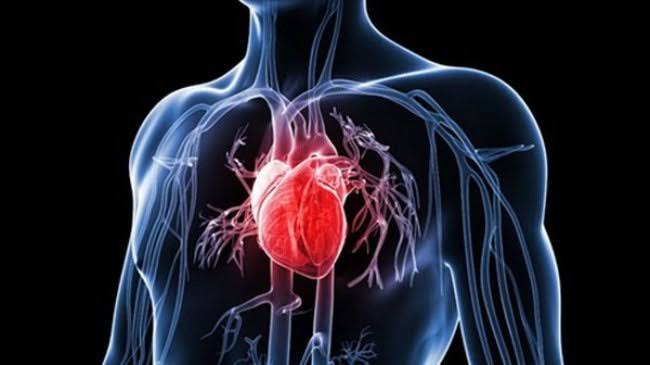
Heart disease is not one condition. It is an umbrella term covering a range of distinct disorders that affect the heart’s muscle, arteries, valves, and electrical system in fundamentally different ways. Coronary artery disease is not the same as heart failure, and heart failure is not the same as atrial fibrillation — even though all three are classified as “heart problems” and all three can affect the same patient simultaneously. Adults who understand what the most common heart problems look like are better equipped to recognize warning signs, engage more effectively with their physicians, and take the right preventive steps for their specific situation.
Coronary Artery Disease — The Most Common Heart Problem
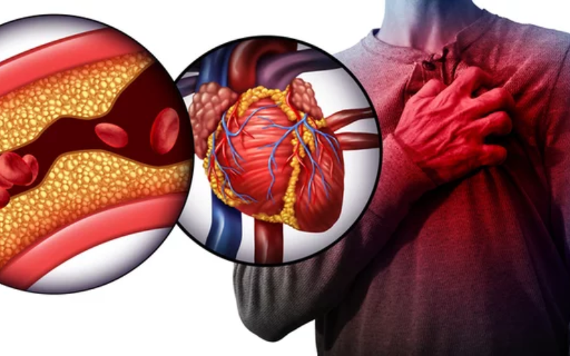
Coronary artery disease (CAD) is the single most common heart problem in adults and the most common cause of death in most high-income countries. It develops when atherosclerosis — the progressive accumulation of cholesterol-laden plaques within artery walls — narrows the coronary arteries that supply blood to the heart muscle. As narrowing increases, the heart muscle receives insufficient blood flow during increased demand, producing ischemia (insufficient oxygen relative to demand).
The characteristic symptom is angina: chest pain, pressure, squeezing, or tightness that typically occurs during physical exertion or emotional stress and resolves with rest or nitroglycerin within minutes. Angina may radiate to the left arm, jaw, neck, or back. Women are more likely to present atypically — with fatigue, shortness of breath, nausea, or jaw pain — without classic chest pressure.
Stable angina is predictable, occurring consistently at similar levels of exertion and resolving reliably with rest. Unstable angina is a medical emergency: angina occurring at rest, new-onset severe angina, or previously stable angina that has worsened significantly in frequency or with less exertion. It represents an acute coronary syndrome requiring immediate evaluation.
A myocardial infarction (heart attack) occurs when a coronary artery is completely blocked — typically by the sudden rupture of an atherosclerotic plaque followed by acute clot formation. Heart muscle begins to die within 20 to 40 minutes of complete occlusion if blood flow is not restored. A STEMI (ST-elevation myocardial infarction) requires emergency reperfusion — either percutaneous coronary intervention (stenting) or thrombolysis — within 90 minutes of hospital arrival.
Treatment for established CAD includes high-intensity statins, antiplatelet therapy (aspirin, often combined with clopidogrel or ticagrelor), blood pressure medications, lifestyle modification, and when appropriate, coronary revascularization.
Heart Failure — When the Heart Can’t Keep Up
Heart failure does not mean the heart has stopped. It means the heart cannot pump enough blood to meet the body’s needs. It is a syndrome that can result from virtually any condition that damages the heart muscle — with coronary artery disease and hypertension being the most common causes.
Heart failure with reduced ejection fraction (HFrEF) occurs when the heart pumps too weakly, with an ejection fraction (EF) below 40 percent. The heart is typically enlarged. This is the form with the most established drug therapy: four medication classes — ACE inhibitors or ARBs (or sacubitril-valsartan), beta-blockers, mineralocorticoid receptor antagonists, and SGLT2 inhibitors — each independently reduce mortality and hospitalizations.
Heart failure with preserved ejection fraction (HFpEF) occurs when the heart pumps with adequate force but is too stiff to fill properly. HFpEF now accounts for at least half of all heart failure in adults over 65 and is strongly associated with hypertension, diabetes, and obesity. SGLT2 inhibitors have the strongest current evidence for reducing hospitalizations in HFpEF.
Characteristic symptoms include progressive breathlessness (initially with exertion, then at rest), orthopnea (difficulty lying flat), paroxysmal nocturnal dyspnea (waking suddenly breathless), ankle and leg swelling, and severely reduced exercise capacity. Diuretics (furosemide) relieve fluid congestion but do not independently reduce mortality.
Atrial Fibrillation — The Most Common Arrhythmia
Atrial fibrillation (AF or AFib) is the most common sustained cardiac arrhythmia, affecting an estimated 6 million Americans. In AF, the atria fire chaotically at 400 to 600 impulses per minute rather than in a coordinated rhythm. The AV node transmits these impulses irregularly to the ventricles, producing the characteristic “irregularly irregular” pulse — a heartbeat with no discernible pattern of regularity.
Symptoms range widely from palpitations, shortness of breath, and fatigue to no symptoms at all. The primary danger of AF is stroke — blood pooling in the left atrial appendage can form clots that embolize to the brain, increasing stroke risk approximately five-fold. Anticoagulation with a direct oral anticoagulant (DOAC: apixaban, rivaroxaban, edoxaban, or dabigatran) substantially reduces this risk and is recommended for most AF patients with a CHA₂DS₂-VASc score of 2 or higher in men (3 or higher in women).
Beyond anticoagulation, management focuses on either rate control (slowing the ventricular response using beta-blockers, calcium channel blockers, or digoxin) or rhythm control (restoring and maintaining normal sinus rhythm through cardioversion, antiarrhythmic medications, or catheter ablation — the most effective option for rhythm control in appropriate patients).
High Blood Pressure and the Heart
Hypertension (blood pressure ≥130/80 mmHg) is the most common cardiovascular risk factor and an important direct cause of heart disease in its own right. The condition it causes — hypertensive heart disease — develops through progressive structural remodeling of the heart in response to chronically elevated afterload.
The primary structural change is left ventricular hypertrophy (LVH): the ventricular wall thickens as the heart adapts to increased pressure work. The hypertrophied ventricle fills more slowly and at higher pressure — producing diastolic dysfunction that can progress over years to HFpEF and predispose to atrial fibrillation. Hypertensive heart disease develops silently, detectable on ECG and echocardiography before any symptoms appear. Blood pressure control before these structural changes become established is the most effective intervention.
Heart Valve Disease
The heart’s four valves ensure blood flows in one direction. Valve disease causes either stenosis (narrowing that impedes forward flow) or regurgitation (leaking that allows backward flow). Both increase the work of specific heart chambers and can cause irreversible structural damage over time.
Aortic stenosis (AS) is the most common valvular heart disease in adults over 65. Calcific degenerative AS progressively narrows the aortic valve over decades. The classic clinical triad — angina (from impaired coronary supply), syncope (from fixed cardiac output failing to meet exercise demand), and dyspnea from heart failure — typically appears late, after severe stenosis has been present for years. Treatment is valve replacement: surgical aortic valve replacement (SAVR) or transcatheter aortic valve replacement (TAVR), now available even for lower-risk patients.
Mitral regurgitation (MR) is the most common valvular disease overall. The mitral valve can leak due to primary degenerative causes (most commonly mitral valve prolapse, where leaflets bulge back into the left atrium during systole) or secondary functional causes (left ventricular dilation distorting valve geometry). Significant MR causes progressive dilation of the left atrium and ventricle, eventually impairing ventricular function. Surgical mitral valve repair — when feasible — is preferred over replacement for superior durability. Any murmur detected on physical examination warrants echocardiography.
Cardiomyopathy — Disease of the Heart Muscle
Dilated cardiomyopathy (DCM) is the most common type: the left ventricle is enlarged and weak, producing significantly reduced ejection fraction. Causes include genetic mutations (25–50% of cases), prior viral myocarditis, alcohol toxicity, cardiotoxic chemotherapy, peripartum cardiomyopathy, and idiopathic causes. Treatment mirrors that of HFrEF.
Hypertrophic cardiomyopathy (HCM) is the most common genetic cardiovascular disease, caused primarily by sarcomere protein mutations (MYH7 and MYBPC3 most commonly). The interventricular septum thickens abnormally, and in obstructive HCM creates dynamic left ventricular outflow tract obstruction. HCM is the leading cause of sudden cardiac death in young athletes. Many patients have few symptoms; others experience dyspnea, syncope, or palpitations. Mavacamten (Camzyos), a cardiac myosin inhibitor, has demonstrated meaningful symptom improvement in obstructive HCM. High-risk patients benefit from ICD placement for sudden cardiac death prevention.
Stress cardiomyopathy (Takotsubo syndrome) is a usually reversible form of acute heart failure provoked by intense emotional or physical stress — a catecholamine surge that transiently stuns the cardiac apex, producing a characteristic “apical ballooning” pattern on echocardiography. It predominantly affects postmenopausal women, closely mimics an acute MI in presentation, but coronary arteries are found to be unobstructed. Ventricular function typically recovers fully within weeks with supportive management.
Pericarditis and Cardiac Tamponade
Pericarditis is inflammation of the pericardium — the fibrous sac surrounding the heart. Most commonly viral or idiopathic, it can also occur after myocardial infarction, in autoimmune disease, after cardiac surgery, or with certain medications. The characteristic symptom is sharp, pleuritic chest pain worse when lying flat and relieved by sitting forward. The ECG classically shows diffuse saddle-shaped ST elevation and PR depression. First-line treatment is NSAIDs combined with colchicine, which significantly reduces recurrence rates.
Large or rapidly accumulating pericardial effusions can produce cardiac tamponade — a life-threatening emergency in which rising intrapericardial pressure compresses the cardiac chambers, preventing adequate filling. Beck’s triad (hypotension, distended neck veins, muffled heart sounds) and pulsus paradoxus are the classic clinical signs. Emergency pericardiocentesis is the definitive treatment.
When to Seek Immediate Medical Attention
Understanding which cardiac symptoms require emergency care rather than a scheduled appointment can be life-saving:
- Heart attack: chest pain, pressure, or tightness lasting more than a few minutes; pain radiating to the left arm, jaw, neck, or back; new breathlessness at rest; cold sweat or nausea with chest symptoms. Call emergency services immediately — do not drive yourself.
- Stroke: FAST — Face drooping, Arm weakness, Speech difficulty, Time to call emergency services. Every minute of delay costs brain cells.
- Arrhythmia emergency: loss of consciousness (syncope), near-syncope, or sustained rapid palpitations with lightheadedness or chest pain.
- Acute heart failure: sudden or rapidly worsening breathlessness, inability to lie flat, new marked ankle swelling, or rapid weight gain (more than 2–3 pounds in 24 hours) in someone with known heart failure.
- Cardiac tamponade: acute dyspnea, lightheadedness, and low blood pressure in someone with known pericardial effusion or recent cardiac procedure.
For a comprehensive overview of cardiovascular conditions, see our article on what is cardiovascular disease. For practical heart health monitoring guidance, see what is heart health. To understand why the 40s are the most important decade for cardiovascular prevention, see why heart health matters after age 40.
Sources: American Heart Association | CDC — Heart Disease | National Heart, Lung, and Blood Institute
Less Common But Important Heart Problems Adults Should Know
Beyond the most prevalent conditions, several heart problems are encountered less frequently but carry significant clinical importance either because of their severity, their characteristic presentations, or because they affect specific populations disproportionately.
Infective endocarditis (IE) is an infection of the heart’s inner lining, most often affecting the valve leaflets. Staphylococcus aureus, the most common causative organism in developed countries, produces an acute and aggressive form that can destroy a valve within days. Streptococcal species tend to produce a more subacute presentation over weeks. The classic features include fever, a new or changed heart murmur, and embolic phenomena — the septic fragments that break off and lodge in distant organs, producing Janeway lesions on the palms and soles, Osler nodes (painful nodules on fingers), and splinter hemorrhages under fingernails. Blood cultures and echocardiography are the cornerstones of diagnosis. Treatment requires prolonged intravenous antibiotics and, in cases of severe valve destruction, uncontrolled infection, or high embolic risk, cardiac surgery.
Aortic aneurysm is the abnormal dilation of the aorta beyond 1.5 times its normal diameter. Abdominal aortic aneurysm (AAA) is the most common and most dangerous type — a ruptured AAA carries a mortality above 80 percent in the community. Current guidelines recommend one-time ultrasound screening for AAA in men aged 65 to 75 who have ever smoked, a population with substantially elevated prevalence. Repair is recommended when the diameter reaches 5.5 cm or more, or when growth exceeds 0.5 cm in 6 months. Aortic dissection — a tear in the inner layer of the aortic wall that allows blood to track between the wall layers — is a cardiovascular emergency. Type A dissections (involving the ascending aorta) require emergency surgery; Type B dissections (descending only) are typically managed medically with blood pressure control — unless complications develop.
Ventricular tachycardia (VT) is a rapid heart rhythm originating in the ventricles, producing a wide-complex tachycardia on ECG. Sustained VT in a patient with structural heart disease (particularly after a myocardial infarction, which leaves a scar that can serve as a reentry circuit) can cause hemodynamic collapse and progress to ventricular fibrillation and cardiac arrest. Implantable cardioverter-defibrillators (ICDs) are the most effective means of preventing sudden cardiac death in patients with sustained VT and structural heart disease — the device detects the arrhythmia and delivers a shock to restore normal rhythm. Catheter ablation can reduce VT burden and ICD shocks but does not replace the device in high-risk patients.
Heart block is a failure of electrical impulse transmission from the atria to the ventricles through the AV node. First-degree heart block (prolonged PR interval) is benign and requires no treatment. Second-degree Mobitz Type II block (intermittent failure of conduction) and complete (third-degree) heart block (complete dissociation of atrial and ventricular activity, with the ventricles beating slowly under an escape rhythm) carry risk of syncope, cardiac arrest, and death — and typically require permanent pacemaker implantation. Heart block can result from aging and degeneration of the conduction system, inferior myocardial infarction (RCA territory, which supplies the AV node in most patients), medication toxicity (beta-blockers, digoxin, calcium channel blockers at toxic levels), or infiltrative disease (amyloidosis, sarcoidosis).
How Common Heart Problems Are Diagnosed
Most common heart problems are diagnosed through a combination of clinical history, physical examination, and targeted testing. The sequence depends on the presenting symptom or sign:
For chest pain: ECG (immediately), cardiac troponin (serial measurements over 1–3 hours), and coronary evaluation (stress testing or CT coronary angiography for stable presentations; urgent cardiac catheterization for ACS). An ECG performed during the pain is far more informative than one obtained afterward — transient ischemia may produce ST changes visible only during the symptomatic episode.
For breathlessness: ECG, chest X-ray (showing cardiomegaly or pulmonary edema), BNP or NT-proBNP (elevated in heart failure), and echocardiography (the most informative single test for distinguishing heart failure from respiratory or other causes of dyspnea).
For palpitations: 12-lead ECG during the episode (if possible), ambulatory Holter monitor (24–48 hours) for frequent palpitations, event monitor (2–4 weeks) for infrequent palpitations, or implantable loop recorder (up to 3 years) for very rare events causing syncope or undiagnosed stroke (where AF is suspected but not yet captured).
For murmurs: all clinically significant murmurs warrant echocardiography to assess the valve anatomy, quantify regurgitation or stenosis severity, and characterize hemodynamic impact on chamber size and function. Murmurs that are soft, systolic, and occur in young adults with no symptoms are often “innocent” (physiological, non-pathological) — but this determination requires at minimum a clinical assessment and, for any doubt, echocardiographic confirmation.
Living Well With Common Heart Problems
A diagnosis of any of the heart conditions described in this article is not the end of the story. For most common heart problems, effective management — combining evidence-based medications, appropriate procedures when indicated, and sustained lifestyle modification — allows people to live full, active, productive lives for decades.
Heart failure with reduced ejection fraction, treated with the four-drug regimen that has emerged from the past 30 years of clinical trials, carries dramatically better prognosis today than it did in the 1990s. Atrial fibrillation, managed with appropriate anticoagulation and rate or rhythm control, no longer carries the catastrophic stroke risk it once did. Aortic stenosis, once a condition with median survival measured in 1 to 3 years after symptom onset, is now effectively treated in even the oldest and highest-risk patients through transcatheter techniques.
The most important predictors of outcome for most common heart problems are not the specific condition, but how well its risk factors are managed, how consistently its medications are taken, and how actively the person with the diagnosis participates in the lifestyle choices — diet, activity, not smoking, blood pressure and glucose management — that determine the pace of progression and the frequency of complications. Cardiac rehabilitation programs, which combine supervised exercise, risk factor education, and psychosocial support for patients following heart attacks, heart failure hospitalizations, and cardiac procedures, produce measurable improvements in survival, quality of life, and functional capacity — yet remain substantially underutilized by patients who would benefit.
The Overlap Between Common Heart Problems
One of the most clinically important aspects of common heart problems is that they frequently coexist in the same patient and are causally linked. Understanding these relationships helps explain why a person diagnosed with one condition requires monitoring and prevention for others:
Coronary artery disease leads to heart failure. A large myocardial infarction that destroys a significant portion of the left ventricular myocardium will reduce ejection fraction and produce HFrEF. Even without a clinically recognized MI, chronic ischemia from multi-vessel coronary artery disease can silently impair ventricular function over years (“ischemic cardiomyopathy”). CAD is the most common cause of HFrEF in developed countries.
Hypertension causes both valve disease and heart failure. Long-standing hypertension accelerates the calcification of the aortic valve (contributing to aortic stenosis), causes left ventricular hypertrophy that progresses to HFpEF, and creates atrial structural changes that promote atrial fibrillation.
Heart failure and atrial fibrillation are mutually reinforcing. Heart failure causes atrial dilation and scarring that promotes AF. AF reduces cardiac output (by eliminating the atrial kick and producing an irregular ventricular rate) and can precipitate or worsen heart failure. This bidirectional relationship means that treating one often requires treating both simultaneously.
Atrial fibrillation can cause cardiomyopathy. Tachycardia-mediated cardiomyopathy — dilated, weakened heart caused by years of uncontrolled rapid ventricular rate in AF — is a reversible form of HFrEF. When rate or rhythm control is achieved, the dilated cardiomyopathy often partially or fully recovers, demonstrating that the cardiomyopathy was a consequence of the arrhythmia rather than an independent disease process.
Valve disease causes heart failure. Severe aortic stenosis imposes pressure overload on the left ventricle; severe aortic or mitral regurgitation imposes volume overload. Both mechanisms, if untreated, eventually impair ventricular function. This is why valve intervention is recommended before irreversible ventricular dysfunction develops, not after — once the ventricle has been severely damaged, valve replacement alone may be insufficient to restore function.
For most people, the prevention, early detection, and effective management of these common heart problems ultimately comes down to the same foundational set of risk factor controls: blood pressure below 130/80, LDL-C at goal for their cardiovascular risk level, blood glucose controlled if diabetic, no smoking, regular physical activity, a healthy diet, and adequate sleep. These interventions reduce the risk of developing most of the conditions described in this article, slow the progression of those already established, and reduce the likelihood of developing the complications — heart failure, arrhythmias, stroke — that convert manageable conditions into life-threatening ones.