
How Walking Supports Circulation and Vascular Health
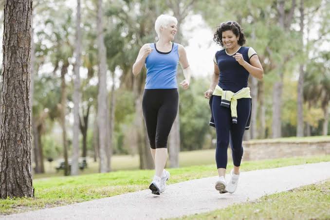
Walking supports circulation through mechanisms that operate simultaneously at every level of the vascular system — from the deep veins of the leg to the coronary arteries of the heart. Among all forms of physical activity, walking is uniquely suited to supporting vascular health: it is accessible to nearly all adults regardless of fitness level, can be performed without equipment or gym membership, has a very low injury rate compared to higher-impact exercise, and produces consistent, measurable improvements in circulatory function with as little as 30 minutes of moderate walking on most days of the week.
The circulatory benefits of walking extend from venous return (improved by the calf muscle pump with each step) through arterial function (improved endothelial nitric oxide production and arterial compliance) to the heart itself (improved cardiac output reserve and reduced resting heart rate with regular training). For patients with specific circulation problems — particularly peripheral artery disease and chronic venous insufficiency — walking is not merely beneficial but is one of the most evidence-based treatments available, producing improvements in symptom severity and walking capacity that rival or equal more expensive interventional therapies.
How Walking Supports Venous Circulation — The Calf Muscle Pump
The most immediate mechanism by which walking supports circulation is the calf muscle pump — the most powerful mechanism the body has for returning venous blood from the legs to the heart against gravity. Understanding how the calf muscle pump works clarifies why prolonged sitting is so harmful to venous circulation, and why even brief walking interruptions are so valuable during sedentary periods.
During walking, the gastrocnemius and soleus muscles of the calf contract rhythmically with each push-off phase of the gait cycle — generating pressures of 50 to 100 mmHg within the deep leg veins and soleal sinuses (the low-flow venous reservoirs within the soleus muscle that are the primary site of DVT formation). This muscular compression propels venous blood upward through the competent venous valves toward the heart, emptying the deep venous system with each step. The result: venous return from the legs during walking is 3 to 4 times higher than during standing still, and venous pressure in the ankle veins drops from approximately 90 mmHg (standing still) to 20 to 30 mmHg (walking) — a hemodynamically dramatic difference that explains why leg symptoms of venous insufficiency consistently improve with walking.
The clinical implications of the calf muscle pump are significant: venous stasis (the stagnation of blood in the deep leg veins during immobility) is the primary mechanism driving DVT formation during prolonged sitting, surgery, hospitalization, and long-haul air travel. Walking eliminates venous stasis. Even brief walking interruptions during prolonged sitting — 2 to 3 minutes of walking every 30 to 45 minutes — significantly reduce the venous stasis that accumulates during continuous sitting, lowering DVT risk and reducing the ankle swelling and leg heaviness that develop during extended desk work or travel.
How Walking Supports Arterial Circulation
Walking improves arterial circulation through both immediate (acute) and long-term (training) adaptations of the arterial system:
Immediate effects on arterial function: During walking, increased cardiac output raises blood flow velocity through all arteries. This increased shear stress on the arterial endothelium (the thin cell layer lining the inner surface of every artery) triggers endothelial nitric oxide synthase (eNOS) to produce nitric oxide — a potent vasodilator that relaxes arterial smooth muscle in arterioles throughout the body, reducing peripheral vascular resistance and further increasing blood flow to working muscles. The vasodilatory response to walking is immediate: peripheral blood flow to working leg muscles can increase 10 to 20-fold from resting values within the first minutes of moderate walking, reflecting the efficiency of exercise-induced vasodilation.
Long-term training adaptations: Regular walking training produces durable structural and functional improvements in arterial health. Endothelial function — as measured by flow-mediated dilation (FMD), the gold standard biomarker of endothelial NO production capacity — improves significantly after 12 weeks of regular moderate exercise training. Arterial stiffness (as measured by pulse wave velocity, a marker of atherosclerosis burden and cardiovascular event risk) decreases with regular aerobic exercise training. Resting heart rate decreases from parasympathetic dominance after training, and blood pressure decreases in hypertensive individuals by 5 to 8 mmHg systolic on average from regular moderate exercise — reducing the arterial pressure load on the heart and reducing stroke risk.
For patients with peripheral artery disease, walking produces an additional arterial benefit: collateral vessel development. In arteries narrowed by atherosclerotic plaque, the ischemic muscle downstream from the obstruction releases angiogenic growth factors (including vascular endothelial growth factor, VEGF) during exercise-induced ischemia. These signals promote the growth of new collateral arteries that bypass the obstruction — providing alternative routes for blood flow to the ischemic territory. This collateral development is one of the key mechanisms behind the dramatic walking distance improvements seen in PAD patients who complete supervised exercise therapy programs.
Walking for Peripheral Artery Disease — How It Improves Circulation
For patients with peripheral artery disease and claudication (calf pain with walking), supervised exercise therapy is a Class I recommendation in ACC/AHA guidelines — with evidence supporting it as equivalent to endovascular revascularization (angioplasty) for symptom improvement in patients with claudication without critical limb ischemia. The mechanisms by which supervised exercise therapy improves walking circulation in PAD are multiple:
Metabolic muscle adaptation: The ischemic calf muscles of PAD patients adapt to repeated exercise-induced ischemia by increasing mitochondrial density, improving the efficiency of ATP production from limited oxygen supply (improved mitochondrial coupling), and upregulating the enzymes of oxidative metabolism. These metabolic adaptations allow the muscle to extract more energy from the available (limited) blood supply — effectively increasing functional walking capacity even without increasing the actual blood flow to the muscle.
Collateral vessel formation: As described above, repeated bouts of exercise-induced ischemia stimulate collateral arteriogenesis — the growth and remodeling of pre-existing arterio-arterial connections into functional collateral vessels that provide alternative perfusion routes around the atherosclerotic obstruction. This is a structural change that persists after the exercise program ends, in contrast to the purely symptomatic benefits of medications.
Improved pain tolerance: The repeated experience of claudication pain during supervised exercise therapy raises the claudication pain threshold — patients learn to walk through mild-to-moderate pain, recognize that it is not dangerous, and develop the psychological confidence to push through the discomfort. This pain tolerance improvement contributes to the walking distance gains measured in clinical trials.
The standard supervised exercise therapy protocol for PAD claudication involves treadmill walking at a speed and grade that induces claudication within 3 to 5 minutes; walking until reaching a 3 to 4 on a 0 to 5 claudication intensity scale; resting until pain subsides; and repeating exercise-rest cycles for 30 to 45 minutes per session, 3 sessions per week, for a minimum of 12 weeks. Studies consistently show 100 to 150 percent improvement in pain-free walking distance and 150 to 200 percent improvement in maximum walking distance in patients completing the full program.

Walking for Venous Insufficiency and Swollen Ankles
For patients with chronic venous insufficiency and varicose veins, walking is both the most immediately effective and the most sustainable intervention for reducing venous stasis, leg edema, and the discomfort of venous poor circulation. The mechanism is the calf muscle pump — already described above — but the clinical evidence specifically supporting walking for venous insufficiency is worth highlighting:
Randomized controlled trials comparing supervised walking exercise programs against usual care in chronic venous insufficiency patients consistently demonstrate significant improvements in venous hemodynamics (measured by air plethysmography — reduction in venous filling index and improvement in venous pump function), reduction in ankle circumference and edema, improvement in CEAP clinical class (the standardized venous disease severity score), and improvement in disease-specific quality of life measures. The venous return improvement from exercise is immediately measurable: duplex ultrasound during treadmill walking versus standing still shows dramatically higher venous flow velocities and lower venous pressures during walking.
The practical walking recommendation for venous insufficiency is consistent moderate walking (30 minutes on most days), wearing compression stockings during the walking activity (compression enhances the calf pump by supporting the dilated superficial veins and directing pump pressure to the deep venous system rather than being lost through superficial vein dilation). High-impact running is not superior to walking for venous pump activation — the rhythmic, sustained gastrocnemius-soleus contraction during walking at moderate pace is biomechanically optimal for venous return, without the joint stress of running.
How Much Walking Is Needed to Support Circulation?
The evidence-based minimum for circulation benefits is the physical activity recommendation common to all major cardiovascular guidelines:
For general cardiovascular and circulation health: 150 minutes per week of moderate-intensity aerobic activity (which includes brisk walking — the pace at which you can speak in sentences but not sing) is the minimum target. This translates to 30 minutes of brisk walking on 5 days per week — achievable for most adults including those with mild-to-moderate cardiovascular disease. This 150-minute weekly target is associated with significant reductions in cardiovascular event risk (approximately 30 to 35 percent relative risk reduction for coronary events compared to sedentary adults), stroke risk, DVT risk, and all-cause mortality.
For venous circulation specifically: Breaking up prolonged sitting with 2 to 3 minutes of walking every 30 minutes is highly effective for preventing venous stasis — even at lower total walking volumes than the 150-minute cardiovascular target. This interruption pattern is specifically valuable for office workers, long-haul travelers, and hospitalized patients for whom full exercise programs are not feasible.
For PAD claudication: The supervised exercise therapy protocol (30 to 45 minutes, 3 times weekly, 12 weeks) requires more structured commitment — but the 100 to 150 percent walking distance improvement justifies the investment for claudication patients who can access a formal SET program.
See our related articles on poor circulation in the legs, peripheral artery disease symptoms, varicose veins vs poor circulation, swollen ankles and circulation, and stroke prevention for adults. The NHLBI physical activity and heart health guide, CDC physical activity guidelines for adults, and Society for Vascular Surgery PAD exercise resources provide additional evidence-based walking and exercise guidance.
- Gardner AW, Poehlman ET. Exercise Rehabilitation Programs for the Treatment of Claudication Pain. JAMA. 1995;274(12):975-980.
- Murphy TP, et al. Supervised Exercise Versus Primary Stenting for Claudication Resulting from Aortoiliac PAD (CLEVER Trial). Circulation. 2012;125(1):130-139.
- Gerhard-Herman MD, et al. 2016 AHA/ACC Guideline on Management of Lower Extremity PAD. J Am Coll Cardiol. 2017;69(11):e71-e126.
- Padberg FT, et al. Structured Exercise Improves Calf Muscle Pump Function in Chronic Venous Insufficiency. J Vasc Surg. 2004;39(1):79-87.
- Haskell WL, et al. Physical Activity and Public Health: Updated Recommendation. Med Sci Sports Exerc. 2007;39(8):1423-1434.
Walking and the Heart — Cardiac Circulation Benefits
The circulation benefits of walking extend beyond the peripheral vasculature to the heart itself. Regular walking training produces measurable improvements in cardiac function that translate directly into reduced cardiovascular event risk and improved quality of life — particularly important for the majority of patients with circulation problems who also carry significant cardiovascular disease burden:
Cardiac output adaptation: During walking, cardiac output (the volume of blood the heart pumps per minute) increases from a resting value of approximately 5 liters per minute to 10 to 15 liters per minute at moderate walking pace. This demand-driven increase in cardiac output is met through both increased heart rate and increased stroke volume (the volume ejected with each heartbeat). With regular training, resting stroke volume increases as the heart adapts to repeated exercise demands — meaning trained individuals can deliver the same cardiac output at a lower heart rate, reducing the myocardial oxygen demand at any given activity level. This resting bradycardia from aerobic training is one of the most reliable physiological markers of cardiovascular fitness and a strong negative predictor of cardiovascular event risk.
Coronary artery benefit: Regular moderate-intensity walking (as in the 150-minute-per-week guideline target) is associated with significant reductions in coronary artery disease event risk in observational epidemiological studies and supported by multiple large randomized trials of cardiac rehabilitation exercise programs in post-myocardial infarction patients. The mechanisms include improvements in coronary endothelial function, reduction in inflammatory markers (CRP, IL-6), modest LDL cholesterol lowering and HDL cholesterol raising, blood pressure reduction, and weight maintenance — each an independent cardiovascular risk modifier.
Heart failure and walking: Even in established heart failure with reduced ejection fraction (HFrEF), regular supervised exercise training (including walking-based programs) improves peak oxygen consumption (VO2 peak), exercise tolerance, and quality of life. The HF-ACTION trial demonstrated that supervised exercise training in heart failure reduced the composite of cardiovascular death and heart failure hospitalization by 11 percent — a clinically meaningful benefit from a non-pharmacological intervention. Walking-based exercise programs are now a Class I recommendation in heart failure management guidelines for patients who are clinically stable.
How Walking Reduces DVT and Clot Risk
Deep vein thrombosis forms when three factors — venous stasis, endothelial injury, and hypercoagulability (Virchow’s triad) — combine within the deep veins of the leg. Walking directly counters the venous stasis component of this triad, making it one of the most effective preventive measures for DVT in ambulatory patients:
Prolonged immobility — sitting for extended periods during desk work, car travel, or long-haul flights — allows venous blood to stagnate in the soleal sinuses and deep calf veins. This stasis reduces the local oxygen tension and concentrates activated clotting factors within the vein, creating conditions favorable for thrombus initiation. Every step taken during walking activates the calf muscle pump, clears the venous stasis, flushes activated clotting factors out of the deep venous system, and resets the clotting risk to baseline.
The DVT risk during long-haul air travel (flights over 4 hours) is well documented — approximately 1 in 200 passengers on very long-haul flights develop a symptomatic or asymptomatic DVT, with risk increasing in passengers with additional risk factors (prior DVT, thrombophilia, active cancer, recent surgery, obesity, oral contraceptive use). The standard prevention recommendation for air travel DVT — walking the aisle every 1 to 2 hours, performing calf raise exercises at the seat, wearing below-knee compression stockings, and maintaining adequate hydration — is entirely built around interrupting venous stasis, reflecting the primacy of the calf muscle pump in DVT prevention.
Hospital-acquired DVT is one of the most common preventable hospital complications, primarily affecting patients immobilized by illness, surgery, or sedation. Mobilization protocols — getting hospitalized patients out of bed and walking within hours of surgery or acute illness if medically safe — are among the most evidence-based DVT prevention strategies in hospital medicine, complementing pharmacological prophylaxis (anticoagulants) and mechanical prophylaxis (sequential compression devices). Early ambulation programs consistently reduce DVT rates in surgical and medical inpatients compared to bed rest.
Walking Pace, Distance, and Circulation — What the Research Shows
Not all walking provides equal circulatory benefit, and understanding the dose-response relationship between walking intensity and circulatory outcomes helps optimize the benefit for specific goals:
Pace matters for arterial health: Studies specifically comparing slow walking (below 2 mph) versus brisk walking (3 to 4 mph) versus fast walking (above 4 mph) for cardiovascular and arterial health outcomes consistently show greater benefit at higher walking speeds, independent of total walking time. A large UK Biobank study (Yates et al., 2021) found that self-reported brisk walkers had approximately 20 percent lower all-cause mortality compared to slow walkers, even after adjusting for total walking volume — suggesting that pace is an independent predictor of cardiovascular benefit beyond simply walking any amount.
The mechanism is that higher walking pace produces greater cardiovascular demand, higher shear stress on the arterial endothelium, more nitric oxide production, and greater improvement in endothelial function per unit time compared to slow strolling. For patients who can tolerate it without musculoskeletal complaints, increasing walking pace toward the brisk end of the range (3 to 4 mph, producing a moderate-intensity cardiovascular response) is the highest-value adjustment to maximize arterial circulation benefits.
Frequency over duration for venous and DVT benefit: For the specific goal of reducing venous stasis and DVT risk during desk work or travel, frequent short walking breaks (2 to 3 minutes every 30 to 45 minutes) are more effective than one longer walk at the end of the day — because venous stasis accumulates within 30 minutes of sitting, and reversing it requires only brief walking. The total step count at the end of the day is less important for DVT prevention than the pattern of movement throughout the day. A step counter showing 8,000 steps all taken in one hour provides less venous protection than the same 8,000 steps distributed across 8 short walking breaks during the work day.
Incline walking amplifies calf pump intensity: Walking uphill on an incline increases gastrocnemius and soleus activation compared to flat walking at the same pace — producing greater calf pump force and more efficient venous emptying per step. Treadmill incline walking at even 5 to 8 percent grade is used in supervised exercise therapy protocols for PAD partly because it provides a more efficient training stimulus (achieving claudication onset earlier with less absolute walking time), improving protocol efficiency for patients with limited walking tolerance.
Starting a Walking Routine for Circulation — Practical Guidance
For patients with poor circulation, peripheral artery disease, or chronic venous insufficiency, starting a walking routine requires addressing both the motivation and the practical barriers to consistent activity:
Starting point for deconditioned adults: Begin with the current comfortable walking duration — even if that is only 5 to 10 minutes — and add 2 to 5 minutes per week. The goal is gradual progression without exacerbating symptoms or causing injury. For PAD patients, this may mean very short initial walks (5 minutes) with rest breaks at home, progressing toward the supervised exercise therapy target of 30 to 45 minutes over several weeks before enrolling in a formal SET program.
Footwear and foot care: Patients with PAD, diabetic neuropathy, or venous insufficiency must wear properly fitted, supportive footwear with cushioned insoles to prevent pressure ulcers, blisters, and foot injuries that may not be perceived due to neuropathy. Inspect feet before and after every walk. A callus, blister, or shoe rub that goes unnoticed in a neuropathic foot can progress to a deep ulcer within days. Walking barefoot — even briefly — is contraindicated in diabetic patients with neuropathy.
Compression stockings during walking for venous disease: Patients with varicose veins, chronic venous insufficiency, or a history of DVT should wear below-knee graduated compression stockings (20 to 30 mmHg for most venous disease; 30 to 40 mmHg for more severe cases as directed by their vascular provider) during all walking activity. Compression during the calf pump cycle enhances venous return efficiency — the compressive stockings support the wall of the dilated superficial veins so that calf pump pressure is directed into the deep venous system rather than being lost through superficial vein dilation.
Walking programs and accountability: Step count targets combined with regular monitoring (wearable fitness tracker, pedometer app) significantly improve adherence to walking programs in cardiovascular and vascular disease populations. Supervised programs — whether formal cardiac rehabilitation, supervised exercise therapy for PAD, or community-based group walking programs — consistently produce better adherence and greater functional improvement than home-walking recommendations alone. Where access to supervised programs is limited, structured walking logs, regular follow-up with a healthcare provider or physiotherapist, and walking partners all improve adherence rates.
Bookmarked this article on how walking supports circulation and vascular immediately — going to reference it regularly. I have tried following advice from several sources but this is the most consistent with what my specialist told me. I wish I had found this article when I was first diagnosed — would have saved a lot of confusion.
This is one of the clearest explanations of how walking supports circulation and vascular I have found. The practical tips at the end made this immediately actionable, not just theoretical. Keep up this kind of thorough health journalism — it genuinely helps patients like me.
As someone dealing with this personally, the how walking supports circulation and vascular section was very helpful. The practical tips at the end made this immediately actionable, not just theoretical. Forwarding this to others in my support group who are dealing with similar issues.
I shared this article on how walking supports circulation and vascular with my doctor and they appreciated the level of detail. What I liked most was that the article didn’t just say what to avoid — it also gave alternatives. Thank you for making complex medical information accessible without dumbing it down.
My doctor recommended I look into how walking supports circulation and vascular and this article covered it perfectly. The specific numbers and thresholds mentioned are exactly what I needed to understand my results. I wish I had found this article earlier — would have saved a lot of confusion.
Really well-written article on how walking supports circulation and vascular. It is refreshing to see an article that acknowledges individual variation rather than one-size-fits-all advice. Shared this with three friends who are dealing with related issues. Very useful resource.