
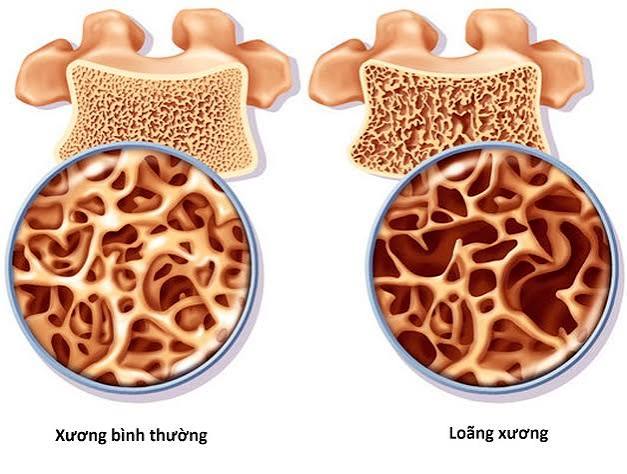
Understanding osteoporosis risk factors is the foundation of fracture prevention. Osteoporosis develops silently over years before a fracture reveals its presence, which means the only opportunity to intervene effectively lies in identifying who is at elevated risk — and why — before that fracture happens. Some risk factors are fixed: age, sex, and genetics cannot be changed. But a significant number are modifiable: smoking, alcohol intake, diet, physical activity, and management of medical conditions that accelerate bone loss can all be addressed. Knowing which risk factors you carry, and which of them can be reduced, is the starting point for a personalised bone health strategy.
How Risk Factors Are Used in Clinical Practice
Osteoporosis risk factors do not operate in isolation — their combined effect on fracture probability is captured by the FRAX tool, a web-based risk calculator developed by the University of Sheffield for the World Health Organization. FRAX takes age, sex, BMI, and a set of clinical risk factors as inputs and outputs the 10-year probability of a major osteoporotic fracture (hip, spine, wrist, or humerus) and hip fracture specifically. It can be used with or without a bone mineral density (BMD) measurement from a DEXA scan, and the result guides whether treatment is warranted.
The clinical importance of risk factor assessment is twofold: first, it identifies who needs a DEXA scan; second, it ensures that the scan result is not interpreted in isolation from the clinical context. A T-score of −2.0 (osteopenia) carries very different fracture probability in a 55-year-old woman with no other risk factors compared with a 68-year-old woman who has had a prior wrist fracture, is on glucocorticoids, and has a family history of hip fracture. FRAX integrates all of these factors into a single probability estimate. Detail on DEXA interpretation is in our guide to bone density: what it means.
Non-Modifiable Risk Factors
Age is the most powerful single predictor of osteoporosis after bone mineral density itself. Bone mineral density declines progressively after peak bone mass is reached in the late twenties, and fracture risk increases steeply with each decade. Prevalence of osteoporosis (T-score ≤ −2.5) rises from approximately 2% in women aged 50–54 to over 25% in women over 80. In men, the trajectory is slower but equally real. The age effect is partly independent of BMD — bone microarchitectural quality (collagen cross-linking, microdamage accumulation) deteriorates with age even when BMD is preserved, making bone more brittle at any given density. FRAX uses actual age as a continuous variable, not age groups, so risk increases incrementally with each additional year. In clinical practice this means that the same T-score at age 65 and age 75 represents a different absolute fracture probability — the older patient is at meaningfully higher risk even with identical bone density.
Female sex approximately doubles lifetime fracture risk compared with men of the same age. This is driven primarily by the accelerated bone loss at menopause — up to 3% per year in the first five to ten years after menopause — which does not have a male equivalent. Women also have smaller bone cross-sections than men, meaning their bones have a lower tolerance for force at any given density. Approximately one in two women over 50 will sustain a fragility fracture in their remaining life.
Menopause is the single most important hormonal event for female bone health. Oestrogen suppresses osteoclast activity; its loss at menopause removes this suppression and dramatically accelerates resorption. Early menopause — before the age of 45 — extends the period of oestrogen-deficient bone loss and significantly increases lifetime fracture risk. Surgical menopause (bilateral oophorectomy) carries the same risk if oestrogen is not replaced. Women who experience early menopause and do not receive hormone replacement therapy have substantially higher osteoporosis rates at any given age than women who reach natural menopause at 50–52.
Family history of hip fracture in a first-degree relative (parent or sibling) is an independent risk factor captured by FRAX. It reflects both shared genetic determinants of peak bone mass and shared lifestyle and dietary patterns. The mechanism is not fully understood, but the risk is robust across multiple large studies. A parental hip fracture approximately doubles the 10-year fracture probability in the offspring, independently of their own BMD.
Previous fragility fracture is the strongest predictor of a subsequent fracture — stronger than any other single risk factor except BMD itself. A fragility fracture (a fracture from a fall from standing height or less) approximately doubles the risk of the next fracture, and multiple prior fractures multiply risk further. This is the rationale for the UK fracture liaison service model, which systematically assesses and treats patients after any fragility fracture.
Modifiable Risk Factors
Smoking is consistently associated with lower bone density and higher fracture risk across all age groups. The mechanisms include direct impairment of osteoblast function by tobacco constituents, earlier menopause in women who smoke, and reduced calcium absorption. The effect is dose-dependent — heavier smokers have lower BMD than lighter smokers. Encouragingly, stopping smoking is associated with partial recovery of bone loss; former smokers have higher BMD than current smokers matched for age and duration of prior smoking. FRAX classifies current smoking as a clinical risk factor.
Alcohol intake above approximately 14 units per week is associated with increased bone loss, lower BMD, and higher fracture risk. Alcohol directly inhibits osteoblast function, impairs calcium and vitamin D metabolism, increases the risk of falls, and is associated with poor nutritional status including low calcium intake. At higher intake levels (≥3 units/day) the effect is incorporated into FRAX as a dichotomous risk factor. Reducing alcohol to within recommended limits (no more than 14 units per week, spread across multiple days) is a relevant intervention for bone health, particularly in patients who are already in the osteopenia or osteoporosis range.
Low body weight and low BMI (below 19 kg/m²) are directly incorporated into FRAX as a continuous variable — lower BMI predicts higher fracture risk independently of BMD. The mechanism involves reduced mechanical loading on the skeleton (less body weight pressing down on bones reduces the osteogenic stimulus), reduced adipose-derived oestrogen in postmenopausal women, and nutritional compromise associated with very low body weight. People with a BMI below 19 are at substantially higher risk than those with a normal or higher BMI.
Physical inactivity is one of the most impactful modifiable risk factors. Weight-bearing and resistance exercise are the primary anabolic stimuli for bone formation — mechanical loading through bone activates osteoblasts. Sedentary adults lose bone more rapidly than active adults at every age. Conversely, regular weight-bearing aerobic exercise (walking, jogging, dancing, tennis) and resistance training (weights, resistance bands) maintain and in younger adults improve BMD. Physical activity also maintains muscle mass and balance, reducing fall risk independently of bone density. More on exercise for bone health is in our guide to osteoporosis: symptoms, causes, and prevention.
Nutritional Risk Factors
Low calcium intake is a directly modifiable risk factor. Calcium is the primary mineral component of bone hydroxyapatite, and chronic inadequate intake increases parathyroid hormone secretion, which drives osteoclast activity to mobilise calcium from the skeleton. The UK recommended daily intake for adults is 700 mg/day, rising to 1,000–1,200 mg/day for postmenopausal women not on HRT and older adults. Most UK adults with osteoporosis or osteopenia benefit from ensuring dietary calcium is adequate, and supplemental calcium is co-prescribed with all pharmacological osteoporosis treatments. Full guidance on calcium sources and supplementation is in our guide to calcium and bone health.
Vitamin D deficiency impairs intestinal calcium absorption — without adequate vitamin D (serum 25-hydroxyvitamin D above 50 nmol/L), dietary calcium cannot be efficiently absorbed regardless of intake. Vitamin D deficiency is extremely common in the UK, particularly between October and March when ultraviolet B light is insufficient to drive cutaneous vitamin D synthesis. Adults who are housebound, have darker skin, cover themselves fully for cultural or religious reasons, or are over 65 are at particularly high risk of deficiency. Supplementation with 400–800 IU/day is recommended for most UK adults. Full detail is in our guide to vitamin D and bone health.
Medical and Medication Risk Factors
Glucocorticoids (oral steroids) are the most important secondary cause of osteoporosis and the medication risk factor most reliably included in FRAX. Prednisolone at 5 mg/day or more for three months or longer directly inhibits osteoblast function and increases osteoclast activity, producing rapid and substantial bone loss — most pronounced in the first 6–12 months of treatment. Any patient starting long-term glucocorticoids should have fracture risk assessed and bone protection with bisphosphonate co-prescribed if the FRAX probability is above the intervention threshold. Higher doses (≥7.5 mg/day) carry higher risk and may warrant treatment initiation without waiting for DEXA. See our guide to bone density test: a simple guide for when and how bone density testing is indicated.
Other medications associated with increased fracture risk include:
- Aromatase inhibitors (anastrozole, letrozole, exemestane) — used for hormone receptor-positive breast cancer; produce severe oestrogen deficiency with 2–3% per year bone loss; DEXA monitoring and bisphosphonate co-prescription recommended
- Androgen deprivation therapy (ADT for prostate cancer) — testosterone suppression removes oestrogen-precursor production; significant bone loss in men on ADT
- Proton pump inhibitors (PPIs) — reduce gastric acid and may impair calcium carbonate absorption; evidence links long-term use with modestly elevated fracture risk
- Anticonvulsants — phenytoin and other enzyme-inducing anticonvulsants accelerate vitamin D catabolism
- Antidepressants (SSRIs) — observational data suggest an association with bone loss; possible mechanism involves serotonin signalling in osteoblasts
Medical conditions associated with secondary osteoporosis include rheumatoid arthritis, coeliac disease, inflammatory bowel disease, hyperparathyroidism, hyperthyroidism, type 1 diabetes, hypogonadism, anorexia nervosa, and chronic kidney disease. Any newly diagnosed osteoporosis should include investigation for secondary causes — particularly when the Z-score is below −2.0, indicating bone density significantly lower than expected for age.
Falls as a Risk Factor
A fracture requires both fragile bone and a fall. While bone density determines whether a fall results in a fracture, the frequency of falls determines how often that threshold is tested. Falls are therefore an independent and clinically important risk factor for fragility fractures — particularly hip fractures, where the vast majority occur as the direct result of a fall onto the greater trochanter.
Falls risk is elevated by: muscle weakness (particularly quadriceps and hip abductors), poor balance, impaired vision, use of sedative or antihypertensive medications that cause orthostatic hypotension, home hazards (loose rugs, poor lighting, lack of grab rails), and inappropriate footwear. Multifactorial falls assessment — addressing each of these factors — reduces fall rates in high-risk older adults. Balance training (tai chi, single-leg standing, proprioceptive exercises) and progressive muscle strengthening reduce falls by approximately 30% in cohort studies. For patients with osteoporosis, fall prevention is as important as pharmacological treatment — a fall that doesn’t happen cannot cause a hip fracture regardless of T-score.
The interaction between falls risk and bone density is multiplicative rather than additive. A person with osteoporosis who rarely falls has a different risk profile from a person with the same T-score who falls several times a year. Hip protectors — padded garments designed to absorb and redirect the force of a fall — reduce hip fracture risk in care home residents who fall onto them consistently, though compliance in community-dwelling adults is often poor. For older adults in particular, a comprehensive falls prevention programme — including medication review, home hazard assessment, strength and balance training, and vision correction — is a clinically recognised and evidence-based component of fracture prevention strategy alongside osteoporosis treatment.
Frequently Asked Questions
Which risk factor for osteoporosis is the most important?
A previous fragility fracture is the strongest individual predictor of future fracture beyond age and bone mineral density. A person who has already sustained a fragility fracture has demonstrated that their fracture threshold has been crossed — their bone strength relative to the forces they encounter is insufficient. Any prior fragility fracture approximately doubles future fracture risk independent of BMD, and multiple prior fractures multiply risk multiplicatively. This is why post-fracture assessment and treatment is such a clinical priority — the fracture liaison service model was specifically designed to ensure that every fragility fracture triggers systematic osteoporosis evaluation and, where indicated, pharmacological treatment.
Can I reduce my osteoporosis risk if I have non-modifiable risk factors?
Yes, substantially. Having non-modifiable risk factors — age, female sex, family history — means your baseline risk is higher, but it does not mean the outcome is fixed. Modifiable risk factors operate independently and additively alongside non-modifiable ones, so addressing them reduces overall fracture probability even in the presence of risks you cannot change. A 60-year-old woman with a strong family history who exercises regularly, maintains adequate calcium and vitamin D intake, does not smoke, and has a healthy BMI has a significantly lower fracture risk than one with the same family history who smokes, is sedentary, and has deficient calcium intake. The value of risk factor modification is greatest, not least, in people who already have elevated baseline risk.
Does taking steroids always cause osteoporosis?
Not necessarily, but long-term glucocorticoid use at therapeutic doses significantly increases the risk, and the risk is dose- and duration-dependent. Short courses of oral steroids (a five to ten day course for an acute illness, for example) are not associated with clinically meaningful bone loss. The risk becomes significant with prednisolone at 5 mg/day or more sustained for three months or longer — the dose and duration range used for inflammatory conditions such as rheumatoid arthritis, inflammatory bowel disease, and chronic obstructive pulmonary disease. In this context, bone protection should be discussed with the prescribing clinician and typically involves co-prescription of a bisphosphonate alongside calcium and vitamin D supplementation. The risk is not inevitable, but it requires proactive management.
Does being overweight protect against osteoporosis?
Higher body weight is associated with higher bone density — mechanical loading from greater body weight stimulates bone formation, and adipose tissue produces oestrogen that provides some bone protection in postmenopausal women. In this narrow sense, a higher BMI is associated with reduced osteoporosis risk. However, obesity is associated with increased fall risk (impaired balance, reduced muscle quality relative to body mass), and in people with very high BMI, standard DEXA measurements can be inaccurate. Obesity is not a clinical strategy for bone protection, and weight management should aim for a healthy BMI range rather than using high BMI as a bone health protection tool. Very low BMI (below 19) is the more concerning end of the weight spectrum for bone health.
How does family history affect my fracture risk?
A parental history of hip fracture approximately doubles the 10-year fracture probability in offspring, independently of their own bone mineral density. This effect is captured in the FRAX calculator, which includes parental hip fracture as a binary risk factor. The mechanism involves shared genetic determinants of peak bone mass, bone microarchitecture quality, and possibly shared falls risk through body habitus and neuromuscular characteristics. Importantly, knowing you have a family history of hip fracture is a reason to discuss bone health with your GP — particularly if you are a woman over 50 or a man over 60 — and to consider earlier or more proactive DEXA assessment. It does not mean fracture is inevitable, but it shifts the risk calculation in a way that justifies earlier preventive action.
Is rheumatoid arthritis a risk factor for osteoporosis?
Yes — rheumatoid arthritis (RA) is one of the most significant secondary risk factors for osteoporosis, and it is directly included as a binary clinical risk factor in FRAX. The mechanism is multifactorial: the chronic inflammatory state in RA (elevated TNF-α and IL-6) drives osteoclast activity systemically; glucocorticoids commonly used to manage RA flares directly suppress osteoblasts; pain and disability reduce physical activity and mechanical loading on bone; and systemic inflammation impairs appetite and nutritional status. People with RA have approximately twice the fracture risk of age-matched controls. Regular bone density monitoring with DEXA, appropriate calcium and vitamin D supplementation, and bisphosphonate prescription when fracture risk is above the threshold are all standard elements of RA management in the UK.
At what age should I start thinking about osteoporosis risk factors?
Bone health begins at birth and is actively shaped throughout childhood and adolescence — peak bone mass, achieved by age 25–30, is the foundation on which all subsequent bone health rests, and it is partly determined by calcium and vitamin D intake, exercise, and hormonal factors during growth. That said, the window of greatest clinical relevance for risk factor identification and management in adults is from around age 40–45, when gradual bone loss has been proceeding for over a decade and is approaching the range where accelerated loss at menopause will bring some individuals close to the osteoporosis threshold. DEXA screening is most relevant from age 50 onward for those with risk factors, and from 65 (women) and 70 (men) routinely. But lifestyle risk factors — smoking, alcohol, physical inactivity, poor calcium and vitamin D intake — are worth addressing at any adult age as part of general health rather than waiting for a bone health diagnosis.
Summary
Osteoporosis risk factors divide into non-modifiable (age, female sex, menopause, family history of hip fracture, prior fragility fracture) and modifiable (smoking, excess alcohol, low calcium and vitamin D intake, physical inactivity, low BMI, glucocorticoid and other bone-depleting medications, and secondary medical conditions). The FRAX tool combines these factors — with or without DEXA BMD — to estimate 10-year fracture probability and guide treatment decisions. Falls risk is an independent and clinically important factor: preventing falls reduces fracture risk as directly as improving bone density. The highest-priority interventions are addressing modifiable risk factors proactively, ensuring calcium and vitamin D adequacy, maintaining regular weight-bearing and resistance exercise, discussing bone protection with a clinician if long-term glucocorticoids or other bone-depleting medications are required, and ensuring falls prevention measures are in place for older adults at elevated fall risk. No single risk factor operates alone — FRAX’s strength is in integrating the full picture into a clinically actionable probability. Acting on that probability early, before a fragility fracture occurs, is the goal of modern osteoporosis risk management. Related guides cover osteoporosis: symptoms, causes, and prevention, osteopenia, and DEXA scanning.
Medical disclaimer: This article is for general educational purposes and does not constitute medical advice. Consult a qualified healthcare professional for personalised fracture risk assessment and management.
References:
NICE CG146. Osteoporosis: assessing the risk of fragility fracture. NICE. 2023.
WHO FRAX. Fracture Risk Assessment Tool. University of Sheffield.
Royal Osteoporosis Society. Risk factors for osteoporosis and fracture. theros.org.uk. 2023.
Kanis JA, et al. FRAX and the assessment of fracture probability in men and women from the UK. Osteoporos Int. 2008;19(4):385–397.
NHS. Osteoporosis. nhs.uk. 2023.
I’ve been on prednisolone for rheumatoid arthritis for four years — 7.5 mg per day — and was started on alendronate from the beginning of steroid treatment after my rheumatologist discussed fracture risk with me. I didn’t really understand at the time why the bone protection was so important alongside the steroid that was managing my RA. This article explains the mechanism clearly: the direct suppression of osteoblasts by glucocorticoids, combined with the RA-related inflammation already driving osteoclast activity, creates a compounding effect on bone loss. I’ve had two DEXA scans since starting steroids — at one year and three years. The first showed osteopenia (−1.8), the second was stable at −1.9, which my rheumatologist said was a good outcome given that untreated, someone on long-term steroids would typically show considerably more rapid decline.
Gillian, your treatment trajectory reflects the correct application of glucocorticoid-induced osteoporosis (GIOP) prevention guidelines. The co-prescription of alendronate from the start of steroid treatment at 7.5 mg/day is aligned with NICE guidance, and your stable T-score at three years (−1.9 versus −1.8 at one year) on combined alendronate and steroid therapy represents a good outcome — the bisphosphonate is effectively opposing the osteoclast-activating effect of the prednisolone. Without bone protection, the expected bone loss at this dose over four years would typically be 5–10% at the lumbar spine, which from a starting point in the osteopenia range would place you in the osteoporosis range. Your case illustrates exactly why automatic bone protection co-prescription at steroid initiation matters. Peter, your management is a model of comprehensive post-fracture care. The fracture liaison service approach — FRAX, DEXA, falls assessment, medication review, physiotherapy referral — addresses both of the determinants of fragility fracture: bone fragility (managed with zoledronate) and falls exposure (managed through medication adjustment and balance training). The orthostatic hypotension from the antihypertensive is a common and modifiable falls contributor that is frequently overlooked in bone health management despite its clinical significance. Your outcome — only one minor fall in two years — is exactly what a well-executed multifactorial intervention should produce.
I’m a 72-year-old man who had a hip fracture two years ago from a fall at home. My GP referred me to a fracture liaison service who assessed my full risk profile — FRAX score, DEXA scan, falls history, medication review. My FRAX score was very high partly because of the prior hip fracture and partly because I’d been on a low-dose steroid for an inflammatory condition for several years. I’m now on zoledronate annual infusion and calcium and vitamin D, and I had a comprehensive falls assessment that identified poor balance and a blood pressure medication that was causing orthostatic dizziness. The medication was adjusted, I was referred to a physiotherapy balance programme, and I’ve only had one minor fall in two years since. The article’s point about falls risk being as important as bone density in fracture prevention reflects exactly the approach the fracture liaison team took with me.