
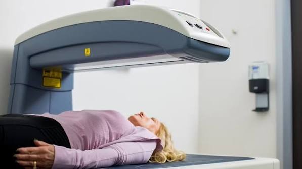
A DEXA scan is the gold standard investigation for measuring bone density and diagnosing osteoporosis. It is a quick, painless, and low-radiation imaging procedure that gives clinicians the bone mineral density measurements they need to calculate fracture risk and guide treatment decisions. Yet despite being one of the most important screening tools in adult preventive health, many people referred for a DEXA scan have limited information about what the procedure involves, what the results mean, and what happens afterward. This guide covers all of it — from the technology and the preparation to interpreting your results and understanding their implications.
What Is a DEXA Scan?
DEXA stands for dual-energy X-ray absorptiometry. It is an imaging technique that uses two X-ray beams at different energy levels — rather than the single energy level used in conventional X-ray — to precisely separate the density of bone from the surrounding soft tissue (muscle and fat). By comparing how much of each energy level is absorbed by bone versus soft tissue, the machine calculates the bone mineral density (BMD) of the area being scanned, expressed as grams per square centimetre (g/cm²).
The DEXA scan is performed at the two sites most relevant for osteoporosis diagnosis and fracture risk:
- The lumbar spine (L1–L4): measures the density of the four lowest lumbar vertebrae, which are predominantly trabecular (spongy) bone — the type most affected by postmenopausal bone loss and most sensitive to early density changes
- The hip: measured at both the femoral neck (the narrowest part of the femur, where hip fractures occur) and the total hip; more cortical bone predominates, making this site more relevant for hip fracture risk specifically and for older patients where spinal measurements may be confounded by arthritis
The lowest T-score across all measured sites drives the diagnosis. DEXA can also be performed at the forearm (one-third radius site) when the hip and spine cannot be measured — for example in patients with bilateral hip replacements or very obese patients who exceed the table weight limit.
How Does DEXA Differ From Other Scans?

DEXA is distinct from other imaging investigations in several important ways:
MRI uses magnetic fields and has no radiation; it shows soft tissue in excellent detail but cannot reliably quantify bone mineral density. MRI is used for spinal fracture assessment and bone marrow disease, not for routine BMD measurement.
CT uses X-rays and can measure volumetric BMD (mg/cm³) in three dimensions — more precise than DEXA’s areal measurement, and able to separate trabecular from cortical bone. However, CT delivers substantially more radiation and is used primarily in research rather than routine clinical practice.
Plain X-rays are insensitive for bone density — approximately 30% of bone mass must be lost before reduced density is visible on a standard X-ray. X-rays are used to identify fractures, not to diagnose osteoporosis or measure BMD.
A nuclear medicine bone scan (scintigraphy) uses a radioactive tracer to show areas of increased bone activity — used for detecting bone metastases, occult fractures, and infection. It does not measure BMD and is not used for osteoporosis diagnosis.
Preparing for a DEXA Scan
DEXA requires minimal preparation. Unlike many imaging investigations, there is no fasting, no injection, and no special diet. The key preparation points are:
- Calcium supplements: do not take calcium supplements for 24 hours before the scan. Residual calcium from supplements in the digestive tract can interfere with the lumbar spine measurement. Regular dietary calcium (from food) and all other medications are taken as normal.
- Clothing: wear comfortable clothing with no metal zips, buckles, or underwired bras in the area of the hip or spine. Metal objects in the scan field cause artefacts and may require repositioning. You will usually be asked to remove belts and any relevant jewellery.
- Recent contrast studies: inform the radiographer if you have had a barium X-ray examination, CT with contrast, or nuclear medicine scan within the preceding week, as residual contrast agents in the bowel can affect the lumbar spine measurement.
- Pregnancy: DEXA is not performed in pregnancy. Inform the radiographer if there is any possibility of pregnancy.
- Hip or spinal hardware: inform the radiographer if you have a hip prosthesis, spinal rods, or other metalwork — the affected site will be excluded from the scan and the forearm site used instead if needed.
What Happens During the Scan?
The DEXA scanner consists of a large flat table with a movable arm above it. There is no enclosed tunnel — the machine is entirely open on all sides. The procedure proceeds in two parts:
Hip measurement: you lie on your back with your legs flat. A triangular wedge is placed between your feet to internally rotate the hips slightly — this standardises the position of the femoral neck for measurement. You remain still while the scanner arm moves slowly over the hip area. This takes approximately three to five minutes per hip (usually one hip is measured, or both if the dominant hip has hardware).
Lumbar spine measurement: the wedge under your feet is removed and replaced with a box that raises your knees — this flattens the lumbar spine against the table, standardising the L1–L4 measurement position. The arm scans from L1 to L4, which takes a further three to five minutes.
The total procedure time is typically 10–15 minutes. You will be asked to breathe normally and remain still during scanning. There is no noise beyond the movement of the scanner arm. No injections, cannulas, or dyes are used. The radiation dose is 1–10 microsieverts (μSv) per scan — to put this in context, the average annual background radiation exposure in the UK is approximately 2,700 μSv, and a transatlantic flight delivers approximately 100 μSv. The dose from DEXA is negligible in any clinical context. More on what the test measures is in our guide to bone density test: a simple guide.
Reading Your DEXA Report
The DEXA report will include several elements:
BMD values (g/cm²): the absolute bone mineral density measurements at each site — femoral neck, total hip, and L1–L4. These values are used for serial comparisons on the same machine; they are not directly comparable between different DEXA machines or manufacturers.
T-scores: the BMD at each site expressed as the number of standard deviations above or below the mean for a young adult reference population. The WHO classification is:
- Normal: T-score ≥ −1.0
- Osteopenia: T-score −1.0 to −2.5
- Osteoporosis: T-score ≤ −2.5
Z-scores: the BMD expressed as standard deviations above or below the mean for an age-matched and sex-matched reference group. Used in premenopausal women and men under 50; a Z-score ≤ −2.0 is clinically significant and should prompt investigation for a secondary cause of low bone density. More on T-scores and Z-scores in context is in our guide to bone density: what it means.
FRAX calculation: some DEXA reports include a FRAX 10-year fracture probability estimate, calculated using the BMD result alongside standard clinical risk factors. This gives the most clinically actionable output — the probability of a major osteoporotic fracture (hip, spine, wrist, or humerus) in the next 10 years. Where FRAX is not included in the report, the referring clinician will calculate it separately.
Vertebral Fracture Assessment (VFA)
Many DEXA machines can also perform vertebral fracture assessment (VFA) — a lateral spine scan that identifies vertebral compression fractures. This is particularly valuable because many vertebral fractures are clinically silent (they occur without a specific injury and may cause only mild back discomfort) and would otherwise be missed. A VFA adds only a few minutes to the standard DEXA procedure and a minimal additional radiation dose.
VFA is recommended by the Royal Osteoporosis Society for any DEXA scan in a patient with height loss of more than 4 cm, back pain, or a prior fragility fracture. Finding a vertebral compression fracture upgrades the fracture risk classification and typically indicates pharmacological treatment is warranted even in patients whose T-score alone would be in the osteopenia range. The presence of a vertebral fracture, by definition, upgrades the WHO classification to “severe osteoporosis” regardless of T-score.
Who Refers and Reports the DEXA Scan?
In the NHS, DEXA scans are requested by GPs and secondary care clinicians. Most referrals come from GPs following a risk assessment using the FRAX tool or clinical criteria from NICE CG146. Secondary care referrals come from orthopaedic surgeons (after a fragility fracture), rheumatologists (for patients on glucocorticoids or with inflammatory conditions), endocrinologists (for secondary metabolic bone disease), and oncologists (for patients on aromatase inhibitors or androgen deprivation therapy).
The scan is performed by a radiographer or DEXA technologist trained in bone densitometry. Reporting is done by a radiologist or bone densitometry specialist. In many NHS trusts, the DEXA result is reviewed through a fracture liaison service (FLS) — a specialist multidisciplinary team (typically a fracture liaison nurse specialist and a consultant) that systematically identifies patients after fragility fracture, arranges bone density testing, and initiates or recommends treatment. The FLS model has been shown to significantly improve the rate of osteoporosis treatment after fracture and reduce re-fracture rates. If you have had a fragility fracture and have not been contacted by a fracture liaison service, asking your GP or hospital whether your trust operates one is worthwhile.
In primary care, the GP or a practice nurse with bone health training reviews the DEXA report alongside the clinical context and FRAX result. Some practices operate a dedicated osteoporosis review clinic. If the result indicates treatment, the GP will typically initiate bisphosphonate therapy with calcium and vitamin D co-prescription, with referral to secondary care for complex cases — prior fracture on treatment, very low T-scores, secondary causes requiring specialist investigation, or consideration of teriparatide.
DEXA Limitations and Confounding Factors
Understanding the limitations of DEXA helps interpret equivocal or unexpected results:
- Lumbar osteoarthritis: osteophytes (bony outgrowths) from lumbar degenerative arthritis increase the apparent areal BMD of the lumbar spine on DEXA, producing a falsely elevated lumbar T-score. This is very common in adults over 65 and is a reason to give more weight to the hip result in older patients, particularly when there is discordance between sites.
- Aortic calcification: calcification of the abdominal aorta, common in older adults, can increase the apparent lumbar spine BMD — another reason to favour the hip result in elderly patients.
- Body size: DEXA measures areal (two-dimensional) BMD rather than true volumetric density. Taller individuals with larger vertebrae may have higher areal BMD than their true density warrants; shorter individuals may have lower areal BMD. This can slightly over- or underestimate fracture risk in patients at the extremes of body size.
- Machine variation: BMD values in g/cm² and T-scores are not directly comparable between DEXA machines from different manufacturers or even between machines of the same model. Serial monitoring must be performed on the same machine. If you change hospitals, ask for the actual BMD values in g/cm² from your previous scan so the new site can calculate the T-score change from a standardised baseline.
After Your DEXA Scan
The scan images and measurements will be reported by a radiologist or trained DEXA operator, with the written report sent to your referring clinician. Results are not usually given on the day of scanning. The clinical pathway following the result depends on the T-score and FRAX probability:
- Normal BMD and low FRAX risk: reassurance, lifestyle advice (calcium, vitamin D, weight-bearing exercise), repeat scan in five to ten years if risk factors change
- Osteopenia with low FRAX risk: lifestyle optimisation, consider calcium and vitamin D supplementation, repeat DEXA in two to three years — see our guide to osteopenia: what adults should know
- Osteoporosis or high FRAX risk: pharmacological treatment (bisphosphonate first-line), calcium and vitamin D co-prescription, monitoring DEXA at three years — see our guide to osteoporosis: symptoms, causes, and prevention
Frequently Asked Questions
Is a DEXA scan safe?
Yes. A DEXA scan is extremely safe. The radiation dose is 1–10 microsieverts per scan — a fraction of the dose from a standard chest X-ray (approximately 10 μSv) and negligible compared with the annual background radiation we are all exposed to from the environment (approximately 2,700 μSv per year in the UK). There is no injection, no contrast dye, and no enclosed space. The procedure is entirely non-invasive. The only absolute contraindication is pregnancy. For any adult who meets the clinical criteria for bone density assessment, the benefits of identifying fracture risk early far outweigh any risk from the minimal radiation involved.
How long does a DEXA scan take?
The scanning procedure itself takes approximately 10–15 minutes from the point of lying on the table to completion. This includes measurement at both the hip and lumbar spine. If vertebral fracture assessment (VFA) is added — a lateral spine scan to check for compression fractures — the total time increases by approximately five minutes. Allowing for arrival, positioning, and any necessary explanation from the radiographer, a typical appointment is 20–30 minutes in total. The waiting time for results will vary between departments and trusts.
Can I have a DEXA scan if I have a hip replacement?
Yes, but the replaced hip cannot be measured — the metalwork from a hip prosthesis would produce artefacts that make the result uninterpretable. The protocol in this situation is to measure the opposite (non-replaced) hip, the lumbar spine (as long as there is no significant spinal hardware), and if needed the one-third radius site of the non-dominant forearm. Patients with bilateral hip replacements are typically measured at the lumbar spine and forearm. The clinical interpretation proceeds with the available measurement sites, and the result is still clinically useful even without the replaced hip’s measurement.
What does a low DEXA score mean for me?
A low DEXA T-score — in the osteopenia or osteoporosis range — means your bone mineral density is below the normal range for a young adult reference population, which increases your risk of fracture from relatively minor forces. What this means practically depends on your overall fracture risk, calculated with the FRAX tool: some people with osteoporosis T-scores have a relatively low 10-year fracture probability when other risk factors are favourable, while others with osteopenia T-scores have a high probability when age, prior fractures, and other risk factors are added in. A low T-score alone does not inevitably mean you need medication — it means your fracture risk needs formal assessment, and management (lifestyle, supplementation, or medication) will be tailored to that overall risk.
Why are T-scores different at the spine and hip?
Discordant T-scores between the spine and hip are very common and expected. The spine (predominantly trabecular bone) and hip (more cortical bone) lose density at different rates and are affected differently by various conditions. Postmenopausal bone loss preferentially affects trabecular bone — making spine T-scores typically lower than hip T-scores in women in the early years after menopause. In older adults, lumbar T-scores may be falsely elevated by osteoarthritis-related osteophytes. Age-related cortical bone loss affects the hip proportionally more in later decades. A T-score discordance of one to two standard deviations between sites is clinically normal; the lower of the two scores drives the diagnosis, and the pattern of discordance gives additional diagnostic information about the likely underlying mechanism.
What is a vertebral fracture assessment and do I need one?
Vertebral fracture assessment (VFA) is an additional lateral spine scan performed on the same DEXA machine immediately after the standard BMD measurement. It uses the DEXA imaging to look for vertebral compression fractures — fractures in which the body of a vertebra has collapsed partially under load. Many vertebral compression fractures are clinically silent (no recognised injury event, only mild back discomfort) and would otherwise be found only incidentally on X-ray or CT. The Royal Osteoporosis Society recommends VFA for patients who have lost more than 4 cm in height, have back pain that may be vertebral in origin, or have had a prior fragility fracture. A confirmed vertebral fracture on VFA upgrades clinical management — it typically indicates pharmacological treatment is warranted even in patients with osteopenia T-scores who might not have qualified for medication on BMD alone.
How accurate is a DEXA scan?
DEXA is highly accurate and reproducible for measuring bone mineral density. Its measurement precision error (the variation seen when the same patient is scanned twice without moving) is approximately 1–2% for the lumbar spine and hip. This means that changes in BMD between sequential scans are only clinically meaningful when they exceed approximately 3% — smaller changes fall within the test’s precision range and may reflect positioning variation rather than true BMD change. DEXA’s main limitation is that it measures areal (two-dimensional) rather than volumetric bone density, which means bone size affects the result. Large bones and osteophytes from arthritis can produce higher apparent BMD than the true bone quality warrants. Overall, DEXA remains the most clinically validated and widely used tool for fracture risk assessment and monitoring treatment response.
Summary
A DEXA scan — dual-energy X-ray absorptiometry — is the gold standard for measuring bone mineral density and diagnosing osteoporosis and osteopenia. It is safe, quick, painless, and requires minimal preparation — no fasting, no injection, and no enclosed space. The scan measures BMD at the hip and lumbar spine, producing T-scores and Z-scores that, combined with the FRAX clinical risk calculator, give an accurate estimate of 10-year fracture probability. Preparation requires only avoiding calcium supplements for 24 hours before the scan. Limitations include false elevation of lumbar T-scores by osteoarthritis and the inability to compare results between different machines. Vertebral fracture assessment (VFA) can be added to identify silent vertebral fractures that significantly affect management. Results should be interpreted with clinical context — the T-score drives the diagnosis, but FRAX drives the treatment decision. Related bone health information is in our guides to bone density test: a simple guide, osteoporosis: symptoms, causes, and prevention, and osteopenia: what adults should know.
Medical disclaimer: This article is for general educational purposes and does not constitute medical advice. Consult a qualified healthcare professional for personalised bone health assessment and DEXA referral.
References:
NHS. DEXA scan. NHS. 2023.
NICE CG146. Osteoporosis: assessing the risk of fragility fracture. NICE. 2023.
Royal Osteoporosis Society. Bone density scanning and VFA. theros.org.uk. 2023.
Kanis JA, et al. The diagnosis of osteoporosis. J Bone Miner Res. 1994;9(8):1137–1141.
International Osteoporosis Foundation. Bone density testing. iofbonehealth.org. 2023.
I had a standard DEXA scan after my GP suggested it at 67, and the radiographer mentioned they would also do a vertebral fracture assessment at the same time. I had never heard of VFA before. The lateral spine scan found two mild vertebral compression fractures at T12 and L1 that I had had no idea about — I had put any occasional back discomfort down to normal ageing and had never had a significant injury. My T-score was −2.1 (osteopenia rather than osteoporosis), but finding the vertebral fractures changed my treatment recommendation — my GP started me on alendronate, which she said wouldn’t have been triggered by the T-score alone. I’m really glad the VFA was done. Without it I would have left that appointment with just an osteopenia result and lifestyle advice.
Caroline, your case is an excellent example of why VFA is so clinically valuable and why it is now recommended as a standard addition to DEXA in anyone with back pain, height loss, or prior fragility fracture. Your T-score of −2.1 would have classified you as osteopenia — below the threshold at which bisphosphonate treatment is routinely recommended in the absence of fractures. The two vertebral compression fractures identified by VFA changed the clinical picture entirely: a confirmed vertebral fracture, regardless of the T-score, meets the WHO criterion for severe osteoporosis and establishes that your fracture threshold has already been crossed. Starting alendronate was correct, and the VFA was the investigation that made that decision possible. Graham, your clinician’s interpretation is precisely right. The discordance between your lumbar T-score (−0.6) and hip T-score (−2.2) is very characteristic of an older adult with lumbar degenerative disease: the osteophytes that form on the vertebral endplates and facet joints in lumbar osteoarthritis increase the apparent areal BMD on the DEXA projection, making the lumbar spine appear denser than it actually is. In adults over 65 with known lumbar pathology, the hip T-score is generally the more reliable measurement. Your management plan — hip T-score as the primary endpoint, two-year follow-up DEXA, lifestyle optimisation — is appropriate for a T-score of −2.2 at the hip in the context you describe.
I found this article useful for explaining something that had confused me about my DEXA results. My lumbar spine T-score was −0.6 (normal), but my hip T-score was −2.2 (osteopenia). I had assumed the lumbar score was the important one and wasn’t very concerned. But my GP said the hip result was more relevant in my case — I’m 71 with known lumbar osteoarthritis — and that the lumbar score was probably artificially high because of osteophytes. The article’s explanation of why lumbar T-scores can be falsely elevated by osteoarthritis in older adults makes the clinical logic clear. I’ve now been recommended to have a follow-up DEXA in two years alongside lifestyle optimisation, with the hip result as the primary measurement.