
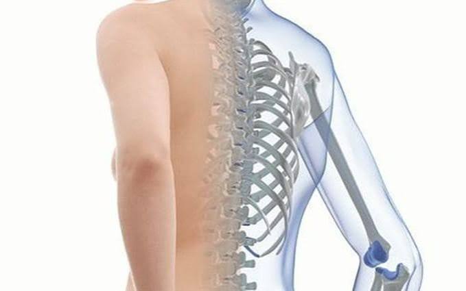
Osteoporosis is a silent disease. Bone is lost gradually over years without pain, without symptoms, and without warning — until a fracture occurs. In the United Kingdom, approximately 3.5 million people are estimated to have osteoporosis, and around half of all women and one in five men over the age of 50 will sustain a fragility fracture — a fracture caused by a force no greater than a fall from standing height — at some point in their remaining life. Hip fractures, the most serious consequence, carry a mortality rate of around 20% within the first year. Understanding what osteoporosis is, who is at risk, how it is diagnosed, and what can be done to prevent it and treat it is therefore relevant to a large proportion of the adult population.
What Is Osteoporosis?
Bone is living tissue, continuously broken down and rebuilt through a process called bone remodelling. Specialised cells called osteoclasts resorb (dissolve) old bone, while osteoblasts form new bone in its place. Under normal circumstances these two processes are balanced — the amount of bone destroyed equals the amount replaced. In osteoporosis, this balance is disrupted: resorption exceeds formation, net bone mass falls, and the microscopic architecture of bone — the fine lattice of trabeculae that give bone its strength — becomes thinner and more porous.
Peak bone mass — the maximum bone density an individual achieves — is reached between the mid-twenties and early thirties. Genetics determine approximately 60–80% of peak bone mass, with the remainder influenced by nutrition, physical activity, and hormonal factors during childhood and early adulthood. After peak bone mass is reached, gradual bone loss begins. In women, the loss accelerates sharply at the menopause because oestrogen normally suppresses osteoclast activity — without it, osteoclasts become more active and bone resorption increases significantly, with losses of up to 3% of bone mass per year in the first five to ten years after menopause.
The World Health Organization defines osteoporosis as a T-score of −2.5 or below on DEXA scanning (dual-energy X-ray absorptiometry) at the hip or lumbar spine, where the T-score compares an individual’s bone mineral density to that of a healthy young adult reference population. A T-score between −1.0 and −2.5 is classified as osteopenia — below normal but not yet in the osteoporosis range. Osteopenia is covered in detail in our companion guide to osteopenia: what adults should know.
Types of Osteoporosis
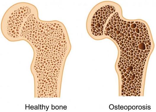
Driven by oestrogen deficiency after menopause. Predominantly affects trabecular (spongy) bone, with vertebral compression fractures and wrist fractures most common. Typically presents between ages 50–70.
Affects both cortical and trabecular bone in adults over 70. Hip fractures predominate. Affects both sexes equally; driven by declining osteoblast function and reduced calcium absorption with age.
Due to an identifiable medical cause. The most common is long-term glucocorticoid (steroid) use, which directly inhibits osteoblast function. Others include hyperparathyroidism, coeliac disease, inflammatory bowel disease, hypogonadism, and rheumatoid arthritis.
Symptoms of Osteoporosis
The most important thing to understand about osteoporosis symptoms is that there usually are none — until a fracture happens. Bone loss is painless and invisible. The disease earns its description as a “silent thief” because it takes years of bone loss to reach the threshold at which fracture risk becomes significant, and throughout that process the person feels entirely well.
The first sign of osteoporosis is often a fragility fracture — a fracture that occurs as a result of a minor fall or, in the case of vertebrae, sometimes with no injury at all. The most common fracture sites are:
- Vertebral compression fractures: sudden onset of back pain, often severe; loss of height over time (more than 4 cm of height loss is clinically significant); progressive kyphosis (forward curvature of the upper spine — “Dowager’s hump”). Many vertebral fractures are subclinical and found only incidentally on imaging taken for another reason.
- Hip fractures: typically from a fall; inability to bear weight; the affected leg may appear shortened and externally rotated. Hip fractures require hospitalisation and surgical repair, and carry significant morbidity and mortality — approximately 20% of people with a hip fracture die within 12 months, and half lose some degree of independence permanently.
- Wrist fractures (Colles’ fracture): from a fall onto an outstretched hand; common in women in their 50s as the first sign of postmenopausal bone loss.
- Humerus (upper arm) fractures: particularly in older adults.
A previous fragility fracture approximately doubles the risk of sustaining another, which is why assessment and treatment after any fragility fracture — the fracture liaison service pathway — is a key element of UK NHS osteoporosis management. If you have had an unexplained fracture, prompt bone density assessment is warranted.
Who Is at Risk of Osteoporosis?
Osteoporosis risk is understood through two categories: factors that cannot be changed, and factors that can be addressed through lifestyle choices and medical management.
Non-modifiable risk factors include:
- Age — bone loss is universal with ageing; risk increases sharply after 50
- Female sex — oestrogen withdrawal at menopause is the single most powerful driver of bone loss
- Early menopause (before age 45) or surgical menopause (oophorectomy)
- Family history of hip fracture — particularly in a parent
- Previous fragility fracture — the strongest predictor of the next fracture
- White or Asian ethnicity — lower baseline bone density compared to Black ethnicity
Modifiable risk factors — those that prevention and treatment programmes can address — include:
- Smoking: directly impairs osteoblast function and accelerates bone loss at all ages
- Alcohol: more than 14 units per week is associated with increased bone loss and fall risk
- Low calcium intake: calcium is the primary mineral component of bone; inadequate dietary intake (below 700 mg/day) accelerates bone loss
- Vitamin D deficiency: vitamin D is essential for calcium absorption in the gut; deficiency is common in older adults in the UK, particularly in winter
- Low body weight or BMI: less mechanical loading on bone; less adipose-derived oestrogen in postmenopausal women
- Physical inactivity: weight-bearing and resistance exercise stimulates bone formation through mechanical loading
- Long-term glucocorticoid use: prednisolone ≥5 mg/day for three months or longer is the most common secondary cause of osteoporosis and should trigger bone protection with bisphosphonate co-prescription
The FRAX tool (developed at the University of Sheffield for the WHO) calculates an individual’s 10-year probability of major osteoporotic fracture and hip fracture, combining clinical risk factors with or without DEXA bone mineral density. It is the standard risk assessment tool used by GPs and specialists in the UK. More detail on the DEXA scan used to diagnose osteoporosis is in our guide to DEXA scan: what adults should know.
How Is Osteoporosis Diagnosed?
DEXA scanning (dual-energy X-ray absorptiometry) is the gold standard investigation for osteoporosis. It measures bone mineral density (BMD) at the hip and lumbar spine using two low-energy X-ray beams, calculates the T-score against a reference population, and provides a definitive diagnosis. The scan involves no injection, takes approximately 10–15 minutes, and delivers a minimal radiation dose — substantially less than a standard chest X-ray. Full information is in our guide to bone density test: a simple guide.
Alongside DEXA, investigations to identify secondary causes of osteoporosis are standard at first diagnosis. These typically include blood tests for serum calcium, phosphate, alkaline phosphatase, liver function, thyroid function, full blood count, and erythrocyte sedimentation rate. In men, testosterone measurement is appropriate, as hypogonadism is a common secondary cause. Myeloma screen (protein electrophoresis, urine Bence Jones protein) is checked when there is any clinical suspicion.
The Z-score — which compares bone density to an age-matched and sex-matched peer group rather than to young adults — is used rather than the T-score in premenopausal women and men under 50, where the T-score is less clinically meaningful. A Z-score of −2.0 or below in these groups indicates bone density significantly lower than expected for age, which should prompt investigation for a secondary cause.
Preventing Osteoporosis
Because osteoporosis develops over decades, the most impactful prevention occurs in two phases: maximising peak bone mass during childhood and early adulthood, and slowing the inevitable bone loss that follows. The latter is achievable through lifestyle measures that remain beneficial at any age.
Calcium is the primary mineral component of bone. The UK recommended daily intake for adults is 700 mg, rising to 1,000–1,200 mg/day for those over 65 or at risk of deficiency. Dietary sources are preferable to supplements — dairy products (milk, cheese, yoghurt), fortified plant milks, green leafy vegetables (broccoli, kale, pak choi — not spinach, which has oxalates that block calcium absorption), canned fish with bones (sardines, salmon), and calcium-set tofu. Supplemental calcium is appropriate when dietary intake is consistently inadequate, but high-dose calcium supplements alone should be taken with care as some data links them with cardiovascular risk when taken without vitamin D. Full detail on dietary calcium and bone health is in our guide to calcium and bone health.
Vitamin D is essential for calcium absorption from the gut. Without adequate vitamin D, dietary calcium cannot be efficiently absorbed regardless of intake. The UK government recommends that all adults consider taking a 400 IU vitamin D supplement, particularly between October and March when sunlight exposure is insufficient to maintain levels. For older adults and those at high risk of deficiency, 800–1,000 IU may be appropriate. More detail on the role of vitamin D in bone health is in our guide to vitamin D and bone health.
Physical activity is the most evidence-based lifestyle intervention for maintaining bone density. The specific type of exercise matters:
- Weight-bearing aerobic exercise — walking, jogging, dancing, tennis, aerobics — applies mechanical load to bone through ground reaction forces, stimulating osteoblasts to form new bone. Swimming and cycling, while excellent for cardiovascular fitness, are non-weight-bearing and provide minimal benefit to bone density.
- Resistance training — using weights, resistance bands, or bodyweight — is particularly effective for hip and spine BMD and is recommended two to three times per week. It also maintains muscle mass and balance, reducing fall risk independently of its bone density effect.
Lifestyle measures: stopping smoking and limiting alcohol to no more than 14 units per week have meaningful effects on bone health independent of all other interventions. Fall prevention — balance exercises, home hazard assessment, appropriate footwear, correction of visual impairment — reduces the risk of the fragility fracture that is the clinical endpoint of osteoporosis.
Treatment Options for Osteoporosis
When osteoporosis is diagnosed — or when fracture risk is high enough to warrant treatment based on FRAX assessment — pharmacological treatment is indicated alongside the lifestyle measures above. All pharmacological treatments for osteoporosis are co-prescribed with calcium and vitamin D supplements.
Bisphosphonates are first-line treatment for most adults with osteoporosis. They inhibit osteoclast activity, reducing bone resorption and slowing bone loss. NICE recommends alendronic acid (alendronate) 70 mg once weekly as the first-choice agent, which reduces the risk of vertebral fracture by approximately 50% and hip fracture by approximately 40% over three years. Bisphosphonates must be taken on an empty stomach with a full glass of water, remaining upright for at least 30 minutes afterward to prevent oesophageal irritation. Intravenous zoledronate (annual infusion) is used for patients who cannot tolerate oral bisphosphonates or have swallowing problems. The most publicised risk associated with bisphosphonates — osteonecrosis of the jaw — is extremely rare at the doses used in osteoporosis (primarily a risk with the much higher doses used in cancer treatment) and should not discourage use in the large majority of patients.
Denosumab (Prolia) is a monoclonal antibody that inhibits RANKL, a key driver of osteoclast activity. Given as a subcutaneous injection every six months, it has efficacy comparable to bisphosphonates for reducing fracture risk and is used when bisphosphonates are not tolerated, contraindicated (including in renal impairment), or ineffective. An important consideration is that bone mineral density falls rapidly on stopping denosumab — transitioning to a bisphosphonate is recommended rather than simple discontinuation.
Hormone replacement therapy (HRT) prevents postmenopausal bone loss effectively and reduces fracture risk. It is appropriate for women under 60 who have menopausal symptoms as well as osteoporosis risk, where the benefits include both symptom relief and bone protection. For women over 60 seeking treatment for osteoporosis alone, bisphosphonates are generally preferred.
Teriparatide (recombinant parathyroid hormone) is an anabolic agent — it builds new bone rather than simply slowing resorption. It is given by daily subcutaneous injection for a maximum of two years and is reserved for severe osteoporosis, multiple vertebral fractures, or failure of or contraindication to antiresorptive therapy. Bone mineral density gains are significant, but a bisphosphonate or denosumab must follow the two-year course to maintain those gains.
Frequently Asked Questions
Can osteoporosis be reversed?
Osteoporosis cannot be fully reversed in the sense of completely restoring the bone architecture lost over years, but treatment significantly reduces further bone loss and fracture risk — and anabolic agents such as teriparatide can produce meaningful increases in bone mineral density. The more realistic and clinically important goal is preventing the fragility fractures that are the disease’s major harm. Bisphosphonates and denosumab reduce fracture risk significantly — by around 30–50% for vertebral fractures — without necessarily restoring T-scores to the normal range. Starting treatment after diagnosis and maintaining it consistently is the most important step available to any individual with confirmed osteoporosis.
At what age should I be checked for osteoporosis?
In the UK, NICE guidance recommends assessing women aged 65 and over for osteoporosis, as well as any woman over 50 with risk factors (early menopause, prior fragility fracture, family history of hip fracture, use of glucocorticoids). Men over 70 or men over 50 with secondary risk factors should also be assessed. Any adult of any age who sustains a fragility fracture should be assessed for osteoporosis. Younger adults on long-term glucocorticoids should begin bone protection therapy (bisphosphonate) without necessarily waiting for a DEXA scan, given the well-established bone-depleting effect of steroids.
Does osteoporosis cause back pain?
Osteoporosis itself does not cause back pain — the process of bone loss is painless. However, vertebral compression fractures, which are the most common consequence of osteoporosis in the spine, often cause sudden severe back pain, and may be followed by chronic back pain as multiple vertebrae fracture over time and kyphosis develops. Many vertebral compression fractures are subclinical — they occur without a specific injury and produce only mild discomfort that is attributed to muscle strain or normal ageing, meaning osteoporosis goes unrecognised. Persistent unexplained back pain in a woman over 55 or a man over 65 should prompt consideration of vertebral fracture assessment — either a lateral spine X-ray or DEXA with vertebral fracture assessment facility.
Is osteoporosis the same as arthritis?
No — osteoporosis and arthritis are entirely separate conditions that happen to both affect the musculoskeletal system. Osteoporosis is a disease of bone density and strength — bone loses mass and becomes more likely to fracture, but the joint surfaces are not primarily affected. Arthritis (both osteoarthritis and rheumatoid arthritis) affects the joints themselves — the cartilage lining, the synovial lining, and the joint mechanics. The confusion is understandable because both are common in older adults, both cause problems with mobility and independence, and rheumatoid arthritis is itself a secondary cause of osteoporosis. It is possible to have both conditions simultaneously.
Can men get osteoporosis?
Yes. Osteoporosis in men is significantly underdiagnosed because it is often perceived as a women’s disease. Around one in five men over the age of 50 will sustain a fragility fracture. Men do not undergo the rapid oestrogen-related bone loss of the menopause, but testosterone decline with age contributes to gradual bone loss, and secondary causes — particularly long-term glucocorticoid use, hypogonadism, and alcohol — account for a higher proportion of osteoporosis in men than in women. Men who sustain a hip fracture have a higher mortality rate than women with the same injury, partly because diagnosis and treatment rates are lower. Any man who sustains a fragility fracture, has significant secondary risk factors, or is over 70 should have his fracture risk formally assessed.
How long do you need to take bisphosphonates?
Current NICE guidance recommends reassessing bisphosphonate treatment at five years (for oral alendronate) or three years (for IV zoledronate). In patients at low ongoing fracture risk, a treatment break (“drug holiday”) of two to five years may be appropriate, taking advantage of the residual effect of bisphosphonates that remain in bone mineral. In patients with ongoing high fracture risk — those with a T-score below −2.5, prior hip fracture, or other major risk factors — continuation beyond five years is recommended, potentially switching to a different agent. Indefinite continuation without review is not appropriate; the decision to continue or pause should be made with a clinician familiar with the individual’s fracture history and current bone density.
Does taking calcium supplements prevent osteoporosis?
Calcium supplements alone are not sufficient to prevent or treat osteoporosis. Calcium is a necessary raw material for bone mineralisation, and ensuring adequate intake (through diet or supplementation) is important, but calcium intake does not directly drive bone density in the way that exercise and hormonal factors do. Calcium supplements should be combined with vitamin D, which is required for their intestinal absorption. As a standalone intervention, calcium supplementation does not substantially reduce fracture risk in women with adequate dietary intake. It is most beneficial as an adjunct to pharmacological treatment (bisphosphonates, denosumab) in women with documented dietary deficiency, and is routinely co-prescribed alongside anti-osteoporosis medication in the UK.
Summary
Osteoporosis causes progressive, silent bone loss that becomes clinically apparent only when a fragility fracture occurs — typically at the spine, hip, or wrist. It affects around half of all women and one in five men over 50 in the UK, and its consequences — particularly hip fracture — carry substantial mortality and morbidity. Risk assessment using the FRAX tool, DEXA scanning for bone mineral density, and exclusion of secondary causes are the pillars of diagnosis. Prevention centres on adequate calcium and vitamin D intake, regular weight-bearing and resistance exercise, smoking cessation, and fall prevention. Pharmacological treatment — bisphosphonates as first-line, with denosumab, HRT, and teriparatide for specific indications — significantly reduces fracture risk when indicated. Related bone health conditions are covered in our guides to osteopenia, bone density, and the DEXA scan.
Medical disclaimer: This article is for general educational purposes and does not constitute medical advice. Consult a qualified healthcare professional for personalised assessment and treatment of osteoporosis.
References:
NHS. Osteoporosis. NHS. 2023.
NICE CG146. Osteoporosis: assessing the risk of fragility fracture. NICE. 2017 (updated 2023).
Royal Osteoporosis Society. Strong, Steady and Straight. theros.org.uk. 2019.
Black DM, Rosen CJ. Postmenopausal Osteoporosis. N Engl J Med. 2016;374(3):254–262.
International Osteoporosis Foundation. Osteoporosis facts and statistics. iofbonehealth.org. 2023.
WHO FRAX. Fracture Risk Assessment Tool. University of Sheffield.
I was diagnosed with osteoporosis at 62 after a wrist fracture from a minor fall — I tripped on a rug and put my hand out to catch myself. Until that fracture I had no idea anything was wrong with my bones. The DEXA scan showed a T-score of −2.8 at the lumbar spine, and my GP started me on alendronate weekly. The article captures exactly what my experience was — you feel entirely well and then suddenly a small fall that wouldn’t have hurt a younger person causes a fracture. I’ve been on alendronate for four years now and my most recent DEXA showed improvement to −2.4, which my GP said was a good response. The instruction about staying upright for 30 minutes after the tablet is something I wish someone had explained more clearly at the start — I had oesophageal discomfort the first few weeks before I understood the importance of that.
Margaret, your trajectory is a very typical osteoporosis presentation: silent bone loss discovered only after a fragility fracture, then DEXA confirmation of a T-score in the osteoporosis range, followed by bisphosphonate initiation. The improvement from −2.8 to −2.4 over four years on alendronate is a meaningful and expected response — bisphosphonates predominantly stop further bone loss and allow normal bone formation to run unimpeded, producing gradual T-score improvement. On the oesophageal tolerability issue: the 30-minute upright rule and the importance of a full glass of water (200 ml minimum) with no other food or drink are critical for patient safety and tolerability, and are not always communicated as clearly as they should be at prescription. Sitting upright — not lying back — and avoiding lying down for at least 30 minutes after the dose prevents pooling of the tablet at the gastro-oesophageal junction. Robert, your case highlights a well-documented diagnostic gap: male osteoporosis is substantially underdiagnosed because clinicians and patients alike default to perceiving it as a female disease. Vertebral fractures in men presenting as back pain are frequently misattributed to muscular or disc causes, delaying osteoporosis diagnosis by months or years. The investigation workup you describe — including testosterone, which was low-normal in your case — is exactly appropriate for a man under 75 with a fragility fracture; hypogonadism is a contributing secondary cause in a significant proportion of men with osteoporosis, and even borderline testosterone values may warrant specialist review in this context.
I’m a 68-year-old man who was diagnosed with osteoporosis two years ago after a vertebral compression fracture — I was lifting something at the allotment and felt a sudden severe pain in my mid-back that turned out to be a T9 fracture. I think this article is valuable specifically for the section addressing osteoporosis in men. My GP initially attributed my back pain to a muscle strain, and it was only when an X-ray was eventually taken that the vertebral fracture was found. I subsequently had a DEXA scan, which showed a T-score of −2.9. My case was referred to a secondary care bone health clinic where testosterone measurement was included in the workup — mine was low-normal — and I was started on zoledronate infusion rather than oral bisphosphonate. The section on secondary causes and the investigation workup is accurate and useful for patients trying to understand why their doctor is ordering multiple blood tests.