
Shoulder pain that worsens with reaching overhead, is worst when lying on the affected side at night, and is accompanied by weakness when trying to lift the arm — this is the classic presentation of rotator cuff problems. The rotator cuff is the most common source of shoulder symptoms in adults over 40, and the spectrum of problems it can develop — from tendinopathy through partial tears to complete rupture — is wider than most people realise. Many rotator cuff tears, including significant ones, are entirely asymptomatic. Understanding where you are on that spectrum, what it means for your shoulder function, and what the treatment options are is the starting point for managing the condition effectively.
What Is the Rotator Cuff?
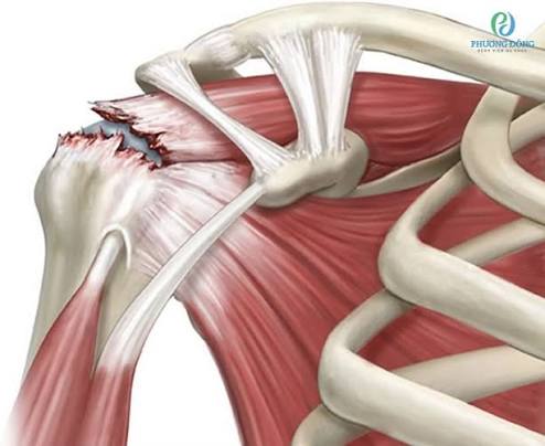
The rotator cuff is a group of four muscles and their tendons that surround the glenohumeral joint — the ball-and-socket shoulder joint — and hold it together. The four muscles are most easily remembered by the SITS mnemonic:
- Supraspinatus — runs above the shoulder joint through the subacromial space; responsible for the first 15–30 degrees of shoulder abduction and for humeral head depression during overhead movement. The most commonly injured and most frequently torn of the four.
- Infraspinatus — runs posteriorly; responsible for external rotation of the arm. Involved in many larger rotator cuff tears.
- Teres minor — posterior; external rotation; rarely torn in isolation.
- Subscapularis — anterior; responsible for internal rotation; torn in anterior shoulder dislocations and iatrogenic injury.
Together, these four tendons act as dynamic stabilisers — compressing the humeral head into the glenoid socket and allowing the precise, coordinated shoulder movement required for overhead activities. When the rotator cuff is damaged or painful, this coordinating function is disrupted, producing not just pain but the characteristic weakness and altered movement pattern that shoulder specialists assess. The subacromial bursa — a fluid-filled sac between the supraspinatus tendon and the acromion above — is anatomically adjacent to the rotator cuff and is almost always inflamed concurrently in supraspinatus tendinopathy and tears. For this reason, subacromial bursitis and rotator cuff tendinopathy are often described together in clinical practice. More detail on bursitis is in our guide to bursitis symptoms and causes.
The Spectrum of Rotator Cuff Problems
Rotator cuff pathology exists on a spectrum, and understanding which point on that spectrum any individual occupies is essential for treatment planning:
Degeneration at the supraspinatus watershed zone without structural tear. Pain with overhead activity and the painful arc sign. Approximately 25% of adults over 50 have some degree of rotator cuff tendinopathy on imaging — most asymptomatic.
A tear involving part of the tendon thickness — either the bursal surface, articular surface, or intratendinous. Can be managed non-operatively in many adults; may progress to full thickness if loading continues without treatment.
A tear through the complete thickness of the tendon — classified as small (<1 cm), medium (1–3 cm), large (3–5 cm), or massive (>5 cm or involving ≥2 tendons). Many are asymptomatic, particularly in older adults.
Irreversible structural damage with superior migration of the humeral head, secondary glenohumeral arthritis, and severe functional limitation. More common in elderly patients; may require reverse total shoulder arthroplasty.
What Causes Rotator Cuff Problems?
Most rotator cuff tears in adults over 50 are degenerative — they develop gradually as the result of age-related collagen deterioration in the supraspinatus tendon, particularly at its watershed zone approximately 1 cm from the bony insertion on the greater tuberosity of the humerus. This zone is poorly vascularised, making it vulnerable to cumulative microtrauma that exceeds the tendon’s repair capacity. The prevalence of full thickness tears rises steeply with age: approximately 10% in the seventh decade, rising to over 35% in the ninth decade. The majority of these are asymptomatic — the shoulder compensates for the tear through dynamic muscle activation, and the person may not be aware of any problem.
Traumatic tears — acute ruptures in previously normal or mildly degenerated tendons — occur at any age but are most clinically significant in active adults under 60. A fall onto an outstretched arm, a sudden forced abduction, or an acute shoulder dislocation can avulse the supraspinatus (and sometimes other cuff tendons) from their bony insertion. Acute traumatic tears in young active adults are generally best treated surgically and early, before tendon retraction and muscle atrophy make repair technically more difficult.
Repetitive overhead loading is the occupational and sporting cause — painting ceilings, throwing sports, swimming, and overhead assembly work all load the supraspinatus in the position of maximum subacromial compression. Chronic impingement — repeated compression of the supraspinatus between the humeral head and the acromion during overhead movements — is both a cause of pain and a mechanism for progressive tendon damage. The shape of the acromion contributes: a curved (Type II) or hooked (Type III) acromion reduces the subacromial space compared with a flat (Type I) acromion, increasing the compressive load on the supraspinatus with overhead movement.
Modifiable risk factors include smoking (which reduces the already limited vascularity of the supraspinatus watershed zone), diabetes (which impairs tendon collagen repair), and hyperlipidaemia (which causes lipid deposits in the tendon that disrupt collagen fibre organisation). Addressing these systemic factors as part of rotator cuff management is appropriate — see our guide to tendinitis in adults for the broader context of metabolic risk factors in tendon disease.
Symptoms of Rotator Cuff Problems
Rotator cuff pain has a characteristic clinical picture that allows clinicians to identify it — and to gauge its likely severity — from the history and examination:
- Anterolateral shoulder pain radiating into the deltoid muscle, exacerbated by reaching overhead, across the body, or behind the back
- Night pain — difficulty sleeping on the affected side; often the most functionally limiting symptom in tendinopathy and partial tears
- Painful arc — pain from approximately 60° to 120° of shoulder abduction, as the supraspinatus tendon passes under the acromion; characteristically painful in this range and easier above and below it
- Weakness — difficulty raising the arm above the shoulder, particularly against resistance; more pronounced in full thickness tears
- Sudden onset weakness after an acute injury suggests acute tendon rupture, which needs prompt assessment
Importantly, pain does not reliably indicate tear size. Some large full thickness tears are relatively painless (the shoulder adapts), while some cases of tendinopathy without any structural tear produce severe night pain and functional limitation. The clinical presentation cannot distinguish reliably between tear and no-tear — imaging is needed. For guidance on when shoulder and joint symptoms need urgent evaluation, see our guide on when joint pain needs medical evaluation.
How Is the Rotator Cuff Assessed?
The clinical examination for rotator cuff problems combines several specific tests with an overall shoulder movement and strength assessment:
- Painful arc: pain from 60° to 120° of abduction — highly suggestive of subacromial impingement
- Neer test: the examiner internally rotates and forward flexes the arm — reproduces impingement pain (sensitivity 72%)
- Hawkins-Kennedy test: arm to 90° forward flexion, then forced internal rotation — reproduces impingement (sensitivity 79%)
- Empty can test (Jobe): resisted abduction with the arm in 30° forward flexion and internal rotation (thumb pointing down) — tests supraspinatus strength; weakness or pain suggests partial or full thickness supraspinatus tear
- External rotation lag sign: inability to maintain passive external rotation suggests full thickness infraspinatus tear
Ultrasound is the most accessible imaging investigation and is highly accurate for full thickness tears when performed by an experienced operator. It can also identify partial tears, subacromial bursitis, and calcific tendinitis, and it enables real-time guided injection into the subacromial space. MRI is the gold standard for preoperative planning — it provides complete information about tear size, the degree of tendon retraction, the quality of the remaining muscle (Goutallier grade of fatty infiltration), and the integrity of the other cuff tendons. Both of these factors — retraction and muscle quality — are important predictors of whether a surgical repair will hold once performed. Related information on joint range of motion assessment is in our guide to limited range of motion in adults.
Treatment Options
Non-Operative Management: First-Line for Most Adults
The majority of rotator cuff problems in adults — including many full thickness tears, particularly in those over 65 — are successfully managed without surgery. Non-operative treatment centres on physiotherapy-led rotator cuff strengthening and scapular stabilisation exercises, combined with appropriate use of subacromial injections for pain relief.
Physiotherapy for rotator cuff problems targets two areas: strengthening the intact or partially intact cuff muscles to compensate for the damaged tendon and maintain dynamic shoulder stability, and correcting scapular dyskinesis — abnormal scapular movement that dynamically narrows the subacromial space during arm elevation and contributes to ongoing impingement. A supervised physiotherapy programme of 8–12 weeks, progressively loading the shoulder through overhead and rotational exercises, is the most evidence-based primary intervention for both tendinopathy and degenerative tears. NSAIDs reduce pain and allow more active participation in rehabilitation.
Subacromial Corticosteroid Injection
Subacromial injection — delivering corticosteroid and local anaesthetic into the subacromial bursa under the acromion — is a highly effective and appropriate intervention for rotator cuff tendinopathy and bursitis. It reduces pain rapidly, often dramatically, and allows patients to engage in the physiotherapy programme that produces the long-term improvement. The injection is distinct from direct tendon injection: steroid delivered into the tendon body increases rupture risk, whereas subacromial bursal injection is safe when correctly placed. Image guidance (ultrasound) significantly improves accuracy of subacromial injection compared with landmark-guided injection. A maximum of three subacromial injections, no more frequently than every three months, is the standard guidance.
Surgical Repair
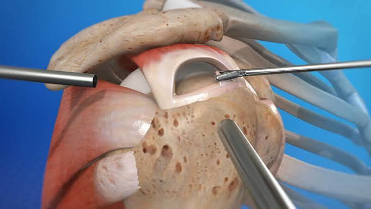
Arthroscopic rotator cuff repair — reattaching the torn tendon to its bony footprint on the greater tuberosity using small anchor devices inserted under arthroscopic vision — is indicated in several scenarios:
- Acute traumatic full thickness tear in an active adult (especially under 60)
- Failed conservative management after 3–6 months in a patient with functional limitation
- Large or massive tear in a patient who needs shoulder function for occupation or sport
- Progressive tear growth documented on serial imaging
Surgical outcomes depend substantially on tear characteristics at the time of repair. Small and medium tears (under 3 cm) repaired arthroscopically show healing rates of 80–90% at two years and consistently high patient satisfaction. Large and massive tears have progressively lower healing rates, though even partial healing produces meaningful functional improvement in many patients. The Goutallier classification of fatty infiltration — assessed on MRI — is the most important prognostic factor: Grade 0–1 (minimal fat) predicts reliable healing; Grade 3–4 (predominantly fat) predicts poor healing and argues against repair, because the replaced muscle cannot generate the tension needed for healing even after the tendon is reattached.
Rotator cuff repair is performed arthroscopically as a day case under general anaesthesia with regional nerve block. Recovery requires a period of sling immobilisation (typically four to six weeks), followed by progressive physiotherapy over four to six months. Full shoulder function — including overhead strength — takes 9–12 months to fully recover.
Massive Irreparable Tears
In patients with massive tears that are not repairable due to tendon retraction and muscle atrophy, or in elderly patients with cuff tear arthropathy, reverse total shoulder arthroplasty (RTSA) produces excellent pain relief and functional shoulder elevation. By reversing the ball-and-socket geometry, RTSA allows the intact deltoid muscle to elevate the arm without requiring the rotator cuff, compensating for the absent cuff function. RTSA has become one of the fastest-growing orthopaedic procedures globally, with high patient satisfaction rates.
Rotator Cuff Problems and Frozen Shoulder
There is an important clinical relationship between rotator cuff tendinopathy and frozen shoulder (adhesive capsulitis). Chronic rotator cuff pain that leads patients to protect the shoulder — moving it as little as possible to avoid pain — can trigger secondary frozen shoulder, in which the glenohumeral capsule becomes fibrotic and globally restricts movement. Conversely, patients with primary frozen shoulder often coexist with supraspinatus tendinopathy because both conditions share the subacromial space.
If you develop progressive global restriction of shoulder movement on top of rotator cuff symptoms — particularly if external rotation becomes markedly limited — frozen shoulder should be assessed in addition to the rotator cuff. Full information on frozen shoulder stages and treatment is in our guide to frozen shoulder symptoms and treatment options.
Frequently Asked Questions
Can a rotator cuff tear heal on its own?
Complete spontaneous healing of a full thickness rotator cuff tear is uncommon — the poor vascularity of the torn tendon ends and the constant mechanical loading of the shoulder work against it. However, many partial tears and even some small full thickness tears stabilise and remain non-progressing, with patients achieving good functional levels through compensatory muscle activation and physiotherapy. The goal of non-operative management is not healing but rather optimising function with the tear that exists. Symptomatic improvement without structural healing is achievable and clinically meaningful in many adults, particularly those over 65 where surgery carries higher risks and recovery expectations are adjusted accordingly.
Do I need surgery for a rotator cuff tear?
Not necessarily. Many rotator cuff tears — including full thickness tears, particularly in patients over 65 — are successfully managed without surgery through physiotherapy, activity modification, and subacromial injection. Surgery is most clearly indicated for acute traumatic tears in young active adults, large tears with significant functional limitation, and tears that have not responded to three to six months of consistent conservative management. The decision should be made jointly by patient and surgeon, taking into account tear characteristics (size, retraction, muscle quality on MRI), patient activity level and functional requirements, age, and medical fitness for surgery. There is no urgency for most degenerative tears — taking two to three months of supervised physiotherapy first does not harm the surgical outcome for most tear sizes.
What is the painful arc and what does it mean?
The painful arc refers to a specific range of shoulder abduction — approximately 60° to 120° — during which pain is reproduced, with relative comfort above and below that range. It occurs because the supraspinatus tendon passes under the acromion precisely in this range of motion, and when the tendon is thickened, inflamed, or the bursa is swollen, the available space is insufficient and compression produces pain. It is one of the most specific clinical signs for subacromial impingement and rotator cuff tendinopathy, and its presence is helpful in distinguishing shoulder pain arising from the cuff from pain arising from the glenohumeral joint or other structures.
How long does recovery from rotator cuff surgery take?
Recovery from arthroscopic rotator cuff repair is a long process — patients should expect 9–12 months before full strength and overhead function is restored. The first four to six weeks involve sling immobilisation to protect the repaired tendon; this phase is often the most frustrating because the repaired tendon cannot be loaded until it has biologically integrated with the bone. Active-assisted exercises begin at six weeks, progressive strengthening at three months, and full overhead loading at six months. Individual variation is significant — larger tears, older patients, and those with higher Goutallier grades of muscle atrophy take longer to recover. Consistent physiotherapy engagement throughout this process is essential for optimising the surgical outcome.
Is the rotator cuff involved in frozen shoulder?
The two conditions can coexist and may be related, but they are pathologically distinct. Rotator cuff tendinopathy involves degeneration of the tendon itself; frozen shoulder involves fibrotic contracture of the glenohumeral joint capsule. The key clinical difference is in the pattern of movement restriction: rotator cuff problems typically produce a painful arc and weakness but relatively preserved passive range of movement (the examiner can move the arm to near-normal positions). Frozen shoulder produces global restriction of passive movement — the examiner cannot move the arm beyond the range the capsule permits. Both conditions can cause significant night pain, which is why a careful examination is needed to distinguish them, and why both conditions are covered in our shoulder health guides.
Can I prevent rotator cuff problems?
Completely preventing the degenerative changes that accumulate in the supraspinatus with age is not possible, but several modifiable factors reduce the rate and severity of that process. Avoiding smoking is the most impactful lifestyle change — smoking significantly reduces vascularity to the watershed zone of the supraspinatus. Maintaining good shoulder and scapular muscle strength through regular exercise supports the dynamic stabilisation of the joint and reduces impingement loads. Technique corrections for overhead sport and work — ensuring the shoulder moves with proper scapular engagement rather than internal rotation — reduce compressive loading on the supraspinatus. Treating early tendinopathy symptoms promptly with physiotherapy, rather than allowing progression to partial or full thickness tear, is achievable with early intervention.
What does a rotator cuff injection involve?
A subacromial corticosteroid injection is delivered into the subacromial bursa — the fluid-filled sac between the supraspinatus tendon and the underside of the acromion — rather than into the tendon itself. The injection is typically performed under ultrasound guidance from a posterior or lateral approach, with local anaesthetic included for immediate pain relief. Most patients feel significant improvement within five to seven days as the corticosteroid effect builds. The injection is safe when correctly placed in the bursa; it is injections directly into the tendon body that carry a rupture risk, which is why image guidance matters. A single well-placed subacromial injection often provides sufficient pain relief to allow full engagement with physiotherapy — which is the long-term treatment.
Summary
Rotator cuff problems cover a spectrum from tendinopathy through partial and full thickness tears to massive irreparable tears — and many are asymptomatic even when structurally significant. The most common presentation is anterolateral shoulder pain worse with overhead activity and at night, with a painful arc on abduction. Most cases in adults over 50 are managed successfully without surgery through physiotherapy-led rotator cuff strengthening, subacromial injection for pain relief, and activity modification. Surgery is most clearly indicated for acute traumatic tears in younger adults, large tears causing significant functional limitation, and cases that have not responded to a supervised conservative programme. MRI before surgery provides the critical information — tear size, retraction, and muscle quality — that determines the likelihood of a durable repair. Related shoulder conditions that commonly coexist with or follow rotator cuff problems are covered in our guide to frozen shoulder symptoms and treatment options.
Medical disclaimer: This article is for general educational purposes and does not constitute medical advice. Consult a qualified healthcare professional for personalised assessment and management of rotator cuff problems or any shoulder condition.
References:
NHS. Rotator cuff injury. NHS. 2023.
NICE Clinical Knowledge Summary. Shoulder pain. NICE CKS. 2022.
Milgrom C, Schaffler M, Gilbert S, van Holsbeeck M. Rotator cuff changes in asymptomatic adults. J Bone Joint Surg Br. 1995;77(2):296–298.
Mather RC, Koenig L, Acevedo D, et al. The societal and economic value of rotator cuff repair. J Bone Joint Surg Am. 2013;95(22):1993–2000.
Versus Arthritis. Rotator cuff disorders. versusarthritis.org. 2023.
NICE. Musculoskeletal conditions guidance. nice.org.uk.
I had a large supraspinatus tear confirmed on MRI — about 3.5 cm — and was told surgery was the right option given my age (54) and the fact that I coach tennis at a club level. What I didn’t expect was how important the pre-operative physiotherapy turned out to be. My surgeon recommended six weeks of physiotherapy before the operation to optimise the strength of the intact cuff tendons and improve my scapular mechanics, so that the shoulder was in the best possible condition going into repair. The outcome was good — I’m now 11 months post-op, back to coaching, and overhead serves are pain-free. The point this article makes about the Goutallier grade being critical for prognosis was something my surgeon explained clearly: my MRI showed Grade 1 fatty infiltration, which he said was a good prognostic indicator for healing.
Michael, your case illustrates several important principles. The decision to proceed with repair at 54 with a large tear was appropriate — at this age, degenerative changes in the muscle (Goutallier Grade 1) are minimal, the tendon is still viable for repair, and your functional requirements as a coach mean the expected benefit of restored overhead strength justifies the surgical risk and rehabilitation investment. The pre-operative physiotherapy your surgeon recommended — optimising intact cuff muscle strength before repair — is supported by evidence that patients who enter repair in better shoulder condition have better post-operative outcomes, partly because the intact muscles provide better tissue perfusion around the repaired tendon during healing. Your 11-month outcome trajectory (pain-free overhead serving) is exactly what a well-executed repair of a large but repairable tear with Grade 1 muscle quality should produce. Patricia, your management course — conservative treatment at 71 for a small degenerative tear — reflects the evidence base precisely. In adults over 65 with small degenerative tears, physiotherapy outcomes are comparable to surgical outcomes at two years in most trials, without the surgical risk or the 9-12 month recovery burden. The goal of non-operative management is not structural healing but functional compensation through intact muscle strengthening — and your functional trajectory at 18 months, with restored sleep and maintained daily activities, represents a successful outcome by that standard.
I’m 71 and was diagnosed with a full thickness supraspinatus tear 18 months ago after an ultrasound for persistent shoulder pain. My orthopaedic consultant recommended conservative management rather than surgery, given my age and the fact that the tear was described as degenerative and small (under 1.5 cm). I had two subacromial injections over the first six months, both of which gave significant temporary relief, and I’ve been doing the physiotherapy exercises consistently. At 18 months I have occasional dull shoulder ache and some weakness with overhead reaching, but I can sleep on that side again and my daily activities are largely unaffected. The article’s explanation of why many full thickness tears in older adults can be managed without surgery — because the intact cuff tendons compensate — matches exactly what my consultant said to me.