
Pancreatic cancer diagnosis is one of the most challenging processes in oncology — not because the tests are unavailable, but because the disease so often stays silent until it has reached an advanced stage. The pancreas sits deep in the retroperitoneum, tucked behind the stomach and surrounded by major blood vessels. Tumors growing there cause few noticeable symptoms until they obstruct a bile duct, press on a nerve, or spread to adjacent structures. By that point, the diagnostic workup begins in earnest: imaging, endoscopy, biopsy, and staging to determine what options are available.
Understanding how pancreatic cancer is diagnosed helps patients and families navigate what can feel like an overwhelming sequence of appointments, scans, and procedures. Each test builds on the previous one, narrowing down the size and location of the tumor, confirming the tissue type, and assessing whether the cancer can be surgically removed. This guide walks through the full diagnostic process — from initial blood tests through molecular profiling and staging — so you know what to expect at each step.
Why Pancreatic Cancer Is Hard to Diagnose Early
The fundamental challenge with pancreatic cancer is that there is no reliable screening test for people at average risk. Unlike colonoscopy for colon cancer or mammography for breast cancer, there is no established method to detect early pancreatic cancer before symptoms appear in the general population.
This matters because the pancreas is an organ where tumors grow silently for months or years before causing problems. Located deep in the abdomen behind the stomach, the pancreas has no pain-sensing surface that gets irritated by early tumor growth. A tumor in the body or tail of the pancreas may reach several centimeters before it compresses any structure that causes symptoms. A tumor in the head of the pancreas may cause obstructive jaundice earlier — because the common bile duct runs directly through the pancreatic head — but even this often occurs when the tumor is already 2 to 3 centimeters.
The result is a disease with a late-presentation problem: approximately 80 percent of patients are diagnosed with locally advanced or metastatic disease. Only about 20 percent of patients have cancer confined enough to be surgically resectable at the time of diagnosis. This is why the five-year survival rate for pancreatic cancer overall remains low, even as outcomes for the subset of patients who undergo surgery have improved significantly.
The symptoms that do prompt a diagnostic workup include: painless jaundice (yellow discoloration of the skin and eyes), new-onset diabetes in someone without risk factors, unexplained weight loss, upper abdominal or back pain, and changes in stool color or fat content. When any of these occur — especially jaundice — prompt evaluation is essential.
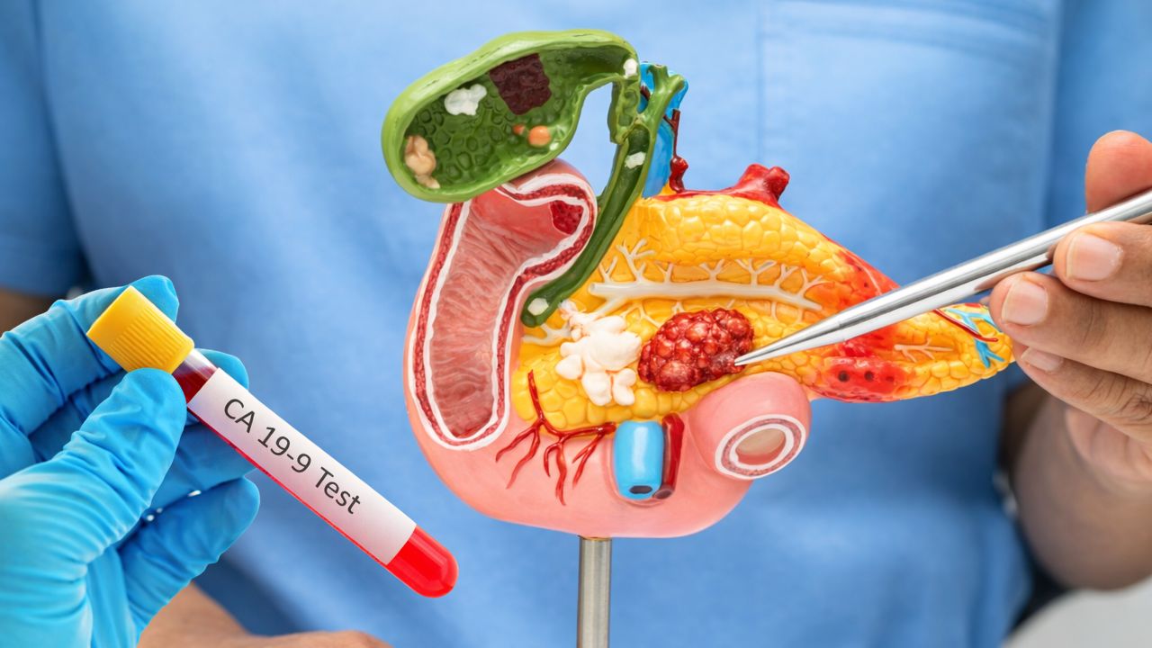
Initial Evaluation — Blood Tests and the CA 19-9 Marker
When a patient presents with symptoms suggesting pancreatic pathology, the initial evaluation includes standard blood work alongside tumor markers. These tests cannot diagnose pancreatic cancer on their own, but they provide critical context for what the imaging will show.
Liver function tests (LFTs) are among the first tests ordered when jaundice or abdominal pain is present. In pancreatic cancer, a tumor in the head of the pancreas compresses the common bile duct, causing obstructive jaundice. This pattern shows up on blood work as elevated total bilirubin, elevated direct (conjugated) bilirubin, elevated alkaline phosphatase (ALP), and elevated gamma-glutamyl transferase (GGT) — the classic cholestatic pattern. Elevated transaminases (AST, ALT) may follow if hepatic damage occurs from prolonged biliary obstruction.
CA 19-9 (cancer antigen 19-9) is the most commonly used serum tumor marker for pancreatic cancer. It is elevated in approximately 80 percent of patients with advanced pancreatic ductal adenocarcinoma (PDAC). However, it has significant limitations. Sensitivity is only about 50 percent in Stage I disease, making it unreliable for early detection. False negatives occur in 5 to 10 percent of the population who are Lewis antigen-negative — a blood type variant in which the body does not produce CA 19-9 regardless of cancer status. False positives occur in biliary obstruction from any cause: a patient with a benign bile duct stone may have a markedly elevated CA 19-9 that normalizes after the obstruction is relieved.
Because of these limitations, CA 19-9 is used as an adjunct to imaging — not as a diagnostic tool on its own. It is most valuable for monitoring treatment response after diagnosis: a falling CA 19-9 during chemotherapy suggests the cancer is responding; a rising CA 19-9 after surgery may signal recurrence before imaging shows it. This is similar to how CA-125 functions in ovarian cancer — another imperfect serum marker that is most useful for monitoring rather than initial screening.
Blood glucose is worth checking because new-onset diabetes can be an early manifestation of pancreatic cancer. The exocrine tumor disrupts insulin-producing beta cells in the islets of Langerhans. New-onset diabetes in someone over 50 without typical risk factors, particularly when accompanied by weight loss, warrants imaging of the pancreas.
CEA (carcinoembryonic antigen) is a secondary marker that is elevated in some mucinous pancreatic tumors but is less specific and sensitive than CA 19-9 for PDAC.
5–10% of the population are Lewis antigen-negative and cannot produce CA 19-9 at all — meaning a normal CA 19-9 result does not rule out pancreatic cancer in these individuals. Always interpret CA 19-9 alongside imaging findings, and recheck after biliary drainage if obstruction was present.
CT Scan with Pancreatic Protocol — The First Imaging Step
The standard first imaging study for suspected pancreatic cancer is a CT scan of the abdomen and pelvis with what is called a “pancreatic protocol.” This is not the same as a routine abdominal CT. The pancreatic protocol uses thin-slice (2 to 3 mm) sections with intravenous contrast given in a specific timed sequence to capture two distinct phases of enhancement.
The arterial phase is captured when contrast first reaches the pancreatic arteries, approximately 40 to 45 seconds after injection. Because the normal pancreas enhances brightly with contrast in this phase, a pancreatic ductal adenocarcinoma — which is characteristically hypovascular — stands out as a relatively dark (hypodense) area against the bright pancreatic background. This is when small tumors are most visible.
The portal venous phase is captured at approximately 65 to 70 seconds, when the portal vein and liver are maximally enhanced. This phase is best for detecting liver metastases and assessing involvement of the portal vein and superior mesenteric vein (SMV), which are critical for determining whether the tumor can be surgically removed.
CT findings that suggest pancreatic cancer include: a hypodense mass within the pancreas; dilation of the main pancreatic duct (greater than 3 mm is concerning); dilation of the common bile duct; the “double duct sign” (simultaneous dilation of both the main pancreatic duct and common bile duct, which is highly suspicious for a pancreatic head mass); vascular encasement or thrombosis involving the SMV, portal vein, superior mesenteric artery (SMA), or celiac axis; liver masses consistent with metastases; and ascites or peritoneal nodules suggesting peritoneal spread.
Sensitivity for detecting pancreatic cancer by CT is greater than 95 percent for tumors 2 cm or larger. Sensitivity drops to 65 to 80 percent for tumors smaller than 2 cm — a critical gap, since small tumors are exactly the ones most likely to be resectable. This is one reason why endoscopic ultrasound is added to the workup when CT is equivocal or when a small mass is suspected.
The CT also provides the first assessment of surgical resectability. Radiologists and surgeons evaluate how much of the tumor is in contact with major vessels: contact of less than 180 degrees of the vessel circumference generally indicates resectable disease, while contact of greater than 180 degrees or encasement of vessels indicates borderline or unresectable disease.
Endoscopic Ultrasound (EUS) and Biopsy
When CT does not provide a clear answer — or when tissue confirmation is needed before starting chemotherapy — endoscopic ultrasound (EUS) is the next step. EUS is also the preferred method for diagnosing small pancreatic tumors that CT may miss.
In an EUS procedure, an endoscope with a high-frequency ultrasound probe at its tip is passed through the mouth, down through the esophagus and stomach, and positioned in the stomach or the first part of the duodenum (small intestine). From these positions, the ultrasound probe is only millimeters away from the pancreas, with no intervening bowel gas or fat to degrade the image. This proximity allows EUS to image the pancreas with much higher resolution than any external imaging modality.
EUS-FNA (fine needle aspiration) involves passing a thin needle through the endoscope and advancing it under ultrasound guidance into the pancreatic tumor to collect cells for cytology. When the needle can reach the tumor, sensitivity for malignancy is approximately 85 to 95 percent.
EUS-FNB (fine needle biopsy) uses a specially designed core biopsy needle to obtain a tissue core for histologic analysis rather than just loose cells. Core biopsy is preferred when the oncologist needs not just a cancer diagnosis but tissue sufficient for molecular profiling and genomic sequencing.
A tissue diagnosis is required before beginning chemotherapy in essentially all cases. The only exception is when surgery is performed immediately based on unequivocal clinical and imaging findings — in which case the surgical specimen itself provides the tissue diagnosis.
EUS has an additional advantage in pancreatic cancer: the celiac plexus — the nerve network that transmits pain from the pancreas — lies adjacent to the celiac artery, which is also visible during EUS. The same session that provides a tissue diagnosis can be used to perform a celiac plexus neurolysis or block, injecting alcohol or local anesthetic around the celiac ganglion to reduce tumor-related abdominal and back pain. This spares patients a separate interventional procedure.
MRI, MRCP, and Other Imaging Tests
MRI with contrast provides complementary information to CT, with superior soft tissue contrast. It is particularly valuable for characterizing liver lesions that appear indeterminate on CT (benign cysts vs. liver metastases), evaluating small periampullary tumors, and assessing patients who cannot receive iodinated CT contrast due to allergy or kidney disease.
MRCP (magnetic resonance cholangiopancreatography) is a specialized MRI sequence that uses water in bile to create images of the biliary tree and pancreatic duct without contrast injection. MRCP is used to evaluate: the anatomy of the bile duct before surgery; cystic lesions of the pancreas such as intraductal papillary mucinous neoplasms (IPMN) and mucinous cystic neoplasms (MCN); and the level and cause of biliary obstruction in jaundiced patients. It can distinguish whether obstruction is from a stone, a stricture, or a mass.
ERCP (endoscopic retrograde cholangiopancreatography) is an endoscopic procedure used primarily for treatment rather than diagnosis in pancreatic cancer. During ERCP, a scope is passed to the ampulla of Vater where the bile duct and pancreatic duct empty into the duodenum, and a wire or catheter is advanced up the bile duct under fluoroscopic (X-ray) guidance. In pancreatic cancer, ERCP is used most often to place a biliary stent — a small plastic or metal tube that props open an obstructed bile duct and relieves jaundice. Brush cytology obtained during ERCP can sometimes yield a diagnosis, but its sensitivity is only 50 to 60 percent — substantially lower than EUS-FNA — so EUS is preferred when tissue diagnosis is the goal.
PET-CT (positron emission tomography with CT) uses a radioactive glucose analog (18F-FDG) that accumulates preferentially in metabolically active cancer cells. Pancreatic cancer is generally FDG-avid, making it visible on PET. PET-CT is not part of the routine initial staging workup but is used selectively when CT and MRI are inconclusive about the presence of distant metastasis, when recurrence is suspected after treatment, or when assessing metabolic response to chemotherapy. An important limitation: elevated blood glucose levels — common in pancreatic cancer patients who have developed diabetes — reduce FDG uptake and can produce false-negative results.
Staging Laparoscopy — Ruling Out Hidden Spread
Even after an excellent pancreatic protocol CT and MRI, a small percentage of patients who appear to have resectable disease on imaging turn out to have peritoneal metastases — tiny tumor deposits on the surface of the abdomen — that imaging cannot detect. These micro-metastases would make surgery futile.
Staging laparoscopy is a minimally invasive procedure performed under general anesthesia, in which a camera is inserted through a small incision in the abdomen to directly inspect the peritoneal surfaces and liver. Peritoneal fluid is collected for cytology. If tumor cells are found in the peritoneal fluid (positive peritoneal cytology), this is classified as metastatic disease (Stage IV) even if no visible implants are seen.
Staging laparoscopy identifies approximately 15 to 20 percent of patients who appear resectable by imaging but actually have occult peritoneal spread. It is particularly useful in patients with borderline resectable tumors, markedly elevated CA 19-9 (greater than 1,000 U/mL), or CT findings that suggest high risk for peritoneal disease. By identifying these patients before a major operation, staging laparoscopy prevents unnecessary Whipple procedures with their significant recovery burden.
Staging — How Doctors Determine the Extent of Disease
Once all diagnostic tests are complete, physicians use the results to assign a formal stage and, more practically for surgical planning, a resectability category. Both classification systems are used simultaneously in pancreatic cancer.
TNM Staging (AJCC)
The formal TNM staging system classifies cancer based on the primary tumor, lymph node involvement, and presence of distant metastasis.
- Stage I: Tumor confined to the pancreas. Stage IA: 2 cm or smaller. Stage IB: more than 2 cm but 4 cm or smaller.
- Stage II: Tumor larger than 4 cm (IIA) or tumor of any size with spread to 1 to 3 regional lymph nodes (IIB).
- Stage III: Tumor has invaded major adjacent vessels (celiac axis, SMA, or hepatic artery) or spread to 4 or more regional lymph nodes. No distant metastasis.
- Stage IV: Distant metastasis present — most commonly to the liver, lungs, or peritoneum.
Surgical Resectability Categories (NCCN)
The National Comprehensive Cancer Network uses a separate surgical staging system that is more directly useful for treatment planning:
Resectable disease means the tumor can be surgically removed with clear margins. The surrounding major vessels — superior mesenteric vein (SMV), portal vein, superior mesenteric artery (SMA), celiac axis, and common hepatic artery — must be free of tumor encasement. Approximately 15 to 20 percent of patients have resectable disease at diagnosis.
Borderline resectable disease means the tumor is in contact with adjacent vessels but has not encased them to a degree that precludes surgery. These patients are often treated with neoadjuvant chemotherapy — chemotherapy given before surgery — to shrink the tumor away from vessels and improve the chance of achieving clear margins at surgery.
Locally advanced (unresectable Stage III) means the tumor encases the major vessels in a way that cannot be surgically reconstructed, but there is no evidence of distant metastasis. Treatment is systemic chemotherapy, sometimes followed by radiation therapy, with the goal of controlling local disease. A minority of locally advanced patients respond well enough to chemotherapy that they become candidates for surgery.
Metastatic disease (Stage IV) means cancer has spread to distant organs, most commonly the liver, lungs, or peritoneum. Treatment is systemic chemotherapy for palliation, with the goal of prolonging survival and maintaining quality of life.
For patients with pancreatic cancer, the stage and resectability category at diagnosis are the most important factors determining treatment options and prognosis.
Molecular Testing and Genetic Profiling
Comprehensive molecular profiling has become a standard recommendation for patients with advanced pancreatic cancer because it identifies a subset of patients who are eligible for targeted therapies that can substantially improve outcomes.
Comprehensive genomic profiling (NGS — next-generation sequencing) of the tumor tissue is recommended for all patients with metastatic PDAC. This test analyzes hundreds of cancer genes simultaneously and identifies specific alterations that may be targetable:
- BRCA1 or BRCA2 mutations: Present in approximately 5 to 7 percent of pancreatic cancers. Patients with BRCA-mutated, platinum-sensitive metastatic PDAC are eligible for the PARP inhibitor olaparib as maintenance therapy after first-line chemotherapy.
- KRAS G12C mutation: Present in approximately 1 to 2 percent of PDAC. KRAS G12C inhibitors have shown activity in this subgroup.
- NTRK gene fusions: Rare but highly targetable; larotrectinib and entrectinib produce responses in any NTRK fusion-positive solid tumor regardless of origin.
- MSI-H (microsatellite instability-high): Present in approximately 1 to 2 percent of PDAC; highly responsive to pembrolizumab (Keytruda), an immune checkpoint inhibitor approved for MSI-H tumors of any type.
Germline (hereditary) genetic testing — testing blood or saliva rather than tumor tissue — is now recommended for every pancreatic cancer patient, regardless of family history or age. Studies have found that 5 to 10 percent of pancreatic cancer patients carry a hereditary cancer syndrome gene: BRCA2 (the most common), BRCA1, PALB2, ATM, Lynch syndrome genes, or CDKN2A. Finding a germline mutation has two major implications: it may qualify the patient for targeted therapy, and it identifies family members who should consider genetic counseling and screening. The BRCA1 and BRCA2 mutations most commonly associated with ovarian and breast cancer also significantly elevate pancreatic cancer risk — underscoring why all patients, not just those with a cancer family history, should undergo germline testing.
What Happens After Diagnosis — The Multidisciplinary Team
Pancreatic cancer diagnosis rarely results in a single physician making treatment decisions alone. The standard of care is a multidisciplinary tumor board that typically includes a pancreatic surgeon, medical oncologist, radiation oncologist, gastroenterologist, interventional radiologist, specialized radiologist, pathologist, and palliative care specialist. This team reviews each patient’s complete diagnostic picture to reach a consensus on the optimal treatment approach.
The path forward depends directly on stage and resectability:
- Resectable disease: Surgery first (Whipple procedure for head/neck tumors; distal pancreatectomy for body/tail), followed by adjuvant chemotherapy with FOLFIRINOX or gemcitabine/capecitabine.
- Borderline resectable: Neoadjuvant chemotherapy for 4 to 6 months, then restaging CT, then surgery if the tumor has responded sufficiently.
- Locally advanced: Systemic chemotherapy, possibly followed by consolidative radiation therapy.
- Metastatic: Systemic chemotherapy; molecular profiling to identify targeted therapy options; clinical trial enrollment strongly encouraged.
Research consistently shows that surgical outcomes for pancreatic cancer are significantly better at high-volume centers that perform 20 or more Whipple procedures per year. Patients who are candidates for surgery should strongly consider evaluation at a center with established pancreatic surgery expertise. If you have been tracking your pancreatic cancer symptoms and recently received a diagnosis, getting a second surgical opinion at a specialized center before committing to a treatment plan is one of the most valuable steps you can take.
Questions to Ask Your Doctor After a Pancreatic Cancer Diagnosis
Knowing what to ask after a pancreatic cancer diagnosis can help you make informed decisions and ensure you are getting the best possible care.
About staging and resectability:
- Is my tumor resectable, borderline resectable, locally advanced, or metastatic? What imaging findings determine this?
- Have my scans been reviewed by a radiologist who specializes in pancreatic cancer?
- Should I get a second surgical opinion at a high-volume pancreatic cancer center?
About molecular and genetic testing:
- Has my tumor been sent for comprehensive genomic profiling (NGS)?
- Should I have germline genetic testing? What mutations should be tested for?
- If I have a BRCA2 or other hereditary mutation, what does that mean for my family?
About treatment and support:
- What are my treatment options at this stage?
- Are there clinical trials I should consider? A searchable database is available at cancer.gov.
- What resources does the Pancreatic Cancer Action Network (PanCAN) offer for newly diagnosed patients?
- Is there a palliative care team I can work with alongside my oncology team?
The NCCN publishes patient-version guidelines for pancreatic cancer at nccn.org/patientresources — these explain staging, treatment options, and questions to ask in accessible language.
Putting the Diagnosis Together
Pancreatic cancer diagnosis is a process that unfolds across multiple tests, specialists, and sometimes multiple weeks. Blood work establishes baseline liver function and tumor marker levels. CT with pancreatic protocol provides the critical first look at the tumor’s size, location, and relationship to surrounding vessels. EUS with biopsy confirms the tissue diagnosis and may characterize small tumors that CT missed. MRI and MRCP clarify biliary anatomy and characterize lesions in the liver and bile ducts. Staging laparoscopy rules out occult peritoneal spread before surgery. Molecular profiling identifies therapeutic targets that can meaningfully change the treatment course.
Each test builds on the last, and the full picture — tumor type, stage, resectability, molecular profile — determines what comes next. For the approximately 20 percent of patients with resectable disease, prompt diagnosis and referral to an experienced surgical center gives the best chance of curative treatment. For the majority with advanced disease, molecular profiling has opened doors to targeted therapies and clinical trials that continue to improve outcomes. Getting an accurate, complete diagnosis — from the right specialists, with the right tests — is the foundation of everything that follows.
Sources: National Cancer Institute — Pancreatic Cancer | NCCN Guidelines for Patients — Pancreatic Adenocarcinoma | PanCAN — Pancreatic Cancer Diagnosis