Stomach cancer is among the deadliest cancers in the world not because it is inherently untreatable, but because it is usually diagnosed at an advanced stage when treatment options are more limited. The five-year survival rate for all stages combined is approximately 35 percent. For stomach cancer diagnosed at stage I, survival exceeds 70 percent — but fewer than 20 percent of US patients are diagnosed at stage I because the early disease produces symptoms so nonspecific that they are easily attributed to gastritis, reflux, or stress. Understanding what causes stomach cancer, what it looks and feels like at different stages, and how modern treatment has evolved is essential for closing this early-detection gap.
What Is Stomach Cancer
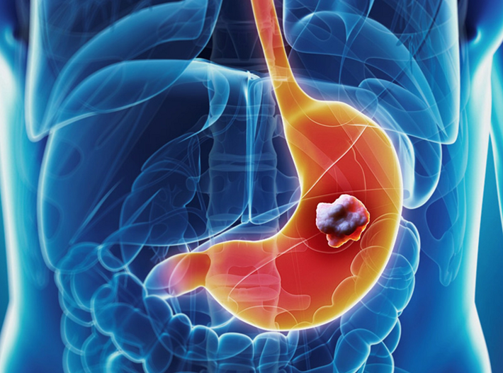
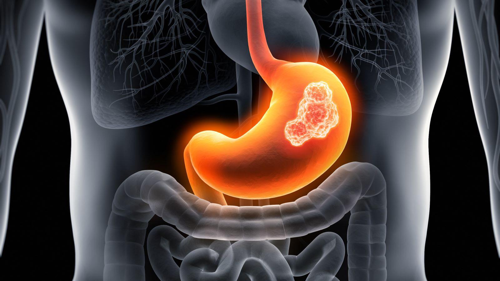
Stomach cancer (gastric cancer) refers to malignant tumors arising from the lining of the stomach. Approximately 95 percent of stomach cancers are adenocarcinomas — tumors originating from the gland cells of the gastric mucosa. Other stomach malignancies include gastrointestinal stromal tumors (GISTs), primary gastric lymphomas (including MALT lymphoma, strongly associated with H. pylori), and neuroendocrine tumors.
Gastric adenocarcinomas are classified by the Lauren system into two types. The intestinal type forms organized glandular structures, is associated with H. pylori and dietary factors, and tends to arise in the antrum and body. Its incidence has declined in Western countries over the past 70 years. The diffuse type consists of poorly cohesive cells infiltrating the stomach wall without forming recognizable glands; it is associated with CDH1 (E-cadherin) gene mutations and is more aggressive, harder to detect, and carries a worse prognosis.
Anatomically, non-cardia gastric cancers (arising in the antrum, body, or fundus) are strongly linked to H. pylori and are declining in the West. Cardia and gastroesophageal junction (GEJ) adenocarcinomas are associated with obesity, GERD, and Barrett’s esophagus and have been increasing in incidence.
Risk Factors for Stomach Cancer
H. Pylori Infection
Helicobacter pylori is the most important modifiable risk factor for stomach cancer — classified by the WHO as a Group I carcinogen. H. pylori triggers chronic gastric inflammation that progresses through a predictable sequence known as the Correa cascade: normal mucosa → chronic gastritis → atrophic gastritis → intestinal metaplasia → dysplasia → carcinoma. This process takes 20–40 years, explaining why stomach cancer predominantly affects older adults. H. pylori is present in approximately 89 percent of non-cardia gastric cancers worldwide. Eradicating H. pylori reduces gastric cancer risk by 33–47 percent — most effectively before intestinal metaplasia develops.
Diet, Smoking, and Lifestyle
High dietary sodium — from salted, pickled, and preserved foods — damages the gastric mucosa and augments H. pylori carcinogenesis. Processed and smoked meats contain nitrosamines that are gastric carcinogens. Diets low in fresh fruits and vegetables lack protective antioxidant micronutrients. Smoking approximately doubles gastric cancer risk. The global decline in stomach cancer over the 20th century correlates with the spread of refrigeration and reduced salt-preservation dependence.
Hereditary Risk
Hereditary Diffuse Gastric Cancer (HDGC) is caused by germline CDH1 mutations and carries a lifetime gastric cancer risk of 70–80%. Women with CDH1 mutations also have a 40–60% lifetime lobular breast cancer risk. Current guidelines recommend prophylactic gastrectomy for CDH1 carriers, typically between ages 20 and 30. Lynch syndrome (MMR gene mutations) confers elevated gastric cancer risk, particularly in Asian populations; Lynch-associated gastric cancers are often MSI-H. A first-degree family history of gastric cancer approximately doubles individual risk.
Early-stage stomach cancer produces symptoms (indigestion, bloating, mild loss of appetite) that are clinically indistinguishable from GERD, peptic ulcer disease, and functional dyspepsia. By the time symptoms are severe enough to prompt endoscopy, most patients are at stage III or IV. Less than 20% of US stomach cancer cases are found at stage I.
Stomach Cancer Symptoms — Early vs. Advanced
Early disease (Stage I–II) often produces no specific symptoms. When present:
- Indigestion or heartburn that is new, persistent, or changing
- Abdominal discomfort or bloating after meals
- Loss of appetite or early satiety (feeling full quickly)
- Mild nausea
Advanced/metastatic disease produces symptoms that are harder to dismiss:
- Significant unintentional weight loss: one of the most consistent advanced gastric cancer signs
- Persistent vomiting: suggests pyloric outlet obstruction from a large antral tumor
- Dysphagia (difficulty swallowing): tumor at or near the GEJ
- Hematemesis (vomiting blood): direct tumor bleeding
- Melena (black, tarry stools): chronic upper GI bleeding
- Palpable abdominal mass: large primary tumor or liver metastases
- Jaundice: liver metastases or biliary obstruction
Physical examination findings in metastatic gastric cancer include Virchow’s node (hard left supraclavicular lymph node from retrograde spread via the thoracic duct), Sister Mary Joseph nodule (periumbilical metastasis), and Krukenberg tumor (ovarian metastasis that may present before the primary tumor is identified).
How Stomach Cancer Is Diagnosed
Upper endoscopy (EGD) with biopsy is the gold standard. The endoscope allows direct visualization and biopsy of any suspicious lesion, mucosal irregularity, or area of color change. Multiple biopsies (6–8 recommended) improve sensitivity. Diffuse-type gastric cancer may appear as subtle mucosal thickening without a discrete mass and requires a high index of suspicion.
Staging evaluation after histological confirmation:
- CT chest/abdomen/pelvis: Liver metastases, lung metastases, peritoneal disease, lymphadenopathy
- Endoscopic ultrasound (EUS): Accurate T-stage (wall invasion depth) and N-stage (regional nodes) for surgical planning
- Diagnostic laparoscopy: Before surgery in locally advanced disease to exclude peritoneal metastases CT may miss (lesions <2–3 mm are below CT resolution)
Molecular biomarker testing is required for all gastric adenocarcinomas:
- HER2: Overexpression/amplification in 15–20%; adds trastuzumab to chemo
- PD-L1 CPS: Guides immunotherapy eligibility
- MSI / MMR status: MSI-H tumors respond exceptionally to pembrolizumab
- EBV status: EBV-positive tumors have high PD-L1 and strong immunotherapy responses
Stomach Cancer Stages and Survival
- Stage I (T1–2, N0): ~70% 5-year survival; confined to inner stomach layers
- Stage II (deeper invasion or limited nodal spread): intermediate survival
- Stage III (more extensive local/regional disease): 20–35% 5-year survival
- Stage IV (distant metastases): ~7% 5-year survival
Treatment of Stomach Cancer
Localized Disease (Stages I–III)
Surgery (total or subtotal gastrectomy with D2 lymphadenectomy) is the treatment cornerstone. The FLOT4 trial established the FLOT regimen (fluorouracil, leucovorin, oxaliplatin, docetaxel) as the perioperative chemotherapy standard — given 4 cycles before and 4 cycles after surgery, it improves survival significantly over older ECF-based regimens. For patients who did not receive perioperative chemotherapy, adjuvant chemoradiation (fluorouracil-based, INT-0116 protocol) reduces recurrence.
Metastatic Disease (Stage IV)
First-line systemic therapy for HER2-negative disease is a platinum-fluoropyrimidine doublet (FOLFOX or XELOX). For HER2-positive tumors, adding trastuzumab (ToGA trial) significantly improves overall survival. For PD-L1 CPS ≥1, nivolumab (CheckMate-649) or pembrolizumab (KEYNOTE-859) added to chemotherapy improves OS. For MSI-H/dMMR tumors, pembrolizumab monotherapy achieves response rates of 40–60% — far exceeding chemotherapy alone. Second-line therapy includes ramucirumab ± paclitaxel (RAINBOW trial).
Life After Stomach Cancer Treatment
Patients who undergo total gastrectomy face lifelong nutritional management. The stomach produces intrinsic factor required for vitamin B12 absorption — after total gastrectomy, B12 must be supplemented indefinitely via injection or high-dose oral forms. Iron, calcium, and vitamin D absorption are also impaired. Dumping syndrome — rapid gastric emptying causing vasomotor symptoms (early; within 30 min) or reactive hypoglycemia (late; 1–3 hours post-meal) — is managed with small frequent meals, avoiding simple sugars, and not drinking fluids during meals.
Surveillance after curative-intent treatment includes CT scans, upper endoscopy, and clinical assessment every 3–6 months for 2 years, then annually for 3 additional years.
Hereditary Stomach Cancer — CDH1 and Lynch Syndrome
Genetic counseling and CDH1 testing are indicated when: two or more first- or second-degree relatives had diffuse gastric cancer (one diagnosed under age 50); three or more cases in the family regardless of age; or personal/family history of both diffuse gastric cancer and lobular breast cancer. Confirmed CDH1 mutation carriers face a choice between intensive surveillance endoscopy — which has limited sensitivity for submucosally distributed signet ring foci — and prophylactic gastrectomy, which eliminates gastric cancer risk but requires lifelong nutritional management. Most major guidelines recommend prophylactic gastrectomy for confirmed CDH1 carriers, typically between ages 20 and 30.
Lynch syndrome patients with elevated gastric risk — particularly those of East Asian descent — may be considered for surveillance upper endoscopy in addition to colon cancer colonoscopy surveillance, reflecting the overlap between these hereditary cancer syndromes.
For broader gastrointestinal cancer context, see our guides on colon cancer and pancreatic cancer. For understanding how molecular testing drives modern cancer treatment decisions, see our overview of targeted cancer therapy.
Sources: National Cancer Institute — Stomach Cancer | American Cancer Society — Stomach Cancer | American Society of Clinical Oncology
H. Pylori Eradication — The Most Effective Stomach Cancer Prevention Strategy
Of all the risk factors for stomach cancer, H. pylori infection is the most actionable. Unlike genetic predisposition or age, H. pylori is a treatable bacterial infection, and its treatment measurably reduces stomach cancer risk. This makes H. pylori test-and-treat one of the most important cancer prevention opportunities currently available in gastroenterology.
Standard H. pylori eradication regimens use a combination of antibiotics with acid suppression. The most widely used approaches include:
- Triple therapy: proton pump inhibitor (PPI) + clarithromycin + amoxicillin or metronidazole for 14 days. This was the historical standard but eradication rates have declined in regions with high clarithromycin resistance.
- Bismuth quadruple therapy: PPI + bismuth subsalicylate + metronidazole + tetracycline for 10–14 days. Preferred in high-clarithromycin-resistance areas and as salvage therapy.
- Concomitant therapy: PPI + clarithromycin + amoxicillin + metronidazole simultaneously. Higher eradication rates than triple therapy in some regions.
Eradication success should be confirmed 4–8 weeks after completing treatment using a urea breath test or fecal antigen test — not serology, which remains positive even after successful eradication. Patients in whom eradication fails require a salvage regimen with different antibiotic choices.
Who should be tested and treated for H. pylori? Current guidelines recommend H. pylori testing in:
- Patients with active peptic ulcer disease, whether or not they are symptomatic
- Patients with a family history of gastric cancer (first-degree relative)
- Individuals of East Asian descent or immigrants from high-prevalence countries (South Korea, Japan, China, much of South America, Eastern Europe)
- Patients with unexplained iron deficiency anemia or immune thrombocytopenic purpura (ITP)
- Patients with gastric intestinal metaplasia found on endoscopy
- Patients with gastric MALT lymphoma
In countries with very high gastric cancer incidence — Japan and South Korea have national screening programs combining upper endoscopy and H. pylori testing starting at age 40 — population-level H. pylori eradication campaigns have contributed to measurable reductions in gastric cancer incidence over the past two decades. Japan has also implemented upper endoscopy screening for early gastric cancer detection, which has shifted the stage distribution of diagnosed cases toward earlier, more treatable disease. These programs have not been adopted in the US due to the lower baseline gastric cancer incidence, but high-risk individuals in the US (East Asian descent, family history, immigrants from high-incidence regions) should be considered for more intensive surveillance.
Screening and Surveillance for Stomach Cancer
In the United States, there is no population-wide stomach cancer screening program — the incidence is too low to justify mass endoscopy screening in the general population. However, certain high-risk groups are candidates for targeted surveillance:
Endoscopic surveillance for intestinal metaplasia: Patients found to have gastric intestinal metaplasia on endoscopy — particularly extensive or severe metaplasia, or metaplasia at the cardia — are at elevated risk for progression to dysplasia and carcinoma. The MAPS II European guideline recommends endoscopic surveillance every 3 years for extensive intestinal metaplasia. In the US, surveillance intervals are not universally standardized but follow similar principles.
Surveillance for pernicious anemia: Autoimmune gastritis leading to pernicious anemia causes achlorhydria and extensive gastric mucosal atrophy — both precancerous states. Patients with pernicious anemia have approximately 2–3 times the gastric cancer risk of the general population. Periodic upper endoscopy surveillance is reasonable in this population.
Post-gastric surgery surveillance: Patients who underwent Billroth II gastrectomy (partial gastric resection with gastrojejunostomy) 15–20 years ago are at elevated gastric remnant cancer risk due to chronic bile reflux and mucosal changes in the gastric remnant. Periodic endoscopy of the remnant stomach is recommended in this population.
Emerging Treatments and Clinical Trials in Gastric Cancer
Gastric cancer treatment has evolved rapidly with the integration of molecular profiling and immunotherapy. Several important developments are changing outcomes for subsets of patients:
Trastuzumab deruxtecan (T-DXd) is an antibody-drug conjugate that links trastuzumab to a topoisomerase I inhibitor payload. In the DESTINY-Gastric01 and DESTINY-Gastric02 trials, T-DXd demonstrated remarkable activity in HER2-positive gastric cancer patients who had progressed after trastuzumab-based first-line therapy, with response rates significantly higher than standard second-line chemotherapy. T-DXd has received FDA approval as a third-line option in HER2-positive gastric cancer and is being studied in earlier lines of therapy.
Claudin 18.2 (CLDN18.2) is a tight junction protein overexpressed in a substantial proportion of gastric cancers. Zolbetuximab, a monoclonal antibody targeting CLDN18.2, combined with chemotherapy demonstrated significantly improved overall survival in CLDN18.2-positive, HER2-negative advanced gastric cancer in the SPOTLIGHT and GLOW trials. CLDN18.2 testing is expected to become part of the standard molecular workup for advanced gastric cancer as these results lead to approvals.
FGFR2 inhibitors: FGFR2b amplification occurs in approximately 5% of gastric cancers. Bemarituzumab, an anti-FGFR2b antibody, combined with chemotherapy improved progression-free and overall survival in FGFR2b-positive gastric cancers in the FIGHT trial. A second larger confirmatory trial is underway.
Immunotherapy combinations: Multiple trials are exploring combinations of PD-1/PD-L1 inhibitors with VEGF/VEGFR inhibitors, HER2-targeted agents, and other checkpoint inhibitors in various biomarker-selected populations. The field is moving toward increasingly precise, biomarker-driven treatment selection based on molecular profiling of each individual tumor.
For patients with newly diagnosed advanced gastric cancer, enrollment in a clinical trial should always be considered — particularly for those with specific molecular profiles (MSI-H, HER2+, CLDN18.2+, FGFR2b+) for which targeted agents are available in trial settings that may exceed standard-of-care options. Clinical trial eligibility can be checked through NCI’s cancer clinical trials database or discussed with an oncologist at a comprehensive cancer center that specializes in upper GI cancers.
Stomach Cancer in Special Populations
East Asian Patients
Stomach cancer incidence is substantially higher in East Asian countries than in the United States. South Korea, Japan, and China have age-standardized incidence rates 3–5 times higher than the US, driven by higher H. pylori prevalence, dietary patterns (high salt and fermented foods), and genetic susceptibility factors. East Asian immigrants to the United States retain higher-than-average gastric cancer risk for at least one generation, declining in subsequent generations as dietary and environmental exposures change. For East Asian patients in the US with new dyspeptic symptoms, H. pylori testing and a lower threshold for upper endoscopy referral are clinically appropriate given this elevated background risk.
Young Patients With Stomach Cancer
Gastric cancer in patients under 45 is uncommon but not rare, and when it occurs it is more likely to be the diffuse histological type — particularly signet ring cell carcinoma — than in older patients. Young-onset gastric cancer is more frequently associated with hereditary causes (CDH1 mutations, Lynch syndrome, BRCA2) than sporadic gastric cancer in older adults. Any patient under 45 with a confirmed gastric adenocarcinoma should be referred for genetic counseling and testing, particularly if there is a family history of gastric cancer, lobular breast cancer, or colorectal cancer.
Women With Stomach Cancer
The male-to-female ratio in gastric cancer is approximately 2:1 in most populations, but this ratio is smaller in diffuse-type gastric cancer. Women with CDH1 mutations face not only the 70–80% lifetime gastric cancer risk but also the 40–60% lifetime lobular breast cancer risk, and their management requires coordination between gastroenterology/surgical oncology (for prophylactic gastrectomy decisions) and breast oncology (for MRI-based breast surveillance). Women with gastric cancer also need to be assessed for ovarian metastases (Krukenberg tumors), which can occasionally be the presenting finding before the primary gastric tumor is identified.
Nutritional Support During Stomach Cancer Treatment
Adequate nutritional support is a central component of stomach cancer management that is sometimes inadequately addressed. Patients with gastric cancer commonly present with some degree of malnutrition at the time of diagnosis, both because the tumor impairs gastric function (early satiety, outlet obstruction, reduced acid production affecting digestion) and because the systemic inflammatory response of cancer increases metabolic demands. Malnutrition at the time of surgery is associated with worse post-operative outcomes, higher complication rates, and longer hospital stays.
Pre-operative nutritional optimization — including high-protein supplementation, enteral nutrition if oral intake is severely impaired, and correction of micronutrient deficiencies (iron, B12, vitamin D) — is recommended for patients with significant weight loss or nutritional impairment before gastrectomy. After gastrectomy, both jejunal tube feeding and oral diet progression protocols have been developed to support recovery.
Patients receiving palliative chemotherapy for metastatic gastric cancer are also at high nutritional risk. Chemotherapy-induced nausea, mucositis, and changes in taste and appetite compound the cancer’s direct effect on eating. Dietitian involvement throughout treatment — not only at diagnosis but at every major transition in treatment — improves quality of life and may support treatment tolerability.
The Bottom Line on Stomach Cancer
Stomach cancer is a disease that rewards early detection and punishes diagnostic delay. The gap between its early-stage cure rate (greater than 70%) and its late-stage survival (approximately 7% at stage IV) represents one of the largest stage-dependent survival gradients in oncology. The tragedy is that the early symptoms — indigestion, early satiety, mild nausea — are so nonspecific that most patients and their physicians attribute them to benign causes for months before pursuing endoscopy. For anyone with a significant H. pylori history, a family history of gastric cancer, or any symptoms that are new, persistent, and changing — particularly in combination with unintentional weight loss — upper endoscopy is the test that makes the difference between an early and a late diagnosis.
For digestive cancer comparisons and broader GI oncology context, see our guides on colon cancer and pancreatic cancer symptoms. For understanding how modern molecular testing drives treatment selection, see our overview of targeted cancer therapy.
New onset of persistent digestive symptoms — especially in individuals over 55, those with a smoking history, those of East Asian descent, or anyone with a family history of gastric cancer — warrants prompt evaluation rather than empirical antacid therapy alone. The decision to proceed to upper endoscopy early rather than late is almost always the right one when the index of suspicion is present.