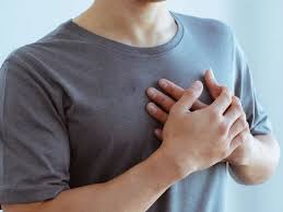
A fast heartbeat — medically called tachycardia — is defined as a resting heart rate above 100 beats per minute. The sensation of the heart beating rapidly is one of the most common reasons people seek medical attention, yet the vast majority of fast heartbeats are physiologically normal responses to identifiable triggers. The challenge — for patients and clinicians alike — is distinguishing the harmless from the hazardous. Understanding what drives a fast heartbeat, which patterns are concerning, and when to seek evaluation is essential for navigating this extremely common complaint.
What Is a Fast Heartbeat?
The normal resting heart rate falls between 60 and 100 beats per minute. Well-trained athletes may have resting rates in the 40s or 50s, reflecting the cardiac efficiency that comes with prolonged aerobic conditioning. A heart rate consistently above 100 beats per minute at rest, or during periods that would not normally provoke tachycardia, is defined as tachycardia — a term covering a spectrum from mild physiologic elevation to life-threatening arrhythmias.
The heart’s natural pacemaker — the sinoatrial (SA) node in the right atrium — sets the heart rate under the influence of the autonomic nervous system. Sympathetic activation (the “fight or flight” response) increases SA node firing rate; parasympathetic (vagal) tone slows it. Most physiologic tachycardias are sinus tachycardias: the SA node accelerates appropriately in response to a trigger, and the rate returns to normal when the trigger resolves. Pathologic tachycardias involve abnormal circuits, ectopic pacemakers, or reentrant mechanisms that generate fast rates independently of the SA node.
Common Harmless Causes of Fast Heartbeat
The majority of fast heartbeats are sinus tachycardias driven by identifiable physiologic demands or triggers.
Exercise is the most common cause. The heart rate increases proportionally to exercise intensity to deliver more oxygenated blood to working muscles. Maximum heart rate is estimated at approximately 220 minus age, and rates near this maximum during vigorous exercise are entirely expected and appropriate. The rate returns to baseline within minutes of stopping, with the speed of recovery reflecting cardiovascular fitness.
Fever raises heart rate because elevated body temperature accelerates metabolic processes and oxygen consumption. Each 1 degree Celsius rise in temperature increases heart rate by approximately 8 to 10 beats per minute. A person with a fever of 39 degrees Celsius may have a resting heart rate of 110 simply as a result of the fever itself — not from any cardiac problem.
Dehydration reduces blood volume and venous return to the heart, causing a reflexive increase in heart rate to maintain cardiac output and blood pressure. The tachycardia resolves promptly with adequate fluid repletion. Anemia reduces the blood’s oxygen-carrying capacity; the heart compensates by beating faster to deliver adequate oxygen through increased cardiac output. Iron deficiency anemia, B12 deficiency, and other causes of significant anemia commonly produce persistent sinus tachycardia that resolves with treatment.
Hyperthyroidism is one of the most important reversible medical causes of tachycardia. Excess thyroid hormone directly accelerates SA node firing and increases myocardial sensitivity to catecholamines, producing persistent sinus tachycardia and — in a significant proportion of patients — triggering atrial fibrillation. Checking TSH is a critical step in any evaluation of persistent unexplained tachycardia.
Anxiety and emotional stress activate the sympathetic nervous system, releasing adrenaline that accelerates the SA node. Panic attacks can produce heart rates of 120 to 140 beats per minute in a normal heart. Caffeine and stimulants — including decongestants (pseudoephedrine), ADHD medications, energy drinks, and illicit stimulants — produce dose-dependent sinus tachycardia through their sympathomimetic or adenosine receptor-blocking effects.
Arrhythmias That Cause a Fast Heartbeat
When a fast heartbeat arises from an abnormal cardiac rhythm — rather than from the SA node responding to physiologic demand — it is classified as a tachyarrhythmia. These are distinguished by their onset pattern, rate, regularity, and clinical context.
Supraventricular tachycardia (SVT) — including atrioventricular nodal reentrant tachycardia (AVNRT) and atrioventricular reentrant tachycardia (AVRT) — produces heart rates of 150 to 250 beats per minute, with the defining clinical feature being abrupt onset and termination. The patient’s heart rate “flips” from normal to 180 or 200 beats per minute in a single beat, and then back to normal just as suddenly. SVT is commonly benign in terms of cardiac risk but the symptoms it causes — racing heart, dizziness, dyspnea, near-syncope — can be highly disruptive. Catheter ablation is curative in more than 95 percent of cases.
Atrial fibrillation (AF) produces a rapid and irregularly irregular ventricular rate, typically 100 to 180 beats per minute when uncontrolled. It is the most common sustained cardiac arrhythmia, affecting more than 6 million Americans, and its importance lies not in the fast rate itself but in its consequences: five-fold increase in stroke risk and risk of tachycardia-induced cardiomyopathy. A fast, irregular heartbeat that is persistent or recurrent requires evaluation for AF.
Atrial flutter produces a very organized atrial circuit firing at 250 to 350 beats per minute with a characteristic 2:1 AV block, creating a regular ventricular rate of approximately 150 beats per minute. Ventricular tachycardia (VT) arises from ectopic foci or reentrant circuits within the ventricular myocardium. Sustained VT — lasting more than 30 seconds or requiring termination — is a serious arrhythmia that can cause hemodynamic collapse and requires urgent treatment. Ventricular fibrillation (VF) — chaotic ventricular activation with no organized cardiac output — causes cardiac arrest and is immediately life-threatening.

When a Fast Heartbeat Is Dangerous
The distinction between a concerning and a non-concerning fast heartbeat depends on multiple factors.
Rate: Higher rates generally indicate more concern. A sinus tachycardia of 110 bpm in a febrile patient is expected. A sustained rate of 200 bpm in a resting patient is an arrhythmia until proven otherwise.
Associated symptoms: A fast heartbeat accompanied by syncope, near-syncope, chest pain, severe shortness of breath, or profound weakness suggests hemodynamic compromise and constitutes a medical emergency. A fast heartbeat with mild palpitations and no other symptoms in an otherwise healthy person is far less likely to represent danger.
Underlying heart disease: The same tachycardia carries vastly different risk depending on the cardiac substrate. Non-sustained VT in a structurally normal heart is usually benign. Non-sustained VT in a patient with a prior myocardial infarction and reduced ejection fraction is a risk factor for sudden cardiac death. Structural heart disease — cardiomyopathy, prior MI, hypertrophic cardiomyopathy, and inherited arrhythmia syndromes (long QT syndrome, Brugada syndrome, arrhythmogenic right ventricular cardiomyopathy) — significantly raises the risk associated with any tachyarrhythmia.
Wolff-Parkinson-White (WPW) syndrome: Patients with an accessory pathway connecting the atria and ventricles are at particular risk if they develop AF. Instead of being filtered by the AV node, atrial impulses can conduct directly to the ventricles through the accessory pathway at very high rates — 200 to 300 bpm — potentially degenerating to ventricular fibrillation. AF in a WPW patient is a medical emergency, and AV nodal blocking agents (including verapamil and digoxin) are contraindicated in this setting.
Family history: A personal or family history of sudden cardiac death before age 50, unexplained fainting during exertion, or a known inherited arrhythmia syndrome significantly raises the clinical stakes for any fast heartbeat presentation.
Postural Orthostatic Tachycardia Syndrome (POTS)
Postural orthostatic tachycardia syndrome (POTS) is a form of autonomic dysfunction in which the heart rate increases abnormally upon standing. The diagnostic criterion is a heart rate increase of at least 30 beats per minute (or at least 40 bpm in patients younger than 19) within 10 minutes of standing, in the absence of orthostatic hypotension. POTS affects an estimated 1 to 3 million Americans and predominantly affects women between the ages of 15 and 50.
Patients with POTS experience rapid heart rate — often reaching 120 to 140 bpm — along with dizziness, lightheadedness, fatigue, brain fog, and near-syncope when upright. Symptoms typically improve significantly or resolve when lying down, which helps distinguish POTS from other causes of chronic tachycardia. Post-COVID POTS — a form of dysautonomia triggered by SARS-CoV-2 infection — has been recognized as a significant component of long COVID syndrome.
Management of POTS includes increased salt and fluid intake (targeting 2 to 3 liters of water per day and 10 grams of sodium), compression stockings and abdominal binders to reduce venous pooling, graduated exercise rehabilitation, and medications including low-dose beta-blockers, ivabradine, fludrocortisone, and midodrine. Most patients improve substantially with a comprehensive management approach.
Inappropriate Sinus Tachycardia
Inappropriate sinus tachycardia (IST) is characterized by a persistently elevated resting heart rate above 100 beats per minute in sinus rhythm, with normal P wave morphology, without an identifiable physiologic cause. It predominantly affects young women and can cause significant symptoms including palpitations, fatigue, exercise intolerance, and anxiety about the elevated rate.
IST is a diagnosis of exclusion — hyperthyroidism, anemia, dehydration, stimulant use, anxiety disorders, and other reversible causes of sinus tachycardia must be systematically excluded before the diagnosis is made. Management includes lifestyle modifications (caffeine reduction, adequate hydration, reduced stress), and pharmacologic therapy with beta-blockers or ivabradine, which reduces the SA node firing rate by blocking the HCN channel (If current).
When to Seek Medical Care
A fast heartbeat warrants urgent or emergency evaluation when it is accompanied by syncope or near-syncope, chest pain or pressure, severe shortness of breath, an ongoing rapid rate that does not resolve, or any of these symptoms in a patient with known structural heart disease or inherited arrhythmia syndrome. A family history of sudden cardiac death before age 50 elevates the urgency of any fast heartbeat evaluation.
A fast heartbeat is more likely to be benign when it has a clear identifiable trigger (exercise, fever, anxiety, stimulants), resolves promptly when the trigger is removed, is not associated with any other symptoms, and occurs in a young, otherwise healthy person with no cardiac history.
How a Fast Heartbeat Is Evaluated
Evaluation begins with a 12-lead ECG, which can identify the rhythm mechanism in real time if the tachycardia is present, and reveals baseline abnormalities (pre-excitation suggesting WPW, prolonged QT, LVH, prior MI) when the heart is in normal rhythm. Blood tests including TSH, CBC, and a basic metabolic panel screen for thyroid disease, anemia, and electrolyte abnormalities. An echocardiogram evaluates structural heart disease and left ventricular function.
For intermittent tachycardia, ambulatory monitoring — 24 to 48-hour Holter monitoring for frequent episodes or a 30-day event recorder for less frequent symptoms — is the most important tool for documenting the rhythm during symptoms. A tilt table test or active stand test evaluates suspected POTS or orthostatic tachycardia. An exercise stress test is used when tachycardia is exertional. For confirmed arrhythmias amenable to catheter ablation, electrophysiology (EP) study characterizes the arrhythmia mechanism and guides definitive treatment.
For heart rate context, see our article on what resting heart rate means. For the broader picture of cardiovascular health numbers, see heart health numbers every adult should know. For more detail on palpitation causes that overlap with fast heartbeat, see our article on heart palpitations: common causes and warning signs.
The American Heart Association offers patient-friendly information on tachycardia and its management. The NIH National Heart, Lung, and Blood Institute provides detailed clinical information on types and treatments of tachycardia. The CDC provides epidemiological data on arrhythmia as a cardiovascular condition.
A fast heartbeat is a symptom, not a diagnosis. The significance of tachycardia depends entirely on its cause, the clinical context, and the patient’s underlying cardiac health. When the cause is a normal physiologic response to a recognizable trigger, reassurance and trigger management are sufficient. When it reflects an arrhythmia — particularly in a patient with structural heart disease or with high-risk features — prompt evaluation and targeted treatment can prevent serious complications.
Treatment Options for Arrhythmia-Related Tachycardia
When a fast heartbeat is caused by a cardiac arrhythmia rather than a physiologic trigger, treatment depends on the specific arrhythmia, its hemodynamic impact, and the patient’s underlying cardiac status.
SVT (AVNRT/AVRT): Acute termination is achieved with vagal maneuvers (modified Valsalva first), adenosine intravenously if vagal maneuvers fail, or synchronized cardioversion for hemodynamically unstable patients. Long-term management options include watchful waiting for infrequent and well-tolerated episodes, rate-control or rhythm-control medications (beta-blockers, calcium channel blockers, flecainide, propafenone), or — the preferred definitive approach — catheter ablation, which achieves cure rates above 95 percent for most SVT subtypes with minimal procedural risk.
Atrial fibrillation management involves two parallel tracks: stroke prevention through anticoagulation (direct oral anticoagulants such as apixaban or rivaroxaban are preferred over warfarin for most patients), and control of the ventricular rate and/or rhythm. Rate control uses AV nodal blocking agents (beta-blockers, diltiazem, digoxin) to keep the ventricular response below 100 to 110 bpm at rest. Rhythm control — restoring and maintaining sinus rhythm — uses antiarrhythmic medications (flecainide, sotalol, amiodarone, dofetilide, dronedarone) or catheter ablation targeting pulmonary vein isolation. Catheter ablation for AF is increasingly used as a first-line rhythm control strategy, particularly in younger patients and those with heart failure with reduced ejection fraction.
Ventricular tachycardia: Acute sustained VT with hemodynamic compromise requires immediate synchronized cardioversion. Stable sustained VT can be terminated with intravenous antiarrhythmic medications (amiodarone, lidocaine, procainamide). Long-term management of VT in patients with structural heart disease includes implantable cardioverter-defibrillator (ICD) therapy for secondary prevention and catheter ablation for VT storm or frequent ICD shocks. Beta-blockers reduce the burden of VT in most cardiomyopathy patients and reduce sudden cardiac death risk in patients with reduced ejection fraction.
Lifestyle Factors That Affect Heart Rate
For patients with physiologic tachycardia — sinus tachycardia driven by modifiable lifestyle factors — non-pharmacologic interventions often substantially reduce resting heart rate and symptom frequency. Regular aerobic exercise reduces resting heart rate through increased vagal tone and improved cardiac efficiency; well-conditioned athletes have resting rates 10 to 20 bpm lower than sedentary individuals. Caffeine reduction benefits caffeine-sensitive patients who notice palpitations or elevated heart rates with high intake. Adequate hydration prevents the dehydration-related reflex tachycardia that many people experience during hot weather or illness. Alcohol moderation reduces sympathetic tone and the risk of holiday heart AF. Stress management — through regular physical activity, adequate sleep, cognitive behavioral approaches, and mindfulness — reduces the sympathetic activation that drives anxiety-related tachycardia.
Weight management is an important cardiovascular risk factor for arrhythmia. Obesity is independently associated with increased AF risk, and significant weight loss — whether through lifestyle intervention or bariatric surgery — has been shown to reduce AF burden and improve rhythm control outcomes. Sleep apnea is another frequently overlooked contributor to fast heart rates and arrhythmia: intermittent hypoxia during apneic episodes triggers sympathetic surges that increase heart rate and predispose to both sinus tachycardia and AF. Treating sleep apnea with continuous positive airway pressure (CPAP) reduces AF recurrence rates.
Frequently Asked Questions About Fast Heartbeat
How high can a heart rate go before it becomes dangerous?
There is no single threshold, because the danger depends on the arrhythmia mechanism and the patient’s cardiac status. During maximum exercise, heart rates of 180 to 200 bpm are physiologically normal for young healthy people. The same rate at rest, arising from VT in a patient with heart failure, is immediately life-threatening. As a practical rule, a resting rate above 150 bpm without an obvious physiologic trigger warrants evaluation. Rates above 200 bpm at rest almost always represent a pathologic arrhythmia requiring urgent assessment.
Can a fast heart rate damage the heart over time?
Yes — persistent or chronic tachycardia at rates above 100 bpm can cause tachycardia-induced cardiomyopathy, a reversible form of reduced left ventricular function that develops when the heart beats too fast for too long. This can occur with uncontrolled AF, persistent inappropriate sinus tachycardia, or incessant SVT. The cardiomyopathy typically reverses substantially or completely once the heart rate is controlled. This is one reason why rate control matters even when AF symptoms are mild — silent heart rate elevation can impair LV function over months to years.
Is it normal to have a fast heart rate when anxious or stressed?
Yes. Anxiety activates the sympathetic nervous system, releasing adrenaline that increases the SA node firing rate. Heart rates of 100 to 140 bpm during acute anxiety or panic attacks are physiologically normal responses. The distinction to be aware of: anxiety-triggered tachycardia is gradual in onset, correlates with the emotional experience, and resolves as anxiety subsides. Arrhythmia-driven tachycardia is often abrupt in onset, may bear no relation to emotional state, and may not resolve spontaneously. When in doubt — particularly with a new or unusual pattern — an ECG during the episode is the most informative diagnostic tool.
What is the relationship between a fast heartbeat and heart attack?
Myocardial infarction (heart attack) can cause tachycardia through several mechanisms: pain-related sympathetic activation, hemodynamic compromise requiring compensatory tachycardia, or direct ischemia triggering ventricular arrhythmias (VT or VF). Ventricular fibrillation is the most common cause of death in the first hour of an acute MI before hospital arrival. A sudden fast heartbeat accompanied by chest pain, jaw or arm discomfort, sweating, nausea, or shortness of breath in a patient at cardiovascular risk should be treated as a potential acute coronary syndrome and evaluated as an emergency.
Heart Rate Monitoring at Home
Consumer-grade heart rate monitoring has become remarkably accessible and accurate. Wearable devices — smartwatches and fitness trackers from major manufacturers — use photoplethysmography (PPG) to measure heart rate continuously throughout the day. Many newer devices also include single-lead ECG capability, allowing users to record a rhythm strip during a symptomatic episode and share it with their physician. These recordings have detected AF and other arrhythmias in patients who previously had no documentation of their symptoms.
The clinical value of wearable monitoring comes with important caveats. Consumer devices are reasonably accurate at rest but less reliable during motion or in patients with arrhythmias that produce irregular rhythms. A detected “high heart rate” alert from a smartwatch can be valuable if it captures an otherwise undocumented tachycardia episode — but it should be followed up with medical evaluation rather than treated as a definitive diagnosis. A clinically validated ambulatory monitor worn for 24 to 30 days remains the standard for documenting and characterizing arrhythmias for medical decision-making. Wearables and medical monitors are complementary tools, not substitutes for one another.
For most people experiencing occasional episodes of a fast heartbeat without warning signs, the probability of a dangerous arrhythmia is low — but the probability is not zero, and the only way to distinguish benign physiologic tachycardia from a rhythm problem is appropriate evaluation. A physician visit, an ECG, and targeted blood tests will reassure the majority and correctly identify the minority who need further workup. When warning signs are present, that evaluation becomes urgent. When they are absent in an otherwise healthy person, the pace of evaluation can be more relaxed — but the evaluation should still happen.