
Angina: Chest Pain from Reduced Blood Flow
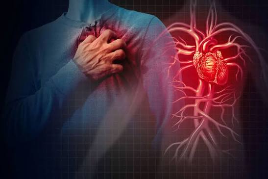
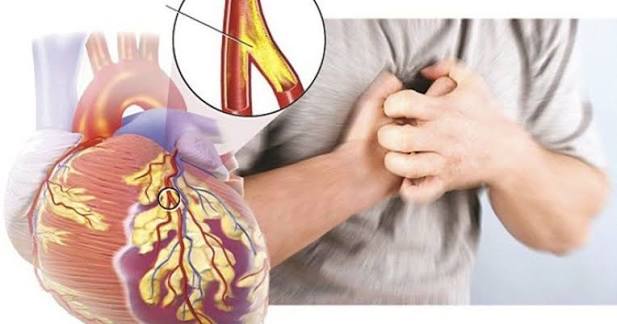
Angina pectoris — most commonly called simply angina — is chest pain, pressure, tightness, or discomfort caused by insufficient blood flow to the heart muscle (myocardium). The word “angina” comes from the Latin for “strangling” or “tightening,” which captures the sensation many patients describe: a squeezing, pressing, or heavy weight on the chest, often accompanied by radiation to the left arm, jaw, neck, or back. Angina is not a disease itself but a symptom — a warning signal that the heart is not receiving enough oxygen to meet its current demands, typically because one or more coronary arteries are narrowed by atherosclerotic plaque and cannot deliver adequate blood flow during periods of increased cardiac work.
Approximately 9 million Americans have stable angina, and it is the initial presentation of coronary artery disease in approximately half of all patients who develop symptomatic coronary heart disease. Understanding angina — its types, triggers, characteristics, and how it differs from heart attack — is essential for the millions of patients who experience it and for recognizing when chest pain requires emergency evaluation versus outpatient management.
Types of Angina — Stable, Unstable, and Variant
Stable angina is the most common type, characterized by predictable, reproducible chest discomfort that occurs consistently at similar levels of exertion or emotional stress and reliably resolves with rest or nitroglycerin within a few minutes (typically 1 to 5 minutes). The “stable” designation reflects a consistent, predictable pattern: the patient knows that climbing two flights of stairs or walking briskly for three blocks will produce angina, and knows that stopping and resting will relieve it. Stable angina reflects a fixed coronary stenosis (a narrowing that does not change acutely) that limits the coronary artery’s ability to increase blood flow in response to increased demand, but does not impair resting flow significantly enough to cause symptoms at rest.
The stability of stable angina is clinically reassuring because it suggests the underlying plaque is not acutely rupturing or rapidly progressing. However, stable angina is not benign — it indicates significant coronary artery disease with meaningful risk of future myocardial infarction, and its presence mandates evaluation, risk stratification, and treatment intensification. New-onset exertional chest pain that has not yet been evaluated represents “new-onset angina” — a subcategory that warrants prompt cardiology evaluation even if the pattern is currently stable, because it reflects new coronary disease that may or may not be fully stable.
Unstable angina is a form of acute coronary syndrome (ACS) — together with NSTEMI (non-ST elevation myocardial infarction) — and represents a medical emergency. Unstable angina is defined by one of three patterns: angina at rest or with minimal exertion (severe enough to limit ordinary activity), new-onset angina that is severe and limits activity, or previously stable angina that has changed in character — becoming more frequent, more severe, occurring at lower exertion thresholds, lasting longer, or responding less well to nitroglycerin. These changes indicate that the underlying atherosclerotic plaque has ruptured or eroded, triggering platelet aggregation and thrombus formation that partially obstructs the coronary artery. Unstable angina requires emergency evaluation and hospitalization because it carries a high short-term risk of progression to complete coronary occlusion and STEMI (ST-elevation myocardial infarction).
Vasospastic (Prinzmetal’s) angina is an atypical form caused by transient coronary artery spasm rather than fixed atherosclerotic obstruction. Unlike stable angina — which characteristically occurs with exertion — vasospastic angina typically occurs at rest, often at night or in the early morning, and may be triggered by cold exposure, emotional stress, hyperventilation, or cocaine use. The underlying coronary arteries may have no significant fixed stenosis, which is why vasospastic angina can occur in young people without traditional coronary risk factors. The ECG during an episode shows ST elevation (as in STEMI) rather than ST depression, because the spasm can produce transmural ischemia; the distinction is that spasm resolves spontaneously or with nitroglycerin, whereas STEMI from plaque rupture does not.
How to Recognize an Angina Episode — Symptoms and Characteristics
Angina has characteristic features that help distinguish it from other causes of chest pain, though its presentation is variable enough that clinical evaluation is always required to confirm the diagnosis.
Quality: Angina is most commonly described as pressure, tightness, squeezing, heaviness, or a band-like sensation across the chest. Patients may make a fist and hold it over the sternum (the “Levine sign”) to describe the sensation — a classic teaching gesture. Sharp, stabbing, or knife-like pain is less typical of angina and more suggestive of musculoskeletal, pericardial, or pleuritic causes. Some patients, particularly women and diabetic patients, experience angina as dyspnea, fatigue, jaw pain, or epigastric discomfort without prominent chest pain — these “atypical” presentations are actually common and should not preclude angina diagnosis in appropriate clinical contexts.
Location: Substernal (behind the breastbone) or left-sided chest discomfort is most common. Radiation to the left arm (particularly the inner forearm and fingers), jaw, neck, or left shoulder is characteristic and reflects the shared embryological origin of cardiac and upper extremity afferent nerves. Right arm or bilateral arm radiation, while less classic, occurs in a minority of angina patients. Interscapular or back pain can represent angina (particularly in inferior ischemia from right coronary artery disease) or — when tearing in quality — aortic dissection, an important diagnostic distinction.
Triggers: Physical exertion is the primary stable angina trigger — walking uphill, stair climbing, carrying heavy objects, sexual activity, or any sustained physical effort. Emotional stress (anger, anxiety, grief), cold exposure (which increases sympathetic tone and afterload), large meals (which divert blood to the splanchnic circulation), and early morning hours (when sympathetic tone and platelet aggregability are highest) are well-recognized non-exertional triggers for stable angina in patients with coronary disease.
Duration and relief: Stable angina typically resolves within 1 to 5 minutes of rest. Chest pain lasting more than 15 to 20 minutes despite rest and nitroglycerin is an acute coronary syndrome (unstable angina or MI) until proven otherwise and warrants emergency evaluation. The Canadian Cardiovascular Society (CCS) classification grades stable angina by functional limitation: Class I (angina only with strenuous or prolonged exertion), Class II (slight limitation — angina with moderate exertion such as climbing more than one flight or walking quickly), Class III (marked limitation — angina with mild exertion, one flight, or 100 to 200 meters on the flat), and Class IV (angina at any level of activity or at rest).
Diagnosing Angina — Tests That Confirm Ischemia
Angina diagnosis involves confirming that chest symptoms are caused by myocardial ischemia and identifying the extent and distribution of coronary disease that underlies it. The evaluation pathway depends on symptom characteristics, pretest probability of coronary disease, and the urgency of the clinical presentation.
Resting ECG is obtained in all patients with chest pain — it may show evidence of prior myocardial infarction (Q waves, ST changes), left ventricular hypertrophy from hypertension, or bundle branch block that could affect downstream testing interpretation. Importantly, the resting ECG is normal in most patients with stable angina — ischemia at rest is typically absent in stable disease, and the ECG only shows ischemic changes (ST depression or elevation) during active ischemia.
Exercise stress testing — the standard treadmill or bicycle ergometry test — provokes ischemia through graded increases in cardiac workload while monitoring ECG, blood pressure, heart rate, and symptoms. ST depression of 1 mm or more during exercise, particularly if accompanied by symptoms, hypotension, or poor exercise tolerance, is a positive test indicating significant coronary ischemia. Exercise stress testing provides important prognostic information beyond binary positive/negative: exercise capacity (measured in METs), heart rate response, blood pressure response, and the Duke Treadmill Score predict long-term cardiac outcomes independently.
Stress imaging tests — stress echocardiography (exercise or dobutamine) or nuclear stress testing (exercise or pharmacological with adenosine, regadenoson, or dipyridamole) — combine provocative stress with imaging to detect regional wall motion abnormalities or perfusion defects that indicate ischemia. Stress imaging is preferred over exercise ECG alone when the resting ECG has abnormalities that limit ECG interpretation (left bundle branch block, pacemaker rhythm, digoxin effect, LVH with repolarization changes), when pharmacological stress is required (patients who cannot exercise adequately), and when greater diagnostic accuracy or localization of ischemia is needed.
Coronary CT angiography (CCTA) is increasingly used as the first-line test for evaluation of chest pain with low-to-intermediate pretest probability of obstructive CAD, particularly following the SCOT-HEART and PROMISE trials that established CCTA’s accuracy and clinical utility in this population. CCTA directly visualizes coronary anatomy, identifying stenoses, plaque burden, and plaque characteristics (calcified vs. non-calcified) with high sensitivity and negative predictive value — a normal CCTA effectively excludes significant obstructive coronary disease.
Treating Stable Angina — Medical Therapy and Interventional Options
Stable angina treatment pursues two simultaneous goals: symptom relief (preventing and treating anginal episodes) and prognosis improvement (reducing the risk of myocardial infarction, cardiovascular death, and disease progression). These goals require different treatments — antianginal medications relieve symptoms but do not necessarily improve prognosis, while secondary prevention medications (statins, aspirin, ACE inhibitors) improve prognosis independently of symptom control.
Beta-blockers are first-line antianginal therapy, reducing angina frequency and severity by lowering resting and exercise heart rate and blood pressure, thereby reducing myocardial oxygen demand at any given level of activity. Beta-blockers also have mortality benefit data in patients with prior myocardial infarction and in those with reduced ejection fraction, making them the preferred initial choice in patients with these comorbidities. Target resting heart rate on beta-blocker therapy for angina management is typically 55 to 60 bpm.
Long-acting nitrates (isosorbide mononitrate, isosorbide dinitrate) are used for angina prevention in patients with frequent episodes not adequately controlled by beta-blocker alone. Nitrates dilate coronary arteries and reduce preload, decreasing angina frequency and improving exercise tolerance. A nitrate-free interval (typically 8 to 12 hours overnight) is required to prevent nitrate tolerance. Calcium channel blockers — both dihydropyridines (amlodipine, nifedipine) and non-dihydropyridines (diltiazem, verapamil) — reduce angina by dilating coronary and peripheral arteries, lowering afterload, and (for non-dihydropyridines) slowing heart rate. Ranolazine is a late-sodium-current inhibitor that reduces angina frequency without significant hemodynamic effects, making it useful in patients with hypotension or bradycardia limiting other antianginal drugs.
Revascularization (PCI or CABG) improves angina symptoms more effectively than medical therapy alone in patients with significantly limiting stable angina — the COURAGE trial showed equivalent MI and mortality outcomes between PCI and medical therapy but superior symptom control with PCI in the first 1 to 2 years, with symptom differences narrowing by 3 to 5 years as medical therapy improves. Revascularization is appropriate for patients with unacceptable symptoms despite optimal medical therapy, high-risk coronary anatomy (left main disease, severe three-vessel disease with depressed ejection fraction), or evidence of large territories of ischemia on stress testing.
The American Heart Association’s angina resources provide comprehensive patient education on recognizing and managing chest pain. The CDC coronary artery disease information covers causes, risk factors, and symptoms including angina. The NHLBI angina guide addresses stable, unstable, and variant angina diagnosis, treatment, and living with chest pain from heart disease.
Related reading: Coronary Artery Disease | Heart Attack Prevention | What Causes Heart Disease? | Major Risk Factors for Heart Disease | How to Lower Heart Disease Risk
Sources
- Fihn SD, et al. 2012 ACCF/AHA/ACP/AATS/PCNA/SCAI/STS Guideline for the Diagnosis and Management of Patients With Stable Ischemic Heart Disease. J Am Coll Cardiol. 2012;60(24):e44-e164.
- Boden WE, et al. Optimal Medical Therapy with or without PCI for Stable Coronary Disease (COURAGE). N Engl J Med. 2007;356(15):1503-1516.
- Newby DE, et al. Coronary CT Angiography and 5-Year Risk of Myocardial Infarction (SCOT-HEART). N Engl J Med. 2018;379(10):924-933.
- Mancini GB, et al. Canadian Cardiovascular Society Guidelines for the Diagnosis and Management of Stable Ischemic Heart Disease. Can J Cardiol. 2014;30(8):837-849.
- Knuuti J, et al. 2019 ESC Guidelines for the Diagnosis and Management of Chronic Coronary Syndromes. Eur Heart J. 2020;41(3):407-477.
When Angina Becomes an Emergency — Recognizing Acute Coronary Syndrome
The most critical skill for any patient with known or suspected coronary artery disease is recognizing when chest pain has transitioned from stable angina — a manageable, predictable symptom — to unstable angina or myocardial infarction, which are medical emergencies requiring immediate emergency medical attention. This distinction can be life-saving, because the window for effective intervention in acute MI is time-critical, and delays in calling emergency services measurably worsen outcomes.
The following changes in a previously stable angina pattern, or any of these features in new chest pain, warrant calling emergency medical services (911) rather than waiting:
- Chest pain at rest or during minimal activity that would not previously have caused angina
- Angina that persists more than 15 to 20 minutes despite rest and nitroglycerin
- Chest pain that is more severe than usual or has changed in character — sharper, more intense, or producing a different sensation
- Angina accompanied by new shortness of breath, sweating, nausea, or lightheadedness
- Angina requiring more than one nitroglycerin dose where previously one dose resolved symptoms
- Angina that has become more frequent over days or weeks, occurring at progressively lower exertion thresholds
- Any chest pain in a patient who has never had angina before — even if the patient believes it is “probably nothing”
These patterns indicate acute coronary syndrome — plaque rupture with partial or complete coronary thrombosis — that requires emergency evaluation including serial ECGs, high-sensitivity troponin measurements, and in the case of STEMI, emergent PCI within 90 minutes of hospital arrival. Driving oneself to the hospital or waiting to see if symptoms improve on their own are both responses that waste critical time and worsen prognosis. Emergency services activation should be immediate.
Secondary Prevention — Reducing MI Risk in Patients with Known Angina
Stable angina is a marker of established coronary artery disease — a condition associated with elevated lifetime risk of myocardial infarction, heart failure, and cardiovascular death. Secondary prevention — the systematic reduction of risk factors and implementation of evidence-based pharmacotherapy — is as important as antianginal therapy in the management of stable angina, because it addresses the underlying atherosclerotic disease that produces both the symptoms and the long-term cardiovascular risk.
High-intensity statin therapy targeting LDL below 70 mg/dL (or below 55 mg/dL in very high-risk patients) is indicated for all patients with angina and established coronary artery disease, regardless of baseline LDL. Statins reduce cardiovascular events not only through LDL lowering but through plaque stabilization (converting vulnerable, lipid-rich plaques to more stable, calcified plaques less prone to rupture) and anti-inflammatory effects. Patients who cannot achieve LDL target on maximum tolerated statin should have ezetimibe added, followed by PCSK9 inhibitors (evolocumab, alirocumab) if still not at target — PCSK9 inhibitors produce additional 20 to 25 percent relative risk reduction in cardiovascular events on top of statin therapy.
Antiplatelet therapy with low-dose aspirin (81 mg daily) is standard for all patients with established coronary artery disease and stable angina, reducing recurrent MI and cardiovascular mortality. Patients who have undergone PCI with stent placement require dual antiplatelet therapy (DAPT) — aspirin plus a P2Y12 inhibitor (clopidogrel, ticagrelor, prasugrel) — for a period determined by stent type and clinical context; premature DAPT discontinuation is associated with stent thrombosis risk and cardiovascular events. Patients with aspirin intolerance or allergy can substitute clopidogrel as long-term antiplatelet monotherapy.
ACE inhibitors or ARBs reduce cardiovascular events in coronary artery disease patients independently of blood pressure effects, through mechanisms including endothelial function improvement, atherosclerosis attenuation, and cardiac remodeling prevention. They are particularly indicated in patients with concurrent hypertension, diabetes, chronic kidney disease, or reduced ejection fraction — common comorbidities in patients with stable angina.
Blood pressure control to below 130/80 mmHg is a secondary prevention priority — hypertension accelerates coronary atherosclerosis and increases myocardial oxygen demand by elevating afterload. Glucose management to target HbA1c in diabetic patients with angina reduces microvascular complications and modestly reduces cardiovascular risk, particularly with SGLT2 inhibitors and GLP-1 receptor agonists, which have demonstrated cardiovascular outcome benefits specifically in patients with established atherosclerotic cardiovascular disease. Smoking cessation is the single most impactful lifestyle modification available to smokers with coronary artery disease, reducing cardiovascular event rates by 35 to 50 percent within 1 to 2 years of cessation — a benefit exceeding most pharmacological interventions.
Living With Angina — Activity, Triggers, and Self-Management
Managing stable angina effectively requires understanding the personal patterns of symptoms — the specific activities, exertion levels, environmental conditions, and emotional states that trigger angina in each individual — and developing a management approach that maintains acceptable quality of life while minimizing ischemic events and cardiovascular risk.
Activity planning for patients with stable angina should be individualized based on the functional threshold at which angina occurs. Activities within the angina threshold (below the level that consistently triggers symptoms) are generally safe to perform without significant concern. Activities that are slightly above the threshold (producing occasional angina) can often be managed by pretreatment with sublingual or oral nitroglycerin 5 to 10 minutes before the anticipated exertion. Activities well above the angina threshold should be avoided or postponed until the coronary disease is better treated — either through optimized medical therapy that raises the ischemic threshold, or through revascularization that improves coronary blood supply.
Regular moderate exercise is strongly recommended for patients with stable angina — the coronary benefit of regular physical activity (which promotes collateral vessel development, reduces systemic inflammatory burden, improves endothelial function, and reduces cardiovascular risk factors) outweighs the concern about exercise-triggered angina for most patients. Cardiac rehabilitation programs provide supervised exercise with continuous monitoring, allowing titration of exercise intensity to just below the ischemic threshold and progressive training that improves exercise capacity and raises the functional angina threshold over time. Patients who complete cardiac rehabilitation after coronary events or revascularization have 20 to 25 percent lower cardiovascular mortality and substantially better functional outcomes than those who do not participate.
Nitroglycerin use should be clearly understood by all patients with stable angina. Sublingual nitroglycerin (tablet or spray) should be taken at the first sign of an anginal episode: one dose at rest, repeated every 5 minutes up to 3 doses if pain does not fully resolve. The absolute rule: if chest pain does not resolve after 3 doses of nitroglycerin over 15 minutes, or if the patient is concerned at any point that the pain feels different or more severe than usual, emergency medical services (911) should be called immediately. Patients should keep nitroglycerin with them at all times; expired nitroglycerin loses potency and should be replaced every 6 months. Common nitroglycerin side effects — headache and lightheadedness from vasodilation — are transient and not dangerous; patients should sit or lie down when taking it to minimize the risk of hypotension-related dizziness.
Emotional stress management is an often underemphasized component of angina management. Mental stress — anger, anxiety, grief, frustration — can trigger angina through sympathetic nervous system activation (which increases heart rate, blood pressure, and myocardial oxygen demand) and coronary vasospasm. Mind-body interventions (mindfulness, relaxation training, yoga, cognitive behavioral therapy for stress and anxiety) have shown measurable reductions in angina frequency and cardiovascular reactivity in patients with coronary artery disease. Regular moderate exercise, adequate sleep, and social support all reduce baseline sympathetic tone and stress-related cardiovascular reactivity — and all are modifiable factors within the patient’s control.
This is one of the clearest explanations of angina: chest pain from reduced blood I have found. What I liked most was that the article didn’t just say what to avoid — it also gave alternatives. Thank you for making complex medical information accessible without dumbing it down.
Bookmarked this article on angina: chest pain from reduced blood immediately — going to reference it regularly. The practical tips at the end made this immediately actionable, not just theoretical. This gave me real confidence going into my next specialist appointment.
As someone dealing with this personally, the angina: chest pain from reduced blood section was very helpful. I especially valued the explanation of why these recommendations exist, not just what they are. Thank you for making complex medical information accessible without dumbing it down.
I shared this article on angina: chest pain from reduced blood with my doctor and they appreciated the level of detail. It is refreshing to see an article that acknowledges individual variation rather than one-size-fits-all advice. This gave me real confidence going into my next specialist appointment.
I never fully understood angina: chest pain from reduced blood until I read this. The connection between lifestyle choices and long-term outcomes is explained clearly here. I wish I had found this article earlier — would have saved a lot of confusion.
This breakdown of angina: chest pain from reduced blood is exactly what patients need before a specialist appointment. I especially valued the explanation of why these recommendations exist, not just what they are. Will definitely be coming back to this site for more health information.