
Heart Attack vs Angina: Key Differences Everyone Should Know
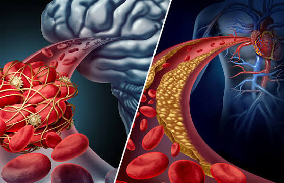
Heart attack vs angina — understanding the difference between these two conditions is one of the most important pieces of cardiovascular knowledge any adult can have. Both produce chest pain or discomfort caused by insufficient blood flow to the heart, and both share the same underlying disease (coronary artery disease). But they differ fundamentally in what is happening to heart muscle cells: in angina, the cells are temporarily oxygen-starved but survive; in a heart attack, they are dying. This distinction determines everything — whether the situation is an emergency requiring 911 activation, or an established condition to be managed in an outpatient setting.
The confusion between the two conditions causes genuine harm each year: patients having heart attacks wait to see if their chest pain resolves on its own, delay calling emergency services, or drive themselves to the hospital rather than calling an ambulance — delays that each worsen outcomes by narrowing the window for effective intervention. Conversely, patients with known stable angina may catastrophize every episode as a heart attack, producing anxiety and unnecessary emergency department visits. Clear understanding of the distinguishing features of each condition reduces both types of error.
The Core Difference — Reversible Ischemia vs Irreversible Necrosis
The physiological distinction between angina and heart attack rests on a single question: are heart muscle cells dying?
In angina, a coronary artery narrowed by atherosclerotic plaque cannot supply enough blood to meet the heart’s oxygen demands during exertion or stress. The heart muscle experiences ischemia (insufficient oxygen delivery) but does not receive enough of a blood flow reduction to kill cells — the narrowing limits flow reserve but not baseline flow sufficiently to cause necrosis. When the demand decreases (with rest) or coronary flow improves (with nitroglycerin), the ischemia resolves completely and the heart muscle recovers without permanent damage. This is the defining feature of stable angina: it is entirely reversible.
In a heart attack (myocardial infarction, or MI), a coronary artery is abruptly and completely (or near-completely) blocked, typically by a blood clot that forms on the surface of a ruptured or eroded atherosclerotic plaque. The territory of heart muscle supplied by that coronary artery is suddenly deprived of all or nearly all blood flow. Within minutes of complete ischemia, cardiomyocytes begin to die — and cardiomyocytes, unlike some other cell types, cannot regenerate. They are replaced by scar tissue (fibrosis) that does not contract. The amount of permanent damage depends entirely on how quickly blood flow is restored: “time is muscle” is the principle that drives the entire emergency cardiac care system, where door-to-balloon time standards (PCI within 90 minutes of first medical contact for STEMI) reflect the exponential relationship between treatment delay and myocardial salvage.
Comparing Symptoms — How They Feel Different
While no symptom feature definitively distinguishes angina from heart attack in isolation, several characteristics significantly influence the probability of each diagnosis and guide the urgency of response.
Duration is the most clinically useful distinguishing feature. Stable angina typically resolves within 1 to 5 minutes of rest and within 1 to 3 minutes of nitroglycerin. Heart attack pain typically does not resolve with rest or nitroglycerin — it persists, may wax and wane in intensity but does not fully resolve, and lasts 20 minutes or longer. The clinical rule: chest pain lasting more than 15 to 20 minutes that does not fully resolve with rest and nitroglycerin should be treated as a heart attack until proven otherwise by medical evaluation.
Intensity and character: Heart attack pain is often — though not always — more severe than typical stable angina. Patients frequently describe MI pain as “the worst chest pain I’ve ever had,” “crushing,” or “like an elephant sitting on my chest.” However, intensity alone is unreliable: some heart attacks produce mild or moderate discomfort, while some angina episodes (particularly with activity) can feel severe. Character overlap is substantial — both produce pressure, tightness, or heaviness — making intensity and character less reliable distinguishing features than duration and response to nitroglycerin.
Associated symptoms are more prominent and more alarming in heart attack. Nausea and vomiting, diaphoresis (cold sweat), profound fatigue, and lightheadedness or presyncope are common accompaniments of MI, reflecting both the hemodynamic impact of myocardial dysfunction and the intense vagal and sympathetic activation that accompanies large myocardial infarctions. New severe shortness of breath accompanying chest pain strongly suggests MI (from acute left ventricular dysfunction causing pulmonary congestion) rather than stable angina. Palpitations with chest pain may indicate the ventricular arrhythmias that commonly complicate acute MI.
Trigger and timing: Stable angina occurs predictably with exertion or emotional stress and resolves with rest. Heart attacks frequently occur at rest, in the early morning hours (when sympathetic tone and platelet aggregability peak), and may occur during or after emotional distress, cold exposure, or physical exertion — but notably also occur in patients at rest watching television or sleeping, without any obvious trigger. Rest-onset chest pain in a patient with coronary risk factors should never be assumed to be musculoskeletal or gastrointestinal without appropriate cardiac evaluation.
Atypical presentations are critically important to recognize. Women, older adults, and diabetic patients are significantly more likely to experience MI with atypical symptoms — jaw pain without chest pain, fatigue and malaise, epigastric pain mimicking indigestion, or shortness of breath without chest discomfort. These atypical presentations contribute to diagnostic delays that worsen MI outcomes in these populations. Any concerning symptom in a patient with cardiovascular risk factors — even if not “classic” chest pain — warrants urgent evaluation.

How Doctors Tell Them Apart — Diagnostic Tests
In the emergency setting, two diagnostic tools rapidly distinguish heart attack from angina: the electrocardiogram (ECG) and cardiac troponin blood tests.
The 12-lead ECG is the first test performed in any patient with chest pain of potential cardiac origin, completed within 10 minutes of hospital arrival. In STEMI (ST-elevation myocardial infarction), the ECG shows characteristic ST segment elevation in the leads corresponding to the affected coronary artery territory — an immediately recognizable pattern that triggers the STEMI activation protocol and the race against the 90-minute door-to-balloon time standard. In stable angina at rest (between episodes), the ECG is typically normal or shows only chronic changes (prior Q waves, ST segment abnormalities from old infarction, left ventricular hypertrophy changes). The ECG during an angina episode shows transient ST depression or T-wave changes that resolve with symptom resolution — demonstrating ischemia without ongoing infarction.
Cardiac troponin (troponin I or troponin T, measured with high-sensitivity assays) is the biomarker that definitively confirms or excludes myocardial cell death. Troponin is a protein released into the bloodstream when cardiomyocytes die — it rises within 1 to 3 hours of myocardial infarction onset and peaks at 12 to 24 hours. A rising troponin level (serial measurements 1 to 3 hours apart showing significant increase) in a patient with chest pain confirms myocardial infarction. Normal troponin levels at presentation and after 1 to 3 hours essentially exclude significant MI. Stable angina, by definition, does not cause troponin elevation — the heart muscle is ischemic but not dying, so no troponin is released into the circulation. This is the biochemical confirmation of the physiological distinction between angina and heart attack.
What to Do in Each Situation — A Practical Action Guide
If you have diagnosed stable angina and experience a typical anginal episode:
- Stop activity immediately and sit or lie down
- Take one sublingual nitroglycerin dose (tablet under the tongue or spray)
- Wait 5 minutes — if pain resolves completely, you have managed a typical anginal episode
- If pain persists after 5 minutes, take a second nitroglycerin dose and wait 5 more minutes
- If pain still persists after the second dose, take a third dose and call 911 immediately
- Do not drive yourself — if emergency services are not available, have someone drive you
If you experience ANY of the following, call 911 immediately (do not attempt to drive or wait):
- Chest pain at rest that is new or different from your usual angina
- Chest pain lasting more than 15 minutes that does not fully resolve with nitroglycerin
- Chest pain accompanied by diaphoresis (cold sweat), nausea, severe shortness of breath, or fainting
- Chest pain that feels more severe or different in character than your typical angina
- Any concerning chest, jaw, arm, or upper back discomfort if you have never been diagnosed with angina
After any emergency evaluation for chest pain, follow up with your cardiologist to reassess your risk, consider whether antianginal therapy adjustment or revascularization evaluation is warranted, and ensure secondary prevention medications (statin, antiplatelet, blood pressure and glucose management) are optimized. A single emergency evaluation is not the end of the management process — it is an inflection point that should trigger reassessment of your overall cardiovascular risk management.
The Overlap Condition — Unstable Angina as a Bridge
Unstable angina is the clinical condition that sits between stable angina and myocardial infarction — it represents acute coronary syndrome without completed myocardial necrosis. In unstable angina, a coronary plaque ruptures or erodes and triggers thrombus formation, but the resulting obstruction is subtotal rather than complete, and troponin does not rise (no myocardial cell death has occurred). The ischemia is severe enough to produce rest pain, minimal-exertion pain, or a dramatically changed angina pattern — but not (yet) severe enough to kill myocardial cells.
Unstable angina is a true emergency because the incomplete obstruction is inherently unstable — the thrombus can propagate to complete occlusion at any moment, converting unstable angina to STEMI. The correct response to suspected unstable angina is identical to MI: immediate 911 activation, not waiting and monitoring. Emergency evaluation with ECG and serial troponins typically distinguishes unstable angina (negative troponin) from NSTEMI (positive troponin) — both require urgent catheterization, but the troponin result affects the timeline and intensity of anticoagulation and antiplatelet therapy during the peri-procedural period.
The American Heart Association’s heart attack resources provide comprehensive guidance on recognizing and responding to heart attacks. The CDC heart attack information covers warning signs, risk factors, and emergency response. The NHLBI heart attack guide addresses symptoms, diagnosis, treatment, and recovery in detail.
Related reading: Angina: Chest Pain from Reduced Blood Flow | Heart Attack Prevention | Coronary Artery Disease | What Causes Heart Disease? | Major Risk Factors for Heart Disease
Sources
- Amsterdam EA, et al. 2014 AHA/ACC Guideline for the Management of Patients With Non-ST-Elevation Acute Coronary Syndromes. J Am Coll Cardiol. 2014;64(24):e139-e228.
- O’Gara PT, et al. 2013 ACCF/AHA Guideline for the Management of ST-Elevation Myocardial Infarction. J Am Coll Cardiol. 2013;61(4):e78-e140.
- Thygesen K, et al. Fourth Universal Definition of Myocardial Infarction. J Am Coll Cardiol. 2018;72(18):2231-2264.
- Hochman JS, Reynolds HR. Stable Ischemic Heart Disease. N Engl J Med. 2020;382(14):1319-1329.
- Mehta LS, et al. Acute Myocardial Infarction in Women. Circulation. 2016;133(9):916-947.
After a Heart Attack — Recovery, Medications, and Long-Term Management
Surviving a myocardial infarction is only the beginning of cardiovascular management — the post-MI period is one of the highest-risk periods for recurrent events, requiring intensive secondary prevention and, increasingly, a structured recovery pathway that begins in the hospital and continues for months to years.
The first hours to days after STEMI or NSTEMI are managed in the cardiac intensive care unit or coronary care unit, with continuous rhythm monitoring (ventricular arrhythmias are most common in the first 48 to 72 hours after MI), hemodynamic stabilization, and initiation of the evidence-based post-MI medication regimen. The “DAPT-STATIN-BB-ACE/ARB” framework captures the core pharmacological components:
- Dual antiplatelet therapy (DAPT): Aspirin 81 mg plus a P2Y12 inhibitor (ticagrelor or prasugrel preferred after STEMI for their superior platelet inhibition versus clopidogrel; clopidogrel used when ticagrelor/prasugrel are contraindicated). DAPT duration after drug-eluting stent placement is typically 12 months for ACS, with decisions about extension or shortening based on ischemic vs. bleeding risk balance.
- High-intensity statin: Rosuvastatin 20 to 40 mg or atorvastatin 40 to 80 mg, targeting LDL below 70 mg/dL (or below 55 mg/dL in very high-risk patients), initiated before hospital discharge regardless of baseline LDL.
- Beta-blocker: Initiated within 24 hours of MI in hemodynamically stable patients, continued indefinitely in patients with reduced ejection fraction (below 40 percent); duration in those with preserved LVEF after MI is more individualized based on recent trial data suggesting limited mortality benefit beyond 1 to 2 years in patients with normal LVEF and no heart failure.
- ACE inhibitor or ARB: Started within 24 hours, particularly important in patients with LVEF below 40 percent, anterior MI, hypertension, or diabetes. ARNIs (sacubitril-valsartan) may be initiated as an alternative in patients who tolerate ACE inhibitors and have reduced ejection fraction.
Echocardiography performed 3 to 5 days after MI (or at discharge) assesses residual LV function — a critical prognostic indicator and determinant of downstream therapy needs (ICD evaluation if LVEF remains below 35 percent at 40 to 90 days post-MI despite optimal medical therapy). Follow-up cardiology visits at 4 to 6 weeks and then at 3 to 6 month intervals allow for medication titration, LVEF reassessment, and evaluation of functional status and symptoms.
Cardiac Rehabilitation After MI — Why Participation Matters
Cardiac rehabilitation (CR) after myocardial infarction is one of the most evidence-supported and most consistently underutilized interventions in cardiovascular medicine. A comprehensive meta-analysis of more than 40 randomized trials demonstrated 20 to 26 percent reductions in cardiovascular mortality and significant improvements in quality of life, functional capacity, and lipid profiles in patients who completed cardiac rehabilitation compared to those who received usual care alone.
Despite this evidence and guideline Class I recommendation for all eligible post-MI patients, CR participation rates in the United States remain below 30 percent of eligible patients — with particularly low rates in women, elderly patients, and patients without referral at discharge. The barriers are real: program availability varies geographically, transportation and scheduling challenges limit access for working adults, and some patients experience post-MI anxiety and fear about exertion that makes initiating exercise feel unsafe despite medical reassurance.
Home-based cardiac rehabilitation — which provides exercise prescription, education, and periodic clinical contact through phone, video, or digital platforms — has demonstrated equivalent cardiovascular outcomes to center-based programs in multiple trials (REACH HF, HOME-HF, RAMIT) and substantially improves access for patients unable to attend a fixed-location program. Patients who cannot access traditional CR should ask their cardiologist about home-based programs, which increasingly integrate wearable monitors, video coaching sessions, and smartphone applications for remote monitoring and support.
Psychological Impact — Depression, Anxiety, and Fear After a Cardiac Event
The psychological aftermath of a myocardial infarction is a clinically significant and frequently underaddressed dimension of post-MI care. Major depressive disorder affects 15 to 25 percent of patients in the year after MI — two to three times the general population prevalence — and post-MI depression is an independent predictor of recurrent cardiovascular events, reduced medication adherence, lower physical activity, and increased mortality. The relationship is bidirectional: depression activates inflammatory and sympathetic pathways that accelerate atherosclerotic progression, while the physiological and life disruption of MI triggers depression through psychological and social mechanisms.
Anxiety — particularly health anxiety, cardiac-focused anxiety, and fear of exertion — is also highly prevalent after MI. Many patients develop a “cardiac invalid” pattern: severely restricting activity because any physical sensation is interpreted as dangerous, avoiding exercise even when medically cleared, and living in constant fear of recurrent MI. This pattern is both psychologically limiting and physiologically harmful — physical deconditioning reduces cardiovascular fitness, raises the resting heart rate, and worsens cardiovascular risk. The paradox is that the most effective intervention for post-MI anxiety is supervised exercise in cardiac rehabilitation, which provides the physical experience of safely reaching moderate heart rates under medical supervision, gradually rebuilding confidence in the heart’s capacity to tolerate exertion.
Evidence-based interventions for post-MI psychological distress include collaborative care models that integrate cardiology and mental health care; cognitive behavioral therapy (CBT) specifically adapted for cardiac patients; selective serotonin reuptake inhibitors (SSRIs) for post-MI depression (SERTRALINE Antidepressant Heart Attack Randomized Trial — SADHART — demonstrated sertraline safety and efficacy in post-MI depression); and participation in cardiac rehabilitation, which consistently reduces anxiety and depression scores alongside its cardiovascular benefits. All post-MI patients should be screened for depression and anxiety at follow-up visits and offered appropriate support.
How Angina and Heart Attack Interact Over Time
For most patients with coronary artery disease, angina and myocardial infarction are not separate conditions but sequential manifestations of a single underlying disease. Stable angina often precedes MI in the same patient — the stable plaque that produced predictable exertional symptoms for years can rupture acutely, converting a managed chronic condition to an acute emergency. This is why stable angina is never truly “benign” — it warrants aggressive secondary prevention and risk factor management regardless of how well-controlled the symptoms are.
Conversely, many heart attacks occur in patients without prior angina — the first symptomatic manifestation of their coronary disease is an acute MI, without preceding stable angina to provide warning. This pattern (often called “silent ischemia”) reflects the fact that large proportions of coronary artery disease is hemodynamically significant but asymptomatic, either because the stenosis has not yet reached the threshold that produces ischemia with ordinary activity, or because the patient has subconsciously reduced activity to avoid symptoms. Preventive cardiovascular risk management — blood pressure control, lipid lowering, diabetes management, smoking cessation — is important for all adults with cardiovascular risk factors, not only those with symptoms, precisely because the first presentation of coronary disease may skip the angina warning and present directly as MI.
The goal of understanding the heart attack vs angina distinction is not academic — it is to give every patient the knowledge and confidence to respond appropriately when symptoms arise: managing predictable stable angina calmly and systematically with established protocols, and recognizing the warning signals that demand immediate emergency response without hesitation or delay.
Key Takeaways — Putting Heart Attack vs Angina Knowledge into Practice
Understanding the difference between heart attack and angina empowers patients to make better decisions under pressure — the moments when clear thinking is most difficult but most consequential. The core principles to hold onto: stable angina is temporary and reversible, always resolves with rest and nitroglycerin within a few minutes, and is managed as an outpatient condition with established protocols. A heart attack involves dying heart muscle, does not resolve with rest or nitroglycerin, and requires immediate emergency response. The overlap condition — unstable angina — should be treated as a heart attack emergency until medical evaluation proves otherwise, because the consequences of waiting are far worse than the consequences of an unnecessary emergency visit.
Patients with established coronary artery disease should review their action plan with their cardiologist annually — confirming the nitroglycerin protocol, clarifying which symptoms warrant 911 versus outpatient evaluation, and ensuring secondary prevention medications are at target doses. Family members and close contacts should also understand the basics: when to call emergency services, that they should not drive the patient themselves in a suspected MI (ambulance response allows treatment to begin en route), and that time is the single most important variable in determining heart attack outcomes. Every minute of delay in restoring coronary flow is muscle that cannot be saved — knowledge that motivates the rapid response that modern cardiac emergency systems are designed to support.
I have been reading about heart attack vs angina: key differences for weeks and this is the most thorough guide I have found. I appreciated how the article addressed both the clinical side and the practical lifestyle adjustments. Keep up this kind of thorough health journalism — it genuinely helps patients like me.
I shared this article on heart attack vs angina: key differences with my doctor and they appreciated the level of detail. It is refreshing to see an article that acknowledges individual variation rather than giving one-size-fits-all advice. Looking forward to reading more articles from this website.
Came across this while researching heart attack vs angina: key differences for a family member. The section on managing this condition day-to-day was especially useful for meal planning. Thank you for making complex medical information accessible without dumbing it down.
I never fully understood heart attack vs angina: key differences until I read this. The specific numbers and thresholds mentioned are exactly what I needed to understand my results. This is going into my health folder that I bring to every doctor’s visit.
My doctor recommended I look into heart attack vs angina: key differences and this article covered it perfectly. This is the kind of evidence-based writing that actually changes how people approach their health. Shared this with three friends who are dealing with related issues. Very useful resource.
Bookmarked this article on heart attack vs angina: key differences immediately — going to reference it regularly. I especially valued the explanation of why these recommendations exist, not just what they are. Looking forward to reading more articles from this website.