
Inflammation and Heart Health
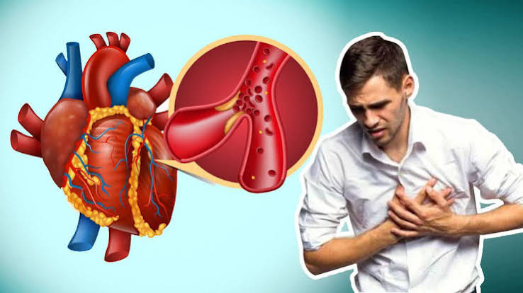
For most of the twentieth century, cardiovascular disease was understood primarily as a lipid deposition problem: cholesterol accumulates in artery walls, forms plaques, narrows vessels, and eventually blocks blood flow to the heart or brain. This model was useful and actionable — it produced statins, dietary fat guidelines, and the entire edifice of LDL-focused cardiovascular risk management. But it was incomplete. The lipid model couldn’t explain why half of all heart attacks occur in people with normal or near-normal cholesterol, why plaques rupture suddenly rather than simply growing until they occlude vessels, or why markers of infection and immune activity so strongly predict cardiovascular events.
The answer, now well-established, is inflammation. Chronic low-grade systemic inflammation is not a bystander in cardiovascular disease — it is a central driver of plaque initiation, progression, destabilization, and rupture. The connection between inflammation and heart health has produced an entirely new understanding of atherosclerosis as an inflammatory disease as much as a metabolic one, and has opened the door to anti-inflammatory therapies that reduce cardiovascular risk independently of any effect on cholesterol.
How Inflammation Drives Atherosclerosis — The Inflammatory Cascade in Arterial Walls
Atherosclerosis begins not with cholesterol deposition but with endothelial injury. The endothelium — the single-cell layer lining all blood vessels — serves as the critical interface between blood and vessel wall, regulating vascular tone, preventing clotting, and controlling the movement of substances into the vessel wall. When the endothelium is injured by oxidative stress, mechanical forces (high shear stress at arterial branch points), elevated glucose, elevated blood pressure, cigarette toxins, or excess modified LDL particles, it responds by upregulating adhesion molecules that attract circulating monocytes and lymphocytes from the bloodstream.
Monocytes that attach to the activated endothelium migrate into the subendothelial space, where they differentiate into macrophages — the tissue-resident immune cells that become the central cellular players in atherosclerotic plaque development. Macrophages engulf oxidized LDL particles, becoming lipid-laden “foam cells” — so named for their frothy appearance under microscopy. As foam cells accumulate, they release inflammatory cytokines including interleukin-1 beta (IL-1β), interleukin-6 (IL-6), and tumor necrosis factor-alpha (TNF-α), which amplify the inflammatory response, recruit additional immune cells, and stimulate the liver to produce C-reactive protein (CRP) as an acute phase reactant — producing the elevated CRP levels measurable in blood tests and associated with cardiovascular risk.
As the inflammatory process continues, smooth muscle cells from the vessel wall media migrate into the developing plaque and produce extracellular matrix proteins that form the fibrous cap — the thin layer of tissue separating the lipid-rich plaque core from the bloodstream. The stability of this fibrous cap determines the clinical behavior of the plaque. A thick, stable fibrous cap produces a stable plaque that may grow slowly over years but is resistant to rupture. A thin, inflamed fibrous cap — actively degraded by matrix metalloproteinases (MMPs) secreted by activated macrophages — produces a vulnerable plaque at high risk of rupture.
Plaque rupture exposes the thrombogenic lipid core to circulating platelets and coagulation factors, triggering rapid thrombus formation that can completely occlude the vessel within minutes. This is the mechanism of the majority of acute myocardial infarctions and many strokes — not gradual progression to complete luminal occlusion, but sudden rupture of an inflamed, vulnerable plaque that may not have been causing significant flow limitation before the event. This explains the clinical reality that many patients have “normal” stress tests and no symptoms until they experience their first MI — because their vulnerable plaques were not hemodynamically significant until they ruptured.
Measuring Inflammation — C-Reactive Protein and the JUPITER Trial
C-reactive protein (CRP) is an acute phase reactant produced by the liver in response to inflammatory signaling, particularly IL-6. In its high-sensitivity measurement form (hsCRP), CRP is one of the most extensively validated cardiovascular biomarkers available, with consistent associations with cardiovascular events across large epidemiological studies regardless of traditional risk factors.
The cardiovascular significance of hsCRP is stratified by level:
- hsCRP below 1 mg/L: low cardiovascular inflammatory risk
- hsCRP 1 to 3 mg/L: intermediate cardiovascular inflammatory risk
- hsCRP above 3 mg/L: high cardiovascular inflammatory risk
- hsCRP above 10 mg/L: likely reflects acute infection or acute inflammatory condition — cardiovascular interpretation requires repeat measurement after resolution
The landmark JUPITER trial (2008) established the clinical utility of hsCRP measurement in a way that directly changed cardiovascular treatment guidelines. The trial enrolled approximately 17,800 patients with normal LDL cholesterol (below 130 mg/dL) but elevated hsCRP (above 2 mg/L) — a population that traditional risk calculators classified as low-to-intermediate cardiovascular risk. Patients were randomized to rosuvastatin 20 mg daily or placebo and followed for a median of 1.9 years before the trial was stopped early due to overwhelming benefit in the statin arm.
The rosuvastatin group experienced a 54 percent reduction in myocardial infarction, a 48 percent reduction in stroke, and a 43 percent reduction in cardiovascular death — benefits substantially larger than most statin trials in patients with elevated LDL. The trial demonstrated that statins reduce cardiovascular risk not only through LDL lowering but through anti-inflammatory effects: hsCRP fell 37 percent in the rosuvastatin arm independent of the LDL reduction, and the magnitude of hsCRP reduction (not just LDL reduction) predicted the degree of cardiovascular risk reduction. The JUPITER findings established hsCRP measurement as a clinically valid tool for identifying statin candidates among patients who would otherwise be considered low risk by LDL-based criteria.

The CANTOS Trial — Direct Anti-Inflammatory Therapy for Cardiovascular Prevention
If the JUPITER trial implicated inflammation in cardiovascular risk through the anti-inflammatory effects of statins, the CANTOS trial (2017) provided the definitive proof that targeting inflammation directly — independent of any lipid-lowering effect — reduces cardiovascular events. This distinction matters enormously: it confirms that inflammation itself, not just its association with lipid disorders, is a causal driver of cardiovascular disease.
CANTOS enrolled approximately 10,000 patients with prior myocardial infarction and persistently elevated hsCRP (above 2 mg/L despite statin therapy) — the highest-risk inflammatory cardiovascular phenotype. Patients were randomized to subcutaneous injections of canakinumab (a monoclonal antibody targeting IL-1β, a key upstream inflammatory cytokine) at doses of 50, 150, or 300 mg quarterly, or to placebo. The trial was designed to test whether reducing inflammation without altering LDL or other traditional risk factors would reduce cardiovascular events.
The 150 mg dose of canakinumab produced a 15 percent reduction in major adverse cardiovascular events (MACE) compared to placebo — without any change in LDL cholesterol, blood pressure, or other traditional risk factors. The anti-inflammatory mechanism alone was responsible for the cardiovascular benefit. Patients in the canakinumab arm whose hsCRP fell below 2 mg/L on treatment had significantly greater cardiovascular benefit than those whose hsCRP remained elevated, establishing the principle of inflammation-guided therapy: achieving the inflammatory target (not just using the anti-inflammatory drug) drives the outcome benefit.
The CANTOS trial also produced an unexpected but important finding: the canakinumab group had significantly lower rates of lung cancer incidence and lung cancer mortality — a striking demonstration that the same inflammatory pathway that drives cardiovascular disease also plays a role in cancer development. This finding has spurred research into IL-1β inhibition for cancer prevention and illustrates the far-reaching consequences of chronic systemic inflammation beyond cardiovascular disease.
A practical limitation of canakinumab as a cardiovascular preventive therapy is cost (over $200,000 per year at the time of the trial) and an increased rate of fatal infections — the immune suppression required to dampen inflammation also reduces pathogen defense. The search for more cost-effective and better-tolerated anti-inflammatory cardiovascular therapies continues, with low-dose colchicine emerging as the most promising near-term alternative.
Colchicine for Cardiovascular Inflammation — The COLCOT and LoDoCo2 Trials
Colchicine — a plant-derived anti-inflammatory agent used for centuries in gout and pericarditis — has emerged as one of the most important new additions to cardiovascular preventive pharmacology, following two landmark randomized controlled trials that demonstrated substantial cardiovascular event reduction at low doses.
The COLCOT trial (2019) randomized approximately 4,700 patients within 30 days of myocardial infarction to colchicine 0.5 mg daily or placebo on top of standard post-MI therapy (including statins, aspirin, and P2Y12 inhibitors). At a median follow-up of 22.6 months, colchicine reduced the composite cardiovascular endpoint by 23 percent, with the most striking reduction in strokes (46 percent) and urgent hospitalizations for angina requiring revascularization (50 percent). The drug’s anti-inflammatory mechanism — inhibiting neutrophil activation, NLRP3 inflammasome activity, and microtubule polymerization required for inflammatory signaling — operates independently of any antiplatelet or lipid-modifying effect.
The LoDoCo2 trial (2020) tested low-dose colchicine in stable coronary artery disease — patients with established CAD not in the acute phase. Approximately 5,500 patients were randomized to colchicine 0.5 mg daily or placebo for a median of 28.6 months. Colchicine reduced the primary composite endpoint (cardiovascular death, MI, stroke, or ischemia-driven revascularization) by 31 percent. Both the COLCOT and LoDoCo2 results were consistent across subgroups, supporting colchicine as effective in both acute and stable CAD settings.
The cardiovascular medicine community has embraced low-dose colchicine based on this evidence. Colchicine 0.5 mg daily is now endorsed in the 2023 AHA/ACC cardiovascular prevention guidelines as a reasonable addition to standard medical therapy in patients with established atherosclerotic cardiovascular disease who remain at elevated residual risk, with hsCRP as one of the biomarkers identifying appropriate candidates. Its low cost (generic colchicine costs pennies per pill) and decades-long safety record make it one of the most accessible anti-inflammatory cardiovascular therapies available.
Sources of Chronic Cardiovascular Inflammation — What Patients Can Modify
Understanding inflammation as a cardiovascular risk driver raises an important practical question: what elevates systemic inflammation in the first place, and what can patients and clinicians do to reduce it?
Visceral adiposity is the single most powerful modifiable driver of chronic low-grade cardiovascular inflammation in the general population. Visceral fat (intra-abdominal fat surrounding the organs) is metabolically active tissue that secretes pro-inflammatory cytokines including IL-6, TNF-α, and leptin — driving persistent elevation of hsCRP and systemic inflammatory tone. Weight loss — particularly the reduction of visceral fat achieved through caloric restriction and aerobic exercise — is among the most effective interventions for reducing hsCRP. A 5 to 10 percent reduction in body weight typically produces a 20 to 40 percent reduction in hsCRP. The connection between obesity, inflammation, and cardiovascular disease is not simply mediated through hypertension, dyslipidemia, and diabetes — the direct inflammatory contributions of visceral fat are an independent pathway.
Chronic infections and periodontal disease represent an often-overlooked source of sustained cardiovascular inflammatory activation. Periodontal disease — chronic bacterial infection of the gums and supporting dental structures — produces persistent low-grade systemic bacteremia and cytokine release that elevates hsCRP and has been associated with elevated cardiovascular risk in multiple observational studies. Treating periodontal disease reduces hsCRP and has been associated with improved vascular function in randomized trials. Regular dental care and periodontal treatment are underappreciated cardiovascular risk reduction strategies.
Physical inactivity promotes inflammatory tone through multiple mechanisms: reduced anti-inflammatory effects of exercise-induced IL-6 from contracting muscles (skeletal muscle during exercise produces anti-inflammatory myokines), increased visceral fat accumulation, reduced antioxidant capacity, and reduced parasympathetic tone. Regular aerobic exercise produces sustained reductions in hsCRP — meta-analyses suggest 10 to 30 percent reductions with consistent moderate-intensity exercise, independent of weight change. Exercise is, in this sense, a form of anti-inflammatory therapy.
Dietary patterns significantly influence systemic inflammatory tone. Ultra-processed foods high in refined carbohydrates, trans fats, and advanced glycation end products promote inflammation; Mediterranean dietary patterns rich in olive oil, fish, vegetables, legumes, and whole grains consistently reduce hsCRP and other inflammatory biomarkers across intervention trials. The PREDIMED trial demonstrated that Mediterranean diet supplemented with extra-virgin olive oil or nuts reduced cardiovascular events by approximately 30 percent — with anti-inflammatory mechanisms likely contributing substantially to this benefit.
The American Heart Association provides guidance on cardiovascular risk factors including inflammation. The Centers for Disease Control and Prevention maintains resources on heart disease risk factors. The National Heart, Lung, and Blood Institute offers patient resources on inflammation and cardiovascular health.
Related reading: What Causes Heart Disease? | Major Risk Factors for Heart Disease | Diabetes and Heart Disease | Obesity and Heart Health | Heart Attack Prevention
Sources
- Ridker PM, et al. Rosuvastatin to Prevent Vascular Events in Men and Women with Elevated C-Reactive Protein (JUPITER). N Engl J Med. 2008;359(21):2195-2207.
- Ridker PM, et al. Antiinflammatory Therapy with Canakinumab for Atherosclerotic Disease (CANTOS). N Engl J Med. 2017;377(12):1119-1131.
- Tardif JC, et al. Efficacy and Safety of Low-Dose Colchicine after Myocardial Infarction (COLCOT). N Engl J Med. 2019;381(26):2497-2505.
- Nidorf SM, et al. Colchicine in Patients with Chronic Coronary Disease (LoDoCo2). N Engl J Med. 2020;383(19):1838-1847.
- Libby P. Inflammation in Atherosclerosis. Arterioscler Thromb Vasc Biol. 2012;32(9):2045-2051.
- Estruch R, et al. Primary Prevention of Cardiovascular Disease with a Mediterranean Diet Supplemented with Extra-Virgin Olive Oil or Nuts (PREDIMED). N Engl J Med. 2018;378(25):e34.
Residual Inflammatory Risk — The Gap That Statins Cannot Close
One of the most clinically important concepts to emerge from the inflammation-and-heart-health literature is the idea of residual inflammatory risk — the cardiovascular event risk that persists in patients who have achieved optimal LDL control on statin therapy but continue to have elevated inflammatory markers. Understanding this concept explains why well-treated patients still experience cardiovascular events, and why inflammation-targeted therapy represents a genuine advance in secondary prevention rather than simply an adjunct to lipid management.
In large statin trials including the PROVE-IT TIMI-22, TNT, and JUPITER follow-up analyses, patients who achieved optimal LDL reduction (below 70 mg/dL) but had persistently elevated hsCRP (above 2 mg/L) had significantly higher rates of cardiovascular events than patients who achieved both low LDL and low hsCRP on statin therapy. This “dual goal” analysis established that LDL-C control and CRP suppression are complementary — achieving one without the other leaves a meaningful residual risk unaddressed.
Paul Ridker, one of the primary investigators of JUPITER and CANTOS, has framed this as two distinct residual risk pathways: residual cholesterol risk (elevated non-HDL, lipoprotein(a), or remnant lipoproteins despite statin therapy) and residual inflammatory risk (elevated hsCRP despite LDL control). Each requires different therapeutic approaches, and clinical risk stratification that measures only LDL will systematically miss patients at high residual inflammatory risk. In practice, this means that patients on maximally tolerated statin therapy who have experienced a cardiovascular event should have hsCRP measured to identify whether residual inflammatory risk is present — and whether low-dose colchicine or other anti-inflammatory therapy is warranted.
The practical clinical message is straightforward: a patient on high-intensity statin therapy with an LDL of 55 mg/dL who has a repeat MI is not simply “statin-inadequate” — they may have high residual inflammatory risk (elevated hsCRP, high neutrophil-to-lymphocyte ratio, elevated IL-6) that requires inflammatory targeting rather than further LDL intensification. Measuring inflammatory biomarkers after optimizing LDL is becoming part of standard secondary prevention workup in academic cardiology centers and is likely to become standard practice more broadly as inflammatory therapies become routine.
Autoimmune and Inflammatory Conditions — Why Rheumatoid Arthritis, Lupus, and Psoriasis Are Cardiovascular Risk Factors
Systemic inflammatory diseases — including rheumatoid arthritis (RA), systemic lupus erythematosus (SLE), psoriasis, and inflammatory bowel disease — substantially elevate cardiovascular risk through mechanisms that are now well-characterized. Patients with these conditions have 1.5 to 3 times the cardiovascular event rates of age- and sex-matched control populations, and this excess risk is not fully explained by traditional cardiovascular risk factors or by the medications used to treat these conditions.
The cardiovascular risk mechanism in autoimmune inflammatory diseases is essentially a sustained, high-level version of the chronic low-grade inflammation that elevates cardiovascular risk in the general population. In rheumatoid arthritis, persistently elevated IL-1β, IL-6, and TNF-α produce the same inflammatory atherosclerotic cascade that drives plaque development in other cardiovascular risk contexts — but at higher intensity and over longer duration. The degree of RA disease activity tracks closely with cardiovascular risk: patients in clinical remission have lower cardiovascular event rates than those with active inflammatory disease, consistent with the causal role of inflammation rather than merely disease-associated confounders.
In systemic lupus erythematosus, cardiovascular risk is dramatically elevated — young women with SLE have 50 times the myocardial infarction rate of age-matched women without lupus, one of the most striking cardiovascular risk elevations associated with any medical condition. The mechanisms in SLE include direct antibody-mediated endothelial injury, immune complex deposition in vessel walls, complement activation, and chronically elevated type I interferons — pathways distinct from but convergent on the same downstream cardiovascular inflammatory damage observed in other contexts. Managing cardiovascular risk in SLE requires both controlling lupus disease activity and aggressively treating traditional cardiovascular risk factors.
In psoriasis — particularly moderate to severe plaque psoriasis — the systemic inflammatory burden generated by the chronic skin disease produces cardiovascular consequences equivalent to those of mild to moderate rheumatoid arthritis. Studies show 25 to 50 percent higher cardiovascular event rates in patients with severe psoriasis compared to matched controls. Biologic therapies that control psoriatic inflammation (particularly TNF-α inhibitors and IL-17 inhibitors) have been associated with reduced cardiovascular event rates in observational studies, with ongoing clinical trials directly testing whether inflammation control in psoriasis translates to cardiovascular benefit — mirroring the CANTOS trial logic in a different inflammatory disease context.
The clinical implication for rheumatology and dermatology practice is that cardiovascular risk assessment and management must be integrated into the care of patients with systemic inflammatory diseases. Traditional risk calculators (Framingham, Pooled Cohort Equations) underestimate cardiovascular risk in RA, SLE, and severe psoriasis patients — and updated guidance recommends multiplying calculated cardiovascular risk by 1.5 to 2 in RA patients to better approximate actual risk. Aggressive lipid management, blood pressure control, smoking cessation, and disease-activity-targeted therapy are all critical components of comprehensive cardiovascular care in these populations.
The Future of Inflammation-Targeted Cardiovascular Prevention
The evidence base for inflammation as a causal cardiovascular risk driver is now substantial and growing. JUPITER established inflammatory risk identification with hsCRP. CANTOS established that targeting inflammation directly reduces cardiovascular events. COLCOT and LoDoCo2 established colchicine as an accessible and affordable anti-inflammatory cardiovascular therapy. Together, these trials have transformed the field and created a new treatment paradigm.
The immediate near-term clinical application is clear: patients with established atherosclerotic cardiovascular disease who have elevated hsCRP despite optimized statin therapy are candidates for low-dose colchicine 0.5 mg daily, as supported by the 2023 AHA/ACC prevention guidelines. This is not a fringe or experimental recommendation — it is guideline-endorsed standard of care for the right patient population.
Looking forward, additional inflammatory targets are under investigation. Ziltivekimab — a more potent and specific IL-6 inhibitor — is being studied in the ZEUS trial for cardiovascular event prevention in high-risk inflammatory cardiovascular patients. Inclisiran (a small interfering RNA targeting PCSK9) and other RNA-based therapeutics are exploring whether gene-silencing approaches to inflammatory pathway targets can produce durable cardiovascular risk reduction. SGLT2 inhibitors and GLP-1 receptor agonists — already established for cardiovascular benefit — also have significant anti-inflammatory effects that may partly explain their cardiovascular outcome improvements beyond glucose lowering.
For patients and clinicians, the most actionable current message is this: inflammation is not just a biomarker of cardiovascular risk — it is a modifiable driver of cardiovascular events, amenable to the same systematic treatment approach applied to LDL cholesterol and blood pressure. Measuring hsCRP in patients at intermediate or high cardiovascular risk, addressing modifiable inflammatory drivers (visceral fat, physical inactivity, poor diet, periodontal disease), and applying guideline-supported anti-inflammatory therapy where indicated are all evidence-based components of contemporary comprehensive cardiovascular prevention.
Thank you for covering inflammation and heart health so thoroughly without being overly technical. I appreciated how the article addressed both the clinical side and the practical lifestyle adjustments. Exactly the kind of evidence-based information that is hard to find in one place.
Came across this while researching inflammation and heart health for a family member. What I liked most was that the article didn’t just say what to avoid — it also gave alternatives. This is going into my health binder that I bring to every doctor’s visit.
This is one of the clearest explanations of inflammation and heart health I have found. The article answered questions I didn’t even know I had until I started reading. This gave me real confidence going into my next specialist appointment.
Came across this while researching inflammation and heart health for a family member. The practical tips made this immediately actionable, not just theoretical. This is going into my health folder that I bring to every doctor’s visit.
Finally a resource that explains inflammation and heart health in plain language. The connection between lifestyle choices and long-term outcomes is explained clearly here. This gave me real confidence going into my next specialist appointment.
Bookmarked this article on inflammation and heart health immediately — going to reference it regularly. The practical tips made this immediately actionable, not just theoretical. Appreciate the effort that went into researching and writing this — it shows.