
How the Body Controls Blood Sugar
Understanding how the body controls blood sugar is one of the most illuminating things you can learn about human physiology — because it reveals a system of extraordinary precision that operates invisibly, continuously, and automatically, keeping glucose within a narrow healthy range across wildly varying circumstances: whether you eat a large meal or fast for twelve hours, run a marathon or sit at a desk all day, sleep peacefully or pull an all-nighter. When this system works perfectly, blood sugar barely fluctuates outside the range of 70 to 140 mg/dL across an entire day. When it begins to fail — through insulin resistance, beta cell dysfunction, or autoimmune destruction — the consequences are both systemic and long-lasting. This guide explains the full architecture of blood sugar regulation: the organs involved, the hormones that drive it, and the feedback loops that keep everything in balance.
The Purpose of Blood Sugar Regulation
Before exploring the mechanisms, it is worth understanding why such tight regulation is necessary. Glucose is the primary fuel for every cell in the body, and the brain — accounting for only about 2 percent of body weight — consumes approximately 20 percent of total resting energy, almost exclusively as glucose. Unlike muscle cells, which can switch to fat for fuel during prolonged fasting, the brain has minimal capacity to store glucose and very limited ability to use alternatives. This means the brain requires a continuous, steady supply of glucose at all times — not too little (which causes rapid cognitive impairment and, if severe, loss of consciousness) and not too much (which causes chemical damage to proteins and blood vessels through glycation and oxidative stress).
The regulatory challenge is significant. A single meal can deliver 50 to 100 grams of glucose into the bloodstream within an hour, which would raise blood glucose dramatically without a rapid hormonal response. Between meals and overnight, the brain’s constant glucose demand would drain blood sugar dangerously low without a compensatory mechanism to release stored glucose. The body’s blood sugar control system solves both problems simultaneously, adjusting glucose availability minute-by-minute through a coordinated network of hormones, organs, and cellular sensors.
The full story of what blood sugar is, what the normal ranges look like, and what happens when levels go too high or too low is covered in our introductory guide — reading it alongside this article builds a complete picture of glucose physiology.
The Pancreas: The Control Center
The pancreas is the central organ of blood sugar regulation, housing the cells responsible for detecting blood glucose levels and secreting the hormones that govern the response. Located behind the stomach, the pancreas is primarily an exocrine gland that secretes digestive enzymes into the small intestine — but embedded within the exocrine tissue are approximately one million clusters of cells called the islets of Langerhans, comprising the endocrine pancreas that controls blood sugar.
The islets contain several distinct cell types, each secreting a different hormone:
- Beta cells (approximately 60–80% of islet cells) produce insulin, the principal blood sugar-lowering hormone. Beta cells continuously monitor blood glucose through specialized glucose-sensing proteins (primarily GLUT2 glucose transporters and glucokinase enzymes) and respond to rising glucose within minutes by secreting insulin
- Alpha cells (approximately 15–20%) produce glucagon, the principal blood sugar-raising hormone. Alpha cells increase glucagon secretion when blood glucose falls, signaling the liver to release stored glucose
- Delta cells produce somatostatin, which modulates the activity of both alpha and beta cells, fine-tuning insulin and glucagon secretion in response to nutrient intake
The interplay between insulin and glucagon — one rising as the other falls, each triggering opposite effects — is the primary feedback mechanism that keeps blood sugar stable across fasting and fed states.
Insulin: The Fed-State Signal
Insulin is released in response to rising blood glucose — primarily after eating — and its fundamental role is to lower blood sugar by directing glucose into cells where it can be used or stored. The insulin response to a meal happens in two phases that operate together to efficiently clear glucose from the bloodstream.
The first phase of insulin secretion begins within two to five minutes of glucose reaching the beta cells. Beta cells release a burst of pre-formed insulin that was stored in secretory granules, waiting for exactly this signal. This first-phase burst suppresses the liver’s glucose release (preventing the liver from adding even more glucose to an already rising supply) and primes the peripheral tissues to receive glucose. First-phase insulin secretion is often the first component of insulin response to deteriorate in the early stages of Type 2 diabetes, which is why post-meal blood sugar spikes in that condition even when fasting levels remain normal.
The second phase begins within 10 to 20 minutes and involves newly synthesized insulin that is released in a sustained, slower wave over the next one to two hours as blood glucose remains elevated. This second phase handles the bulk of the post-meal glucose load, driving glucose into muscle, fat, and liver cells throughout the absorption period.
Insulin acts by binding to insulin receptors on the surface of target cells — primarily muscle cells, fat cells, and liver cells. This binding triggers a cascade of intracellular signaling that causes GLUT4 glucose transporter proteins to move from inside the cell to the cell membrane, creating channels through which glucose molecules can enter. In muscle cells, this glucose is used immediately for energy or stored as glycogen (a branched polymer of glucose) for later use. In fat cells, glucose is converted to fatty acids and stored as triglycerides. In the liver, insulin both stimulates glycogen synthesis and simultaneously suppresses the liver’s glucose output — an important double action that prevents the liver from fighting against the insulin-driven glucose lowering taking place in peripheral tissues.
Glucagon: The Fasting-State Signal
Between meals and overnight, blood glucose would fall continuously as the brain and other tissues consume glucose, without a mechanism to replenish it. Glucagon, secreted by pancreatic alpha cells in response to falling blood glucose, prevents this by signaling the liver to release stored glucose into the bloodstream.
Glucagon acts on the liver through specific receptors, triggering two main processes:
- Glycogenolysis: the breakdown of stored glycogen back into glucose molecules, which are then released into the blood. The liver stores approximately 100 grams of glycogen — enough to sustain blood glucose for 8 to 12 hours of fasting — making it the immediate reservoir for between-meal glucose maintenance
- Gluconeogenesis: the synthesis of new glucose from non-carbohydrate precursors including amino acids (from protein), glycerol (from fat breakdown), and lactate. Gluconeogenesis becomes increasingly important during prolonged fasting (beyond 12 to 24 hours) when glycogen stores are depleted
The beautiful elegance of the insulin-glucagon system lies in their mutual inhibition: insulin suppresses glucagon secretion from alpha cells, and glucagon’s effect on the liver is blocked when insulin levels are high. This means the two hormones cannot fully operate simultaneously — one or the other dominates depending on whether blood glucose is rising (insulin dominates) or falling (glucagon dominates), creating a self-correcting oscillating system.
- Pancreas: Detects blood glucose and secretes insulin (lowers sugar) and glucagon (raises sugar)
- Liver: Stores glucose as glycogen after meals; releases glucose between meals via glycogenolysis and gluconeogenesis
- Muscle: Largest site of insulin-stimulated glucose uptake; stores glucose as glycogen for exercise fuel
- Fat tissue: Takes up and stores glucose as fat; releases fatty acids for fuel during fasting
- Brain: Consumes 20% of resting energy as glucose; monitors blood sugar via hypothalamic sensors and triggers counter-regulatory responses when levels fall
- Kidneys: Filter and normally reabsorb all glucose from blood; contribute to gluconeogenesis during prolonged fasting
The Liver: The Central Glucose Buffer
While the pancreas is the regulatory center, the liver is the primary glucose buffer — the organ that smooths out the dramatic glucose fluctuations that would otherwise occur between eating and fasting. Understanding the liver’s role is essential to understanding how the body controls blood sugar across the full 24-hour cycle.
After a meal, glucose absorbed from the intestine travels through the portal vein directly to the liver before reaching the general circulation. The liver acts as a gatekeeper: it takes up approximately 20 to 30 percent of the post-meal glucose load for its own energy needs and glycogen storage, and allows the remainder to pass into the general circulation where it triggers the pancreatic insulin response. Under insulin signaling, the liver shifts into storage mode — activating glycogen synthase (the enzyme that builds glycogen) and suppressing glycogen phosphorylase (the enzyme that breaks it down) — storing as much glucose as possible for later use.
Between meals, as insulin levels fall and glucagon rises, the liver reverses: glycogen phosphorylase becomes active, glycogen is broken down, and glucose is released steadily into the blood. The liver continuously monitors both portal glucose levels and hormonal signals to calibrate exactly how much glucose to release — a dynamic process that adjusts second by second to keep blood sugar stable.
The liver also performs gluconeogenesis during overnight fasting: manufacturing glucose from amino acids (supplied by overnight protein turnover), glycerol (from triglyceride breakdown in fat cells), and lactate (recycled from anaerobic metabolism in red blood cells and exercising muscle). This nocturnal glucose production sustains blood sugar through the night, which is why fasting blood sugar in the morning reflects not primarily what was eaten the night before, but how much glucose the liver produced overnight — a function directly regulated by insulin sensitivity.
Muscle: The Largest Glucose Consumer
Skeletal muscle is quantitatively the most important site of insulin-stimulated glucose uptake, responsible for approximately 75 to 80 percent of the glucose cleared from the bloodstream after a meal. This makes muscle mass and muscle insulin sensitivity central determinants of overall glucose regulation — which explains why physical activity is such a powerful tool for improving blood sugar control.
Muscle cells have two mechanisms for absorbing glucose: insulin-dependent uptake through GLUT4 transporters (the primary post-meal pathway) and insulin-independent uptake through GLUT4 and GLUT1 transporters activated by muscle contraction itself. The second pathway — exercise-stimulated glucose uptake — is particularly important because it allows working muscle to absorb large amounts of glucose without requiring insulin. This is why exercise lowers blood sugar even in people with significant insulin resistance: contracting muscles can bypass the resistance and take up glucose directly.
Resistance training builds additional muscle mass, increasing the total body capacity for glucose storage and the number of GLUT4 transporter proteins available for insulin-stimulated uptake. Aerobic exercise maintains and improves the insulin signaling pathways within existing muscle cells. A combination of both exercise types produces the most comprehensive improvement in glucose regulation — which aligns with current guidelines recommending both resistance and aerobic activity for people managing blood sugar conditions.
Counter-Regulatory Hormones: The Safety Net
Insulin and glucagon are the primary regulators of blood sugar, but they operate within a broader context of counter-regulatory hormones that can override or supplement the insulin-glucagon system under specific circumstances — particularly stress, illness, prolonged exercise, and hypoglycemia.
Cortisol, the primary stress hormone released by the adrenal glands, raises blood sugar by stimulating gluconeogenesis in the liver, reducing peripheral glucose uptake in muscle and fat, and promoting the breakdown of protein into amino acids that fuel gluconeogenesis. Cortisol’s blood sugar-raising effect is the physiological basis for the “dawn phenomenon” — the natural morning rise in blood sugar driven by the cortisol surge that occurs in the hours before waking, preparing the body for the metabolic demands of the day. It is also why stress (psychological, physical, or illness-related) consistently raises blood sugar in people with diabetes.
Epinephrine (adrenaline) is released during acute stress and hypoglycemia, producing a rapid rise in blood sugar by stimulating glycogen breakdown and gluconeogenesis, and simultaneously reducing insulin secretion. Epinephrine’s hypoglycemia response explains the shakiness, racing heart, and sweating that occur when blood sugar drops — these are symptoms of the adrenaline surge, not of the low glucose itself.
Growth hormone, released in pulses during sleep and in response to exercise and hypoglycemia, promotes glucose availability by reducing peripheral glucose uptake and stimulating lipolysis (fat breakdown), providing alternative fuels and preserving glucose for the brain. Growth hormone’s nocturnal surge contributes to the dawn phenomenon alongside cortisol.
Together, these counter-regulatory hormones form a safety net that protects the body — especially the brain — against dangerous hypoglycemia. In people without diabetes, this system responds rapidly and reliably. In people with Type 1 diabetes, deficient glucagon responses to hypoglycemia develop over time (alpha cells appear to lose their glucose-sensing function), making hypoglycemia more dangerous and harder for the body to self-correct.
The Incretin Effect: How the Gut Amplifies Insulin
One of the most important — and perhaps least known — components of blood sugar regulation after eating is the incretin system. Incretins are hormones released by specialized cells in the intestinal wall in response to food, which travel to the pancreas and amplify insulin secretion before glucose has even been fully absorbed into the bloodstream. This anticipatory response explains why the same amount of glucose administered orally (as food) produces a larger and faster insulin response than the same glucose administered directly into a vein — a difference called the “incretin effect,” which accounts for approximately 50 to 70 percent of the total post-meal insulin response.
The two primary incretin hormones are GLP-1 (glucagon-like peptide-1), secreted by L cells in the small intestine and colon, and GIP (glucose-dependent insulinotropic polypeptide), secreted by K cells in the duodenum and jejunum. Both are released within minutes of food reaching the intestine and amplify beta cell insulin secretion in a glucose-dependent manner — meaning they only amplify insulin release when blood glucose is elevated, not when it is normal or low. This glucose-dependency makes the incretin effect inherently safe: it cannot cause hypoglycemia on its own.
GLP-1 also slows gastric emptying (prolonging the absorption period and blunting glucose peaks), reduces appetite by signaling satiety to the brain, and suppresses glucagon secretion from alpha cells. These combined effects make GLP-1 a powerful regulator of post-meal glucose, which is why GLP-1 receptor agonists — medications that mimic GLP-1 — have become among the most effective and widely used treatments for Type 2 diabetes and obesity.
The incretin effect is significantly impaired in Type 2 diabetes — partly because GLP-1 secretion is reduced and partly because the beta cells’ response to GIP is blunted. This impairment contributes meaningfully to the excessive post-meal glucose spikes characteristic of early Type 2 diabetes. Understanding why blood sugar spikes after meals and how different foods affect this response is an important extension of understanding how the body’s regulatory systems work.
What Happens When Blood Sugar Regulation Fails
The blood sugar control system can fail in several distinct ways, each leading to a different metabolic condition:
In Type 1 diabetes, autoimmune destruction of beta cells eliminates insulin production entirely. Without insulin, blood sugar rises continuously, cells are starved of fuel, and the body begins breaking down fat into ketone bodies — an emergency fuel that, in excess, acidifies the blood (diabetic ketoacidosis). Glucagon secretion continues unchecked without insulin’s suppressive effect, worsening the hyperglycemia. Treatment requires lifelong exogenous insulin.
In Type 2 diabetes, two failures develop in parallel: cells become resistant to insulin’s signal (requiring more insulin to achieve the same glucose uptake), and beta cells gradually lose their capacity to compensate with higher insulin output. First-phase insulin secretion deteriorates early, causing excessive post-meal spikes; later, sustained second-phase secretion fails as well. The incretin effect is impaired. The liver’s overnight glucose output rises due to reduced insulin suppression. The result is fasting hyperglycemia combined with exaggerated post-meal peaks. Understanding insulin resistance in depth explains the metabolic origins of Type 2 diabetes and why lifestyle factors matter so profoundly.
In prediabetes, these same failures are present in early, partial form — sufficient to raise blood sugar above normal but not yet to diagnostic thresholds. This intermediate state is the window where intervention is most effective. See our guide on what is prediabetes for a detailed exploration of this critical stage and the evidence for reversing it.
In hypoglycemia (in people using insulin or sulfonylureas), the regulatory system is overwhelmed in the opposite direction — too much glucose-lowering force relative to available glucose. The counter-regulatory response (glucagon, epinephrine) kicks in to raise blood sugar, but if hypoglycemia is severe enough, or if the counter-regulatory response is impaired, blood sugar can fall to levels that impair brain function.
Supporting the Body’s Blood Sugar Control System
While the blood sugar regulation system is largely automatic, the choices made around sleep, diet, exercise, and stress management profoundly influence how well it functions. Adequate sleep — seven to nine hours per night — preserves insulin sensitivity and prevents the cortisol and growth hormone dysregulation that worsens glucose control. Regular physical activity, particularly the combination of aerobic and resistance exercise, maintains muscle insulin sensitivity and GLUT4 transporter activity. A diet emphasizing fiber-rich carbohydrates, adequate protein, and healthy fats reduces the amplitude of post-meal glucose spikes and the insulin demand placed on beta cells at every meal.
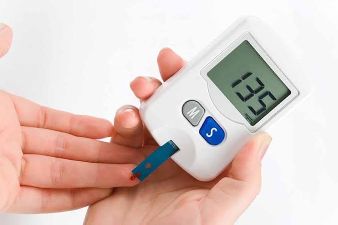
Monitoring blood sugar — whether through periodic fasting tests, A1C checks, or continuous glucose monitoring — provides direct feedback on how well the regulatory system is functioning and how specific lifestyle choices are affecting it. For guidance on home monitoring tools and how to interpret the readings, see our guide on home blood sugar monitoring. For a detailed look at what blood sugar levels mean diagnostically, including the thresholds for normal, prediabetes, and diabetes, see our comprehensive diabetes overview.
The blood sugar control system is one of the most sophisticated and resilient regulatory networks in the human body. Understanding how it works — in detail — is the foundation for understanding why it matters so much when it starts to fail, and why the steps taken to support it can make such an extraordinary difference to long-term health.
Sources: American Diabetes Association. Standards of Medical Care in Diabetes — 2024. Diabetes Care. 2024;47(Suppl 1):S20–S42. • Drucker DJ. The biology of incretin hormones. Cell Metabolism. 2006;3(3):153–165. • Saltiel AR, Kahn CR. Insulin signalling and the regulation of glucose and lipid metabolism. Nature. 2001;414(6865):799–806.
Really well-written article on how the body controls blood sugar. The section on managing this condition day-to-day was especially useful for planning. Shared this with three friends who are dealing with related issues. Very useful resource.
I shared this article on how the body controls blood sugar with my doctor and they appreciated the level of detail. I especially valued the explanation of why these recommendations exist, not just what they are. Forwarding this to others in my support group who are dealing with similar issues.
Bookmarked this article on how the body controls blood sugar immediately — going to reference it regularly. The article answered questions I didn’t even know I had until I started reading. Appreciate the effort that went into researching and writing this — it shows.