
When Kidney Stones Need Medical Treatment: A Guide to Your Options
Most kidney stones are managed without surgical intervention — approximately 85% of stones that enter the ureter and are below 5 mm in diameter will pass spontaneously with adequate fluid intake, pain management, and time. But a significant fraction of stones require medical or urological intervention because of their size, location, composition, the degree of obstruction they cause, or the clinical context (infection, solitary kidney, bilateral obstruction, intractable pain). Knowing when a kidney stone has crossed the threshold from a condition that can be managed conservatively at home to one that requires medical evaluation, hospitalization, or a urological procedure is critical both for patient safety and for avoiding unnecessary emergency department visits and procedures for stones that would pass on their own.
The decision to intervene — and the choice of which intervention to use — depends on factors that include stone size, stone location in the urinary tract, stone composition when known, the degree of ureteral obstruction and whether the kidney is draining adequately, the presence of infection proximal to the obstruction, the patient’s renal function and anatomy, and the patient’s preferences and occupational requirements. A 4 mm stone in the distal ureter with well-controlled pain and no fever in a healthy patient can be managed expectantly. The same stone in a patient with a solitary kidney, or accompanied by fever and rigors suggesting sepsis, is a urological emergency. Understanding these distinctions allows patients to engage more effectively with their urological care and to recognize when symptoms that might seem like a kidney stone actually require immediate emergency evaluation.
When Immediate Emergency Care Is Required
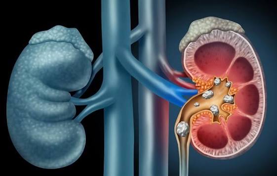
Certain clinical presentations associated with a kidney stone constitute medical emergencies that require immediate evaluation in an emergency department, not monitoring at home or a scheduled outpatient appointment. Failure to recognize these presentations can lead to life-threatening complications including urosepsis, irreversible kidney damage, and in extreme cases, loss of the kidney.
Fever or chills with flank pain is the most critical red flag presentation. When a kidney stone obstructs the ureter, the urine above the obstruction becomes stagnant and provides an environment in which bacteria can proliferate rapidly. If bacteria are present — from a pre-existing urinary tract infection, bacteriuria, or colonization of the stone itself (particularly in patients with struvite stones or a history of recurrent UTIs) — bacterial toxins can enter the bloodstream from the obstructed, infected kidney, producing urosepsis. Urosepsis from an obstructed infected kidney progresses extremely rapidly and can be fatal within hours if not treated. Any patient with fever (temperature above 38°C or 100.4°F) and symptoms of a kidney stone must be evaluated in an emergency department immediately. The emergency treatment is drainage of the obstructed kidney — typically by urgent ureteral stent placement or percutaneous nephrostomy — to relieve the pressure and allow infected urine to drain, followed by intravenous antibiotics. Attempting to treat this with oral antibiotics or pain medication at home while waiting for the stone to pass is dangerous.
Obstruction of a solitary kidney — either because the patient has only one kidney (congenital, from prior nephrectomy, or from a non-functioning contralateral kidney) or because a stone is causing bilateral ureteral obstruction — is a urological emergency requiring urgent evaluation and typically urgent ureteral stent placement or nephrostomy to preserve renal function. A solitary functioning kidney obstructed by a stone cannot produce adequate urine output, and if obstruction persists beyond 24 to 48 hours without intervention, significant irreversible renal injury begins. Complete bilateral ureteral obstruction causes anuria (no urine output) and acute kidney failure that requires urgent intervention.
Intractable pain — severe pain that cannot be controlled with oral analgesics at home, or pain that is escalating despite adequate medication — also warrants emergency department evaluation. Patients who cannot achieve adequate pain relief at home may require parenteral (intravenous or intramuscular) analgesics and antiemetics, adequate hydration, and further imaging to assess whether urgent intervention is indicated. Uncontrolled vomiting that prevents oral medication and fluid intake from being maintained is another indication for emergency evaluation and intravenous management.
Medical Expulsive Therapy: Helping Stones Pass
For stones below 5 to 10 mm in the ureter that do not require urgent intervention, medical expulsive therapy (MET) — the use of medications to relax the ureteral smooth muscle and facilitate spontaneous stone passage — is standard of care alongside adequate fluid intake and analgesics. Alpha-1 adrenergic receptor blockers (tamsulosin, silodosin, alfuzosin) are the most widely used MET agents; they relax the smooth muscle of the distal ureter and ureterovesical junction, reducing ureteral spasm, lowering intraureteral pressure, and increasing the frequency and effectiveness of peristaltic contractions that propel the stone toward the bladder. Randomized controlled trials demonstrate that alpha-blocker MET increases the rate of spontaneous stone passage by 10 to 20% for stones in the 4 to 10 mm size range, and reduces the time to stone passage by several days. The most benefit is seen for distal ureteral stones (in the lower third of the ureter, nearest the bladder), where the alpha-1 receptor density is highest. MET is typically continued for 4 to 6 weeks or until stone passage is confirmed.
The decision to pursue MET versus proceed directly to urological intervention depends primarily on stone size and location. Stones below 5 mm have a spontaneous passage rate above 80% without MET; MET is often offered for stones in the 5 to 10 mm range where passage is uncertain. Stones above 10 mm are unlikely to pass spontaneously regardless of MET and generally proceed to active intervention. During the expectant management period, patients are typically asked to strain their urine using a fine mesh urine strainer to catch any passed stone fragments for analysis, and follow-up imaging is scheduled to confirm stone passage or assess for growth or progressive obstruction.
Shock Wave Lithotripsy (SWL)
Shock wave lithotripsy (SWL) is a non-invasive procedure that uses focused acoustic shock waves — generated externally by a lithotripter machine and targeted at the stone using fluoroscopic or ultrasound guidance — to fragment the stone into small pieces that can then pass spontaneously through the ureter and urethra. SWL is performed on an outpatient basis, typically with intravenous sedation or general anesthesia, and lasts approximately 45 to 60 minutes. The patient lies on a table or in a tub of water (depending on the lithotripter design), and the shock wave generator focuses acoustic energy on the stone location, delivering 2,000 to 3,000 shock pulses over the course of the procedure. Bruising of the skin at the shock wave entry site and blood in the urine for several days after the procedure are common side effects; significant complications are uncommon but include incomplete fragmentation requiring repeat treatment, renal hematoma, and, rarely, steinstrasse (a “stone street” of fragments that obstruct the ureter together).
SWL is most effective for stones in the kidney or upper ureter that are: below 15 to 20 mm in size, below approximately 1,000 Hounsfield units on non-contrast CT (softer stones fragment more readily), in a location accessible to shock wave targeting, and in a patient with no obstruction to passage of fragments below the stone. SWL is less effective for stones in the lower pole calyx (where fragments must travel uphill against gravity to reach the ureteropelvic junction), for stones in the distal ureter (where the intervening pelvic bones can absorb shock wave energy), and for stones with high CT density (calcium oxalate monohydrate, brushite, and cystine stones are harder and more resistant to fragmentation). Obesity also reduces SWL efficacy, because the increased skin-to-stone distance required for focal shock wave delivery reduces the effective energy at the stone. Stone type identification — as reviewed in the types of kidney stones guide on Horizon Health Guide — directly informs the choice between SWL and ureteroscopy for individual patients.
Ureteroscopy with Laser Lithotripsy
Ureteroscopy (URS) with holmium laser lithotripsy is the most versatile surgical option for kidney stones and has become the most commonly performed kidney stone procedure in the United States. A thin flexible or semi-rigid fiberoptic or digital scope — ranging from 4 to 8 French (approximately 1.3 to 2.7 mm) in outer diameter — is passed transurethrally (through the urethra, into the bladder, and up the ureter) under general or spinal anesthesia without any skin incisions. The ureteroscope provides direct visualization of the stone, and a holmium:YAG or thulium fiber laser fiber (typically 200 to 365 micrometers in diameter) is passed through the working channel of the scope to the stone surface, where laser energy is delivered to fragment or dust (pulverize) the stone. Fragments can be extracted using small nitinol basket devices passed through the scope’s working channel, or, if sufficiently small, left to pass spontaneously.
Ureteroscopy achieves stone-free rates of 90 to 95% for ureteral stones and 80 to 90% for renal stones in most series — higher than SWL for ureteral stones and comparable or superior to SWL for most renal stones, particularly hard stones and lower pole stones where SWL is least effective. The advantages of ureteroscopy over SWL include: higher single-session stone-free rates, the ability to retrieve stone material for composition analysis, applicability to stones that are poor SWL candidates (hard stones, lower pole stones, obese patients), and no dependence on the skin-to-stone distance that limits SWL in obesity. The disadvantages include the requirement for general anesthesia (SWL can be performed with sedation), the potential need for a ureteral stent placed at the end of the procedure (a temporary stent left in place for several days to weeks to allow ureteral healing and prevent edema-related obstruction — stents are associated with urinary frequency, urgency, and discomfort that some patients find difficult to tolerate), and, rarely, ureteral injury from the scope.
Percutaneous Nephrolithotomy (PCNL)
Percutaneous nephrolithotomy (PCNL) is the standard treatment for large kidney stones (generally above 20 mm, or “staghorn” calculi that fill multiple calices of the kidney), complex stone burdens, and stones in locations not accessible by ureteroscopy. The procedure involves creating a small puncture wound through the skin of the flank (typically 1 to 2 cm), under fluoroscopic or ultrasound guidance, into the kidney’s collecting system — a nephrostomy tract through which progressively larger dilators are passed until a working sheath (typically 18 to 30 French outer diameter) allows access for a nephroscope and stone fragmentation instruments. Laser, ultrasonic, or pneumatic energy devices fragment the stone through the nephroscope, and fragments are aspirated or removed directly through the tract. PCNL requires general anesthesia and typically a 1 to 2 day hospitalization for recovery.
PCNL achieves the highest stone-free rates of any kidney stone procedure — above 85 to 90% in a single session for appropriately selected patients — and is the only procedure capable of addressing large stone burdens efficiently. Mini-PCNL, ultra-mini PCNL, and micro-PCNL variants use smaller nephrostomy tracts (12 to 16 French or smaller) that reduce the size of the flank puncture and associated morbidity while maintaining access for laser or ultrasonic fragmentation; these smaller-tract variants have lower bleeding risk and shorter recovery than standard PCNL and are increasingly used for moderate-size renal stones (10 to 20 mm) in centers with the appropriate equipment and expertise.
Medical Dissolution for Uric Acid Stones
Uric acid stones occupy a unique category in kidney stone management because they are the only common stone type amenable to complete dissolution by medical therapy alone. As detailed in the uric acid stones guide on Horizon Health Guide, raising urinary pH to 6.5 to 7.0 using oral potassium citrate dramatically increases uric acid solubility (by 15-fold between pH 5.0 and 7.0), allowing existing stones to dissolve over weeks to months without any surgical or urological intervention. For a patient with a radiolucent stone on CT (a strong indicator of uric acid composition), persistently acidic urine, and no urgent obstruction requiring immediate drainage, dissolution therapy is initiated and the stone monitored by CT every 4 to 6 weeks until dissolution is confirmed. Even stones that initially require urgent stenting for obstruction can undergo dissolution of residual fragments after the stent is removed, using this approach.
After any kidney stone episode — whether managed conservatively, with MET, or with a urological procedure — metabolic evaluation and prevention planning are essential to reduce the high recurrence risk. The kidney stone risk factors guide explains the factors that predispose to stone formation. The complete kidney stones overview on Horizon Health Guide covers the full spectrum of symptoms, causes, and initial management. Clinical guidance on procedure selection is available from the AUA surgical management of upper urinary tract calculi guidelines and the NIDDK kidney stone treatment resource.
Sources: AUA Surgical Management Guidelines · NIDDK — Kidney Stone Treatment · StatPearls — Nephrolithiasis
The Ureteral Stent: What to Expect After a Procedure
For many patients who undergo ureteroscopy or who require urgent drainage of an obstructed kidney, a ureteral stent is placed at the end of the procedure. A ureteral stent is a thin, hollow, flexible plastic tube — typically 22 to 30 centimeters in length — that spans from the renal pelvis (the urine-collecting space at the center of the kidney) to the bladder, with a curl at each end (the “pigtail” or “J” shape) that prevents it from migrating out of position. The stent passively drains urine from the kidney to the bladder around any residual edema, stricture, or stone fragments that might otherwise obstruct the ureter in the recovery period.
Ureteral stents are associated with a distinctive and often underestimated symptom burden that patients should be prepared for before their procedure. The most common stent symptoms include urinary urgency (a sudden, intense need to urinate), urinary frequency (needing to urinate every 20 to 30 minutes or more often during waking hours), flank discomfort with urination (as the pigtail curl in the kidney transmits pressure when the bladder contracts during voiding), and hematuria (blood in the urine that typically gives the urine a pink or light red color). Bladder spasms — sudden intense cramping in the lower abdomen and pelvis — occur in some patients and can be severe. These symptoms are the direct result of the stent’s presence: the lower pigtail in the bladder irritates the bladder trigone and contributes to urgency and frequency, while the upper pigtail in the kidney contributes to flank pain during bladder contractions through vesicoureteral reflux of urine up the stent into the renal pelvis during voiding.
Stent symptoms are managed with alpha-1 blocker medications (tamsulosin) that relax the ureteral smooth muscle and reduce reflux-associated discomfort, anticholinergic or beta-3 agonist medications (oxybutynin, solifenacin, mirabegron) that reduce bladder spasm and urgency, and adequate hydration to dilute the urine and reduce irritation. Most stents are removed in the urologist’s office within 1 to 6 weeks after placement using a cystoscope (a thin scope passed through the urethra into the bladder to grasp the stent’s lower loop) — a procedure that takes less than 5 minutes and requires no anesthesia for most patients. Some stents have a string that exits the urethra and allows the patient to remove the stent themselves at home by gently pulling the string at the scheduled time; this option is particularly common for stents placed after uncomplicated ureteroscopy for small ureteral stones.
Imaging for Kidney Stone Diagnosis and Follow-Up
Accurate imaging is the foundation of kidney stone management — it identifies the stone, characterizes its size and location, assesses the degree of obstruction and hydronephrosis (dilation of the kidney collecting system from backed-up urine), and guides procedure selection. Non-contrast computed tomography (CT) of the abdomen and pelvis — often called a “CT KUB” (for kidney, ureter, bladder) or simply a “stone protocol CT” — is the gold standard for kidney stone diagnosis, detecting essentially all kidney stones regardless of composition (including uric acid and cystine stones that are not visible on plain radiographs), measuring stone size and density in Hounsfield units, and providing information about the skin-to-stone distance relevant to SWL planning, all in a scan that takes less than a minute to acquire and requires no intravenous contrast. CT scanning is the preferred initial imaging for a patient presenting with suspected renal colic with unknown stone history.
Ultrasound is increasingly used as an alternative to CT in specific settings — particularly for pregnant patients (where radiation exposure is a concern), for pediatric patients, and for monitoring known kidney stones during follow-up when the primary concern is assessing hydronephrosis rather than characterizing stone density. Ultrasound detects most stones in the kidney and at the ureterovesical junction but misses a significant proportion of mid-ureteral stones (which are obscured by bowel gas). Point-of-care ultrasound in emergency departments has been validated as a safe first-line imaging choice in patients with prior documented stone disease and typical renal colic presentations, reserving CT for patients with atypical presentations or negative or indeterminate ultrasound findings.
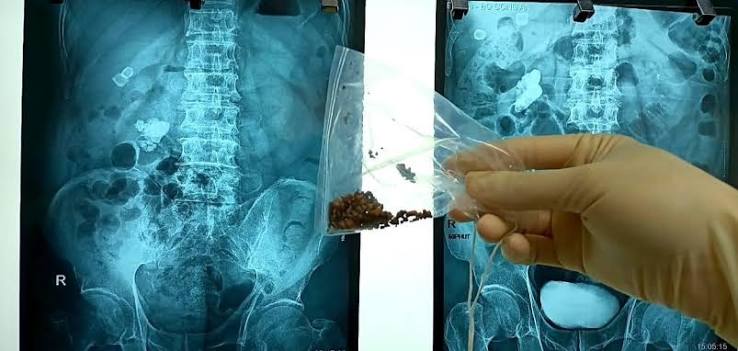
Plain abdominal radiographs (KUB X-ray) were historically the primary tool for kidney stone monitoring but have largely been supplanted by CT and ultrasound. They remain useful for detecting and monitoring radiopaque stones (calcium oxalate and calcium phosphate stones are typically visible on X-ray) in patients with known stone composition and for postoperative surveillance where radiation minimization is desired. Radiolucent stones (uric acid, pure matrix stones) are not visible on plain radiographs. Following urological treatment, stone-free status is confirmed by CT or by a combination of ultrasound and KUB X-ray, depending on the clinical context and the urologist’s preference. Understanding the imaging pathway — from initial emergency CT through treatment to post-procedure stone-free confirmation — gives patients a clearer picture of what to expect at each stage of their care, particularly for the early signs of kidney stones that prompt the initial evaluation.
After the Stone: Metabolic Evaluation and Prevention
A first kidney stone episode — particularly in a younger patient or a patient with a family history of stones — carries a lifetime recurrence risk of 50% within 5 years and 80% within 10 years without preventive intervention. This high recurrence rate makes metabolic evaluation and prevention planning a critical part of kidney stone management that should begin at or shortly after the initial stone episode, not only after a second or third recurrence. The standard metabolic evaluation includes a 24-hour urine collection measuring urinary calcium, oxalate, urate, citrate, pH, sodium, volume, and creatinine (the last serving as a quality check on collection adequacy), along with serum studies for calcium, uric acid, creatinine, potassium, bicarbonate, and parathyroid hormone if hypercalciuria is found. These measurements identify specific urinary risk factors — hypercalciuria, hyperoxaluria, hypocitraturia, hyperuricosuria, low urinary volume — that can be targeted with dietary modification or medication. As detailed in the kidney stone risk factors guide, addressing these metabolic contributors is the most effective long-term strategy for reducing recurrence risk.
Pharmacological prevention is indicated when dietary measures alone are insufficient to normalize urinary risk factors. Potassium citrate reduces urinary calcium, raises urinary citrate, and alkalinizes the urine — it is the most broadly useful preventive medication for calcium oxalate, calcium phosphate, and uric acid stone formers. Thiazide diuretics (hydrochlorothiazide, chlorthalidone, indapamide) reduce urinary calcium by enhancing tubular calcium reabsorption and are first-line pharmacotherapy for idiopathic hypercalciuria. Allopurinol reduces uric acid synthesis and is used for uric acid stone formers and for calcium oxalate stone formers with hyperuricosuria. The choice of preventive medication is guided by the 24-hour urine findings and the stone composition, and effectiveness is monitored by repeat 24-hour urine collections performed 4 to 6 weeks after each medication or dietary change to confirm that the target urinary parameters have improved.
I had no idea that fever with flank pain from a kidney stone was a true emergency until I read this. I actually had that exact presentation two years ago — I thought I was just having a bad stone episode and waited overnight before going to the ER. The ER doctor told me I had an infected obstructed kidney and that another few hours could have led to sepsis. The speed at which things escalated once they found the obstruction was shocking. I wish something like this article had been available to me before that night — understanding that fever plus stone pain equals emergency might have gotten me there six hours sooner.
This is a well-organized and clinically accurate overview of kidney stone intervention thresholds and procedure selection. The discussion of ureteral stent symptom burden is particularly valuable — patients are frequently underprepared for stent-related symptoms, and the gap between expectation and reality is one of the most common sources of post-procedural distress calls in our practice. The distinction between SWL candidacy factors (stone density on CT, lower pole location, obesity effect on focal distance) and ureteroscopy candidacy is also accurately represented. I’d add that flexible ureteroscopy with a ureteral access sheath has expanded the reachable stone locations compared to semi-rigid ureteroscopy alone, making URS an effective option even for most upper pole and interpolar stones.
Dr. Lindqvist, thank you for that clinical addition — the ureteral access sheath point is important and reflects how significantly flexible ureteroscopy technology has advanced over the past decade. The combination of improved flexible scope resolution, smaller scope outer diameters, and the access sheath’s passive dilation effect on the ureter has made URS viable for stone locations that previously required PCNL or SWL with lower success rates. Thomas, your experience is exactly the clinical scenario the fever-plus-flank-pain section is meant to prevent — the fact that urosepsis from an obstructed infected kidney can progress from discomfort to critical illness within hours makes it genuinely different from a simple painful stone that can be managed at home while waiting for spontaneous passage. We’re glad you got to the ER when you did.