
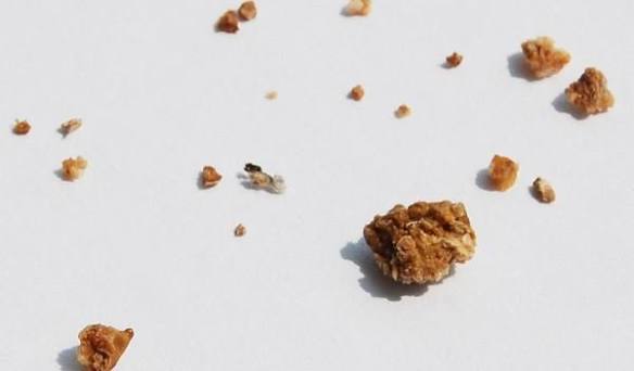
Calcium Oxalate Stones: Causes, Symptoms, and Prevention
Calcium oxalate stones are the most common type of kidney stone, accounting for approximately 70 to 75% of all kidney stone cases in the United States. They form when calcium and oxalate — a naturally occurring compound found in many plant foods and produced by the liver — bind together in the urine at concentrations that exceed their combined solubility, precipitating as crystals that can aggregate into stones of varying sizes. Despite being the dominant kidney stone type, calcium oxalate stones are widely misunderstood by patients and even by some non-specialist clinicians: the most persistent myth is that they are caused by eating too much calcium, when in fact the opposite is true in most cases. Understanding the actual mechanisms behind calcium oxalate stone formation is the foundation of accurate prevention, and this article provides a thorough review of those mechanisms, the symptoms calcium oxalate stones cause, and the evidence-based strategies that reduce recurrence risk.
Calcium oxalate stones affect approximately 1 in 11 Americans over their lifetime, with the incidence increasing over the past several decades — a trend attributed to rising rates of obesity, type 2 diabetes, and dietary patterns characterized by high sodium, animal protein, and fructose consumption. Men are approximately twice as likely as women to develop kidney stones, though the gap has narrowed as women’s rates have increased. The peak age of onset is 40 to 60 years, and once a person has had one calcium oxalate stone, the recurrence rate without preventive intervention is approximately 50% at 5 years and 70% at 10 years. This high recurrence rate makes prevention not just desirable but essential for any patient who has been affected.
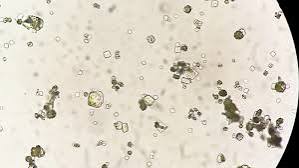
The Biochemistry of Calcium Oxalate Stone Formation
Calcium oxalate crystallization in the urine depends on the balance between the factors that promote crystallization (elevated calcium and oxalate concentrations, low urine volume, acidic urine pH, and insufficient inhibitor concentrations) and the factors that prevent it (adequate urine volume, normal citrate and magnesium levels, and an appropriate urinary pH). When the product of urinary calcium concentration multiplied by urinary oxalate concentration — called the supersaturation ratio — exceeds the crystallization threshold for calcium oxalate, crystals begin to nucleate. These initial microscopic crystals can pass harmlessly in urine; the problem arises when they adhere to the renal tubular epithelium, aggregate with other crystals, and grow into macroscopic stones over days to weeks.
Oxalate is 10 to 20 times more potent than calcium in driving calcium oxalate supersaturation — meaning that for any given increase in urinary concentration, an increase in oxalate elevates crystallization risk far more than an equivalent increase in calcium. This is the chemical basis for a key dietary insight: reducing dietary calcium — while it might seem logical for “calcium” stones — actually worsens calcium oxalate stone risk in most patients, because dietary calcium normally binds oxalate in the intestinal lumen and carries it out in the stool before it can be absorbed. When dietary calcium is restricted, more free oxalate remains in the intestine for absorption into the bloodstream, raising urinary oxalate and increasing the supersaturation ratio. This mechanism, confirmed in clinical trials including the landmark Borghi study, demonstrates that a normal-calcium, low-oxalate, low-sodium diet reduces stone recurrence more effectively than a low-calcium diet.
Risk Factors for Calcium Oxalate Stones
Multiple interacting metabolic, dietary, and anatomical risk factors contribute to calcium oxalate stone formation. The most clinically important are:
Hypercalciuria (elevated urinary calcium) is present in 40 to 60% of calcium oxalate stone formers and is the most common metabolic abnormality identified on 24-hour urine evaluation. It can result from absorptive hypercalciuria (the intestine absorbs calcium too efficiently, raising serum and subsequently urinary calcium), renal hypercalciuria (the kidney fails to adequately reabsorb filtered calcium, allowing excess calcium to enter the urine), or resorptive hypercalciuria (excess bone calcium mobilization, most commonly from primary hyperparathyroidism or vitamin D excess, raises serum calcium and urinary calcium). Distinguishing these subtypes helps guide targeted therapy — thiazide diuretics work best for absorptive and renal hypercalciuria, while primary hyperparathyroidism requires parathyroid surgery.
Hyperoxaluria (elevated urinary oxalate) may be dietary (consuming large amounts of high-oxalate foods such as spinach, nuts, chocolate, tea, and beets), enteric (from gastrointestinal fat malabsorption in inflammatory bowel disease, after small bowel resection, or following Roux-en-Y gastric bypass — unabsorbed fat binds calcium in the colon, leaving oxalate free for colonic absorption), or primary (rare autosomal recessive disorders of oxalate metabolism that cause massive overproduction of oxalate, sometimes requiring liver transplantation). Even mild degrees of hyperoxaluria significantly elevate crystallization risk given oxalate’s potency in driving supersaturation.
Hypocitraturia (low urinary citrate) is one of the most underappreciated risk factors for calcium oxalate stones. Citrate is a natural inhibitor of calcium crystallization — it forms soluble complexes with calcium in the urine, effectively reducing the concentration of free calcium available to bind oxalate, and it directly inhibits crystal growth and aggregation. When urinary citrate is low, these protective effects are lost. Hypocitraturia is caused by systemic acidosis (from high animal protein diets, chronic diarrhea, renal tubular acidosis, or certain medications), potassium depletion (hypokalemia causes intracellular acidosis that reduces citrate excretion), and, in some patients, idiopathic mechanisms not fully understood.
Low urine volume is the most universal risk factor across all kidney stone types and is particularly important for calcium oxalate stones. When urine output is low — below 1.5 to 2 liters per day — the concentration of all urinary solutes (calcium, oxalate, uric acid) increases, elevating the supersaturation ratio even when absolute daily excretion amounts are normal. Hot climates, strenuous physical activity, and inadequate fluid intake all reduce urine output; populations in hotter geographic regions have consistently higher kidney stone rates, and seasonal peaks in stone incidence track with summer heat exposure.
Dietary factors beyond oxalate and calcium intake also contribute to calcium oxalate stone risk. High sodium intake increases urinary calcium by reducing renal tubular calcium reabsorption — for every 100 mEq increase in daily sodium excretion, urinary calcium increases by approximately 25 mg/day, a clinically significant increment. High animal protein intake generates an acid load that reduces urinary citrate, increases urinary calcium (from bone buffering of the acid load), and increases urinary uric acid (which can seed heterogeneous calcium oxalate crystallization). High fructose intake — from sweetened beverages, high-fructose corn syrup, and ultra-processed foods — independently increases urinary calcium and uric acid. Vitamin C supplementation above 1,000 mg per day is metabolized to oxalate and raises urinary oxalate; supplementation at doses above 2,000 mg per day has been shown in prospective cohort studies to significantly increase kidney stone risk in men.
Symptoms of Calcium Oxalate Kidney Stones
Small calcium oxalate stones — those under 3 to 4 mm in diameter — often pass through the ureter without causing noticeable symptoms, appearing only as hematuria (blood in the urine) on a routine urinalysis or discovered incidentally during imaging for another indication. Larger stones, particularly those above 5 to 7 mm, have a progressively lower probability of spontaneous passage and are more likely to cause obstruction and the classic symptom pattern of renal colic.
Renal colic caused by a calcium oxalate stone in the ureter typically begins as sudden onset flank pain — in the back below the ribcage — that rapidly escalates to severe intensity within minutes. The pain is characteristically colicky (waxing and waning in intensity) and moves as the stone migrates: flank and upper back for a stone near the ureteropelvic junction, lateral abdomen and periumbilical region for a stone in the mid-ureter, and lower abdomen and groin — with possible radiation to the labia or testis — for a stone at the ureterovesical junction. Nausea, vomiting, and diaphoresis accompany the pain in most patients. Hematuria is present in over 80% of patients with acute stone passage, visible either grossly (pink, red, or brown urine) or detected by dipstick. Urinary urgency and frequency often occur as the stone reaches the distal ureter near the bladder. For a detailed clinical description of the kidney stone pain experience, including what distinguishes it from other causes of abdominal and flank pain, see the kidney stone pain guide on Horizon Health Guide.
Large calcium oxalate stones that remain within the kidney — not attempting to pass through the ureter — may be completely asymptomatic for months to years. They may be discovered incidentally on imaging performed for other reasons, such as abdominal CT for gastrointestinal symptoms. Asymptomatic renal stones still warrant evaluation and monitoring, because they can eventually pass (causing acute pain), grow to a size that damages the kidney collecting system (hydronephrosis), or, rarely, develop bacterial superinfection. The management of asymptomatic renal stones — active surveillance versus intervention — depends on stone size, location, composition when known, and patient factors including occupation (pilots, for example, have stricter criteria for stone management than the general population).
Diagnosis and Evaluation of Calcium Oxalate Stones
The initial diagnostic evaluation of a patient with suspected kidney stone involves imaging to confirm the stone, assess its size and location, and evaluate for obstruction or complications. Non-contrast CT of the abdomen and pelvis (NCCCT) is the gold-standard imaging study: it identifies virtually all calcium oxalate stones regardless of size (calcium oxalate is radiodense and appears clearly on CT), provides the density of the stone in Hounsfield units (which correlates with composition and predicts treatment response), and simultaneously evaluates for other causes of the patient’s symptoms. Plain abdominal X-ray (KUB) identifies calcium oxalate stones in approximately 60 to 70% of cases but misses smaller stones; ultrasound is radiation-free and identifies hydronephrosis but is less sensitive for small ureteral stones. For the early recognition of symptoms that warrant imaging, the early signs of kidney stones article provides a systematic review of the warning features that distinguish a forming kidney stone from other causes of back and urinary symptoms.
After the acute episode is managed, metabolic evaluation is indicated for all patients with calcium oxalate stones, especially those with recurrence, bilateral stones, stones in childhood or early adulthood, or identifiable risk factors. The cornerstone of metabolic evaluation is the 24-hour urine collection: two separate collections (to average day-to-day variation) that measure total urine volume, urinary pH, and the specific risk factors for calcium oxalate stones — calcium, oxalate, citrate, uric acid, sodium, creatinine, and phosphate. The results of 24-hour urine evaluation directly guide targeted dietary and pharmacological prevention. Serum evaluation includes calcium (to screen for primary hyperparathyroidism), uric acid, creatinine, and in selected patients intact parathyroid hormone (PTH). Stone composition analysis, performed by infrared spectroscopy on retrieved stone material, confirms the stone type as calcium oxalate and often distinguishes between the monohydrate (harder, more resistant to shock wave lithotripsy) and dihydrate (more friable, more amenable to lithotripsy) forms.
Dietary Prevention of Calcium Oxalate Stones
Evidence-based dietary recommendations for calcium oxalate stone prevention rest on several well-established principles, some of which contradict the generic “kidney stone diet” advice patients often receive. The core dietary approach for most calcium oxalate stone formers includes:
Adequate fluid intake is the single most important dietary intervention for any kidney stone type. The goal is a urine output of 2.0 to 2.5 liters per day, which requires drinking approximately 2.5 to 3 liters of fluid daily (depending on climate, activity level, and insensible losses). Water is the preferred fluid; lemon juice added to water provides citrate (from its citric acid content) and may modestly increase urinary citrate. Coffee and moderate amounts of tea appear to reduce stone risk in epidemiological studies despite tea containing oxalate — probably because the net fluid effect outweighs the modest oxalate contribution. Avoid sugar-sweetened beverages and high-fructose drinks, which increase stone risk rather than preventing it.
Maintain adequate dietary calcium — do not restrict it. The recommended intake for calcium oxalate stone formers is 1,000 to 1,200 mg of calcium per day from food sources, ideally spread across meals so that calcium is present in the intestine simultaneously with oxalate. Taking calcium supplements, by contrast, produces a more concentrated calcium load not timed with meals; prospective studies suggest that calcium supplements (particularly when taken between meals) may modestly increase stone risk, while dietary calcium clearly reduces it. Dairy products, fortified plant milks, and calcium-rich vegetables (cooked broccoli, bok choy) are good food sources.
Moderate high-oxalate foods, particularly those with the highest oxalate content: spinach (the highest-oxalate common food, containing approximately 750 mg oxalate per 100g raw weight), rhubarb, beet greens, Swiss chard, almonds, cashews, peanuts, chocolate, bran cereals, and strong steeped teas (green and black). It is not necessary to eliminate all oxalate-containing foods — many vegetables and healthy whole foods contain moderate oxalate levels that do not substantially elevate urinary oxalate when consumed in normal portions alongside adequate calcium. The goal is to avoid the highest-concentration sources rather than to adopt an extreme low-oxalate diet that would be nutritionally inadequate.
Reduce sodium intake to below 2,300 mg per day (the general population guideline), and ideally to 1,500 to 2,000 mg per day for high-risk stone formers. The primary mechanism is sodium’s effect on urinary calcium — lower sodium intake means lower urinary calcium, directly reducing the calcium component of supersaturation. Reducing sodium is also cardioprotective and widely applicable, making it one of the most justified dietary modifications for calcium oxalate stone prevention.
Moderate animal protein intake. High animal protein consumption generates an acid load that reduces urinary citrate, increases urinary calcium, and increases urinary uric acid — all unfavorable for calcium oxalate stone prevention. The recommended approach is not to eliminate animal protein but to limit it to approximately 0.8 to 1.0 g/kg of body weight per day — the standard dietary protein reference intake — rather than the high-protein dietary patterns common in Western diets. Plant protein sources (legumes, tofu) do not carry the same stone-forming risk and can substitute for some animal protein in the diet.
Medications for Calcium Oxalate Stone Prevention
When dietary modification alone is insufficient to normalize 24-hour urine risk factors — or when 24-hour urine evaluation identifies specific metabolic abnormalities that exceed what diet alone can correct — pharmacological prevention is indicated. The choice of medication for calcium oxalate stone prevention depends on which urinary risk factors are elevated or deficient.
Thiazide diuretics (hydrochlorothiazide, chlorthalidone, or indapamide) are the first-line pharmacological therapy for calcium oxalate stones associated with hypercalciuria. By enhancing calcium reabsorption in the distal convoluted tubule, thiazides reduce urinary calcium excretion by 30 to 50%, directly addressing the supersaturation driving stone formation. Randomized controlled trials confirm that thiazide therapy reduces calcium stone recurrence rates by approximately 45 to 50% compared to placebo. Side effects include mild hypokalemia (potassium supplementation or addition of potassium citrate is often needed), impaired glucose tolerance, and orthostatic hypotension; monitoring of electrolytes and renal function is recommended during long-term thiazide therapy.
Potassium citrate is the preferred treatment for calcium oxalate stones associated with hypocitraturia. It raises urinary citrate, raises urinary pH, and provides an alkaline load that corrects the systemic acidosis underlying citrate deficiency. Potassium citrate also directly inhibits calcium oxalate crystal growth and aggregation, adding a crystallization-inhibition effect beyond its pH and citrate effects. Available in oral tablet and liquid formulations, it is generally well tolerated; gastrointestinal upset and the mild laxative effect of the high potassium load are the primary side effects. Serum potassium monitoring is required, particularly in patients with any degree of renal impairment.
Allopurinol is indicated for calcium oxalate stone formers with elevated urinary uric acid — hyperuricosuria — which promotes heterogeneous nucleation of calcium oxalate crystals on a uric acid seed crystal. Allopurinol inhibits xanthine oxidase, reducing uric acid production and lowering urinary uric acid. It reduces calcium stone recurrence in patients with hyperuricosuria even when serum uric acid is not markedly elevated, distinguishing its role in stone prevention from its use in gout management. It is generally well tolerated at the doses used for stone prevention (100 to 300 mg daily); rare but serious hypersensitivity reactions (allopurinol hypersensitivity syndrome) require patients to be counseled about symptoms.
When to Seek Medical Evaluation
Anyone who has passed a calcium oxalate stone should be evaluated by a urologist or nephrologist to assess for metabolic risk factors and develop a prevention plan. First-time stone formers with a single uncomplicated calcium stone and no obvious identifiable risk factors may undergo a simplified initial evaluation — serum calcium, creatinine, uric acid, and a spot morning urine for calcium and creatinine — before proceeding to full 24-hour urine evaluation. Patients with recurrent stones, bilateral stones, stones in childhood or adolescence, nephrocalcinosis (calcium deposits diffusely in the kidney), or a family history of kidney stones should receive complete metabolic evaluation including 24-hour urine collections without waiting for a second stone episode.
Urgent evaluation is required when a stone episode is accompanied by fever or chills (suggesting infection proximal to the obstruction — a urologic emergency), when bilateral obstruction is suspected, or when a patient with a solitary kidney develops obstructive symptoms (any obstruction of a solitary kidney requires prompt urological evaluation). The complete kidney stones treatment guide on Horizon Health Guide covers the full range of treatment options for stones that cannot pass spontaneously, from oral hydration and alpha-blockers for passage facilitation to shock wave lithotripsy, ureteroscopy, and percutaneous nephrolithotomy for larger or more complex stones.
For a broader understanding of how calcium oxalate stones compare to the other kidney stone types — uric acid, struvite, cystine, and calcium phosphate — the types of kidney stones article on Horizon Health Guide provides a side-by-side comparison of composition, risk factors, radiographic characteristics, and management approaches for each type. For clinical guidelines, the American Urological Association kidney stone management guidelines, the NIDDK kidney stone nutrition guidance, and the StatPearls nephrolithiasis review provide evidence-based reference information for patients and clinicians.
Sources: AUA Kidney Stone Guidelines · NIDDK — Kidney Stones · StatPearls — Nephrolithiasis
Hydration Strategies for Calcium Oxalate Stone Prevention
While all kidney stone prevention guidelines emphasize high fluid intake, the practical implementation matters as much as the target volume. The goal of 2.0 to 2.5 liters of daily urine output requires distributed fluid consumption throughout the day — not drinking large volumes at a single sitting. The most evidence-backed approach is to drink a glass of water with each meal and between meals, to drink before bed (nocturnal concentration of urine is when much crystallization occurs, as urine volume falls and solute concentrations rise during sleep), and to increase intake proportionally when exercising, in hot weather, or during illnesses involving fever or gastrointestinal fluid loss. Monitoring urine color provides a simple real-time check: urine should be pale yellow (straw-colored) to near-colorless; dark yellow or amber urine indicates under-hydration and rising stone risk. A urine dipstick used at home periodically can confirm urine specific gravity — a value below 1.010 indicates adequate dilution for most stone formers.
Lemonade — made from real lemon juice and water without sugar — has been studied as a citrate supplement for calcium oxalate stone prevention. Lemon juice contains citric acid, which contributes to urinary citrate; studies show that lemonade therapy modestly increases urinary citrate and may complement, though not substitute for, pharmacological citrate supplementation in patients with hypocitraturia. The effect is smaller than potassium citrate supplementation, but lemonade is a practical option for patients who prefer dietary over pharmacological approaches and who have mildly reduced urinary citrate. Similarly, orange juice raises urinary citrate but also raises urinary oxalate — the net effect on stone risk is less favorable than lemonade, which does not add oxalate to the diet. Grapefruit juice, by contrast, has been associated with increased kidney stone risk in epidemiological studies and is best avoided by stone formers.
This article answered something I’ve been confused about for two years. After my first kidney stone, my doctor told me to cut back on calcium — I dutifully switched to almond milk and stopped eating dairy. Then I had a second stone. My urologist finally sent me for a 24-hour urine test and told me I had high oxalate and low calcium, and that my avoiding dairy had probably made things worse. The explanation in this article about why dietary calcium actually binds oxalate in the intestine and prevents it from being absorbed is exactly the information I needed two years ago. I think so many people get the wrong advice because the word ‘calcium’ in ‘calcium oxalate’ makes everyone assume you need to cut calcium.
Excellent review of calcium oxalate stone pathophysiology. The Borghi study reference is appropriate — the 2002 NEJM trial demonstrating superiority of the normal-calcium, low-protein, low-sodium diet over the traditional low-calcium diet was practice-changing and still not universally implemented in primary care. I’d also highlight the vitamin C supplementation point: it is underappreciated that megadose vitamin C converts to oxalate, and patients taking 2-3 grams daily for immune health without knowing their stone history are creating meaningful risk. The 24-hour urine collection remains underutilized in primary care — many first-time stone formers are discharged with ‘drink more water’ without ever having one.
Thank you for those clinical insights, Dr. Nguyen — the point about vitamin C supplementation is one we included because it consistently surprises patients. Many people taking 1,000 mg or more of vitamin C daily for immune support have no idea that vitamin C is metabolized to oxalate, and for a patient who already has borderline urinary oxalate levels, adding 500–1,000 mg of supplemental vitamin C can push them over the crystallization threshold. Sandra, your experience is unfortunately very common — the ‘avoid calcium’ advice persists in part because it seems logical on its face, and correcting it requires time that a busy primary care visit often doesn’t accommodate. The 24-hour urine collection is the tool that puts numbers on these abstract risks and allows targeted prevention rather than generic advice. We hope this article helps more patients ask for that evaluation by name.