
Early Signs of Kidney Stones: What to Watch For
Most people associate kidney stones with the dramatic, incapacitating pain of renal colic — the severe flank pain that often sends patients to the emergency department. What is less well known is that kidney stones frequently give earlier warning signs before acute pain begins, and that recognizing these early signals can mean earlier evaluation, earlier treatment, and in some cases avoidance of the emergency presentation altogether. The challenge is that early kidney stone symptoms are often subtle, nonspecific, or easily attributed to other conditions — back pain, a minor UTI, digestive discomfort. Understanding what these early signs look like and why they occur gives people the knowledge to act before a stone becomes a crisis.
A kidney stone can exist entirely without symptoms for months or even years while it sits in the renal pelvis or a calyx of the kidney, growing slowly as minerals continue to crystallize onto its surface. This silent phase ends when the stone moves — either within the kidney into a position that irritates the renal collecting system, or into the ureter, where it encounters resistance that triggers obstruction, ureteral spasm, and the full syndrome of renal colic. The early signs described in this article correspond to the transition from silent stone to a stone that is beginning to interact with the urinary tract — a window during which prompt evaluation can change the clinical course.
Dull, Intermittent Flank or Back Discomfort
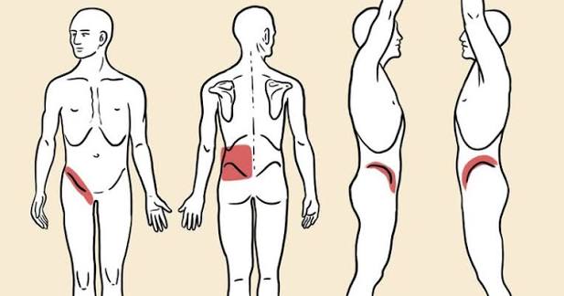
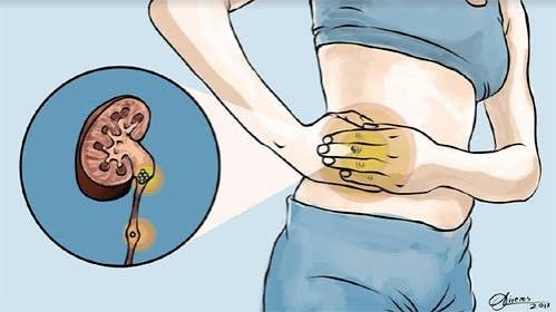
One of the earliest and most commonly overlooked signs of a kidney stone is a dull, aching discomfort in the flank — the area of the lower back or side, between the lowest rib and the hip — that comes and goes rather than being constant. This pain differs in important ways from the classic renal colic of active stone passage: it tends to be lower intensity (an ache rather than a severe cramp), it may not radiate to the groin or genitalia, and it may be easily dismissed as a muscular strain from physical activity, a prolonged sitting position, or other benign causes.
This dull discomfort arises when a stone in the kidney or uppermost ureter intermittently obstructs urine flow or irritates the renal pelvis mucosa during the patient’s movements. It may be positional — worse when lying on one side, or when physically active — and may shift in quality or intensity over days to weeks as the stone moves within the collecting system. Patients who have had previous kidney stones often recognize this prodromal discomfort as familiar; for first-time stone formers, it is easily attributed to something else. A key distinguishing feature: persistent or recurring unilateral flank discomfort that does not follow the mechanical pattern of musculoskeletal pain (worsened by palpation, movement, or specific positions of the spine) warrants evaluation with urinalysis and possibly imaging.
Blood in the Urine: Visible or Microscopic
Hematuria — blood in the urine — is present in approximately 85% of patients with kidney stones at some point during their clinical course, and it often appears before severe pain develops. It may be visible (gross hematuria), producing pink, red, or brown-tinged urine, or it may be microscopic (detectable only on urinalysis but not visible to the naked eye). Microscopic hematuria is frequently an incidental finding on a urine test ordered for another reason — a routine physical, a workup for fatigue, or evaluation of urinary frequency — and when it appears in a person without a history of kidney disease or bladder pathology, kidney stones should be among the first conditions evaluated.
Blood in the urine from a kidney stone results from the stone abrading the mucosal lining of the renal pelvis or ureter as it moves. Even a stone sitting relatively still in the kidney can cause minor bleeding when the patient is physically active — this is one reason hematuria may be intermittent rather than constant in early stone disease. A key clinical point: hematuria is never “normal” and should always be investigated — while kidney stones are one of the most common causes, other causes include bladder cancer, kidney cancer, bladder infections, and glomerulonephritis. Any adult with unexplained hematuria, even microscopic, deserves evaluation rather than watchful waiting.
Urinary Urgency and Frequency Without Infection
A kidney stone that has migrated to the lower third of the ureter — the segment near the bladder — irritates the ureteral wall adjacent to the bladder trigone, producing sensations that closely mimic those of a bladder infection: frequent urges to urinate, a feeling that the bladder is not fully empty despite just voiding, and sometimes a mild burning quality to urination. These symptoms can appear before the stone causes significant obstruction or pain, and they frequently lead patients (and sometimes clinicians) to diagnose and treat a UTI when the actual cause is a stone.
The clinical distinction is important: a pure lower urinary tract infection should produce a positive urine culture with a significant bacterial count, while a stone causing lower ureteral irritation may produce pyuria (white blood cells) and hematuria on the urinalysis but grow no bacteria on culture — or grow bacteria at low counts consistent with contamination. Any patient who is treated for a UTI and whose symptoms do not fully resolve with antibiotics within 48 to 72 hours should be evaluated for the possibility that the symptoms are attributable to a stone rather than or in addition to infection. A urinalysis that shows blood without bacteria in a patient with urinary symptoms is a particularly strong signal to pursue stone evaluation.
Nausea and Vague Gastrointestinal Discomfort
Kidney stones commonly produce nausea — sometimes before significant pain begins — through visceral pain pathways and vagal stimulation from the renal capsule and ureter. The nausea associated with early or mild stone activity can be subtle: a persistent queasiness, reduced appetite, or feeling of upper abdominal fullness that comes and goes over days. Because nausea is such a nonspecific symptom with many possible causes — gastroenteritis, gastritis, medication side effects, anxiety — it is almost never attributed to a kidney stone early in its course.
The combination of nausea with concurrent flank discomfort, even at low intensity, is a meaningful clinical pattern that increases the probability of kidney stone activity. Patients who notice nausea occurring repeatedly alongside ipsilateral (same-side) flank or back discomfort — particularly if accompanied by any change in urine color or urinary frequency — should report this constellation of symptoms rather than treating the nausea in isolation. Gastrointestinal symptoms from kidney stones arise because the nerves supplying the kidney and ureter overlap significantly with those supplying the bowel, which is why mid-abdominal cramping, bloating, and reduced bowel motility can also accompany stone passage.
Urine Color and Odor Changes
Changes in urine appearance — beyond the frank hematuria described above — can be early indicators of kidney stone activity or the metabolic conditions that predispose to it. Consistently dark yellow or amber urine indicates insufficient hydration — a direct risk factor for stone formation, as concentrated urine has less solvent capacity to keep stone-forming minerals dissolved. Patients who chronically produce dark, concentrated urine are not only at increased risk of forming a new stone but are actively promoting the growth of an existing one.
Cloudy urine can indicate either a urinary tract infection (white blood cells and bacteria causing turbidity) or, less commonly, phosphaturia or uricosuria — precipitation of phosphate or urate crystals in the urine that produces a milky or hazy appearance. Neither is normal, and both warrant urinalysis evaluation. Persistently foul-smelling urine suggests infection — in the context of stone disease, this may indicate struvite stone activity or a concurrent UTI, both of which warrant prompt evaluation. Any patient with known kidney stones who develops a change in urine color or odor alongside any of the other early signs described here should contact their urologist or primary care provider promptly rather than waiting for pain to develop.
When Early Signs Appear in Patients With Known Stone Risk
Patients who have had a previous kidney stone, or who have been told they have stone-forming risk factors (hypercalciuria, hyperoxaluria, gout, a family history of stones), should have a lower threshold for reporting and evaluating early signs. For these patients, early evaluation is not overreaction — it is the rational response to a condition known to recur. Evaluation typically begins with urinalysis and may proceed to point-of-care ultrasound or CT-KUB depending on clinical suspicion. Catching a stone early — before it has caused significant obstruction or migrated into a position that makes spontaneous passage unlikely — may allow for earlier medical expulsive therapy (alpha-blockers to relax the ureter and facilitate passage) and avoids the emergency presentation with renal colic.
Patients on kidney stone prevention protocols — 24-hour urine monitoring, potassium citrate, thiazide diuretics, or allopurinol — who experience any of the early signs described above should contact their urologist. A change in symptoms during established prevention therapy may signal a new stone formation despite treatment (indicating the prevention protocol needs adjustment), a stone that was pre-existing and is now becoming symptomatic, or a complication such as concurrent infection. None of these should be managed by waiting for acute pain to develop.
The Difference Between Early Signs and the Acute Emergency
Early kidney stone signs are distinct from the acute emergency presentations that require immediate emergency department evaluation. Early signs — dull intermittent flank discomfort, microscopic hematuria, mild urinary frequency, occasional nausea — warrant evaluation with a scheduled same-day or next-day appointment rather than an emergency visit in most cases. The presentation that requires emergency evaluation is different in quality: severe, escalating, often incapacitating pain (renal colic that cannot be controlled with oral analgesics), fever combined with flank pain (suggesting infected obstruction), inability to keep fluids or medications down, anuria (no urine output), or bilateral flank pain suggesting bilateral ureteral stones or a stone in a solitary kidney.
Understanding this distinction helps patients make appropriate triage decisions — avoiding the unnecessary delay of dismissing genuine early signs while also avoiding the congestion of emergency departments for presentations that can safely be seen in clinic. For a detailed description of what the full pain of kidney stone passage feels like, the kidney stone pain guide on Horizon Health Guide covers the phenomenology of renal colic in clinical detail. For a broader overview of kidney stone causes, types, and treatment, see the kidney stones overview article on this site. External resources including the NIDDK kidney stone patient guide, the National Kidney Foundation stone resource, and the StatPearls nephrolithiasis review provide comprehensive clinical reference material.
Early Detection Through Routine Screening
In some patients, kidney stones are detected incidentally before any symptoms develop — found on imaging ordered for another indication, such as a CT scan for abdominal pain from a different cause, or an ultrasound for evaluation of the liver or gallbladder. These asymptomatic stones — particularly if small and located in a calyx remote from the ureteropelvic junction — may be managed with observation and annual imaging rather than immediate intervention. Monitoring allows the clinician to track stone growth, identify any migration toward a symptomatic position, and time elective intervention if the stone grows or the patient’s anatomy suggests it is unlikely to pass without help.
For patients with recurrent stones, periodic imaging — either annual ultrasound or low-dose CT — allows early detection of new stone formation and monitoring of existing stones, enabling proactive treatment planning before acute episodes occur. This approach, combined with ongoing 24-hour urine monitoring to assess the effectiveness of dietary and pharmacological prevention, represents the most systematic way to manage recurrent stone disease. Patients who understand their own stone-forming risk, recognize early signs, and maintain an ongoing relationship with a urologist who specializes in stone disease have substantially better long-term outcomes than those who engage with the healthcare system only during acute episodes.
Sources: NIDDK — Kidney Stones · National Kidney Foundation · StatPearls — Nephrolithiasis
Pain That Comes and Goes: Understanding Intermittent Renal Discomfort
One of the features that distinguishes early kidney stone discomfort from an established kidney infection or musculoskeletal back pain is its intermittent and often position-dependent quality. A patient with a small stone in the renal pelvis or upper ureter may experience periods of dull aching that appear to come and go over days or weeks — present in the morning after lying still through the night, then subsiding during the day when upright and active, then returning in the evening. This variability reflects the stone’s movement within the kidney’s collecting system and its intermittent engagement with the renal mucosa or ureteropelvic junction.
The character of early stone discomfort differs from renal colic in important ways. Full renal colic — the pain of a stone actively obstructing the ureter — typically reaches maximum intensity within 20 to 30 minutes of onset, remains severe for 20 to 60 minutes per episode, and may recur multiple times over hours. Early stone discomfort, by contrast, is typically moderate in intensity (a 3 to 5 on a 10-point pain scale rather than the 8 to 10 of active colic), does not radiate to the groin, and does not cause the extreme restlessness and inability to find a comfortable position that characterizes full renal colic. Patients describe early stone discomfort variously as “a deep ache in my lower back,” “a heaviness in my side,” or “something that feels like a bruised rib but without any injury.” These descriptions should prompt urological evaluation in any patient without an established musculoskeletal diagnosis to explain them.
Changes in Urination: Frequency, Urgency, and Incomplete Emptying
As a kidney stone descends through the ureter toward the bladder, it passes through three anatomically narrow points where it is most likely to become temporarily lodged: the ureteropelvic junction (where the renal pelvis meets the ureter), the crossing of the ureter over the iliac vessels, and the ureterovesical junction (where the ureter enters the bladder). Each of these locations has distinct symptom profiles when a stone is present.
When a stone reaches the distal ureter near the bladder — the most common location for a stone to become lodged — it stimulates the trigone of the bladder (the triangular region at the base of the bladder where the ureters enter), producing symptoms that are clinically indistinguishable from an overactive bladder or a lower urinary tract infection: urgency (a sudden compelling need to urinate that is difficult to defer), increased frequency (urinating every 30 to 60 minutes rather than the normal 2 to 4 hours), and a sensation of incomplete bladder emptying despite just voiding. These symptoms can precede the onset of significant pain by hours to days.
A patient who presents with these lower urinary tract symptoms and a urinalysis showing red blood cells (hematuria) without bacteria should be evaluated for a distal ureteral stone before being treated empirically for a UTI. A point-of-care bladder ultrasound or renal ultrasound — which can detect hydronephrosis (ureteral dilation from obstruction) — is a valuable and rapid first-line test in this scenario. The absence of significant bacteriuria and the presence of hematuria in a patient with irritative voiding symptoms is a classic clinical pattern for a distal ureteral stone.
When to Act on Early Signs: A Practical Guide
Recognizing early kidney stone signs is only valuable if it leads to appropriate action. The following framework helps patients determine when to seek evaluation and how urgently.
Schedule a same-day or next-day appointment with a primary care provider or urologist if you experience: dull, recurring unilateral flank or back discomfort without a clear musculoskeletal explanation; visible or reported microscopic hematuria; urinary frequency and urgency without fever or vaginal symptoms and with a negative or equivocal urine culture; or any combination of two or more of the early signs described in this article. A urinalysis, urine culture, and renal ultrasound or CT-KUB provide the diagnostic information needed to confirm or rule out a stone within one clinical encounter.
Go to an urgent care clinic the same day if the discomfort is moderate (interfering with normal activities but controllable with over-the-counter analgesics), if visible hematuria is present, or if urinary symptoms are severe enough to be significantly disruptive. Urgent care clinics can perform urinalysis, basic blood work, and in many cases arrange same-day imaging that will confirm or rule out a stone and guide next steps.
Go directly to the emergency department if: pain becomes severe and uncontrollable with oral medications; fever develops in association with flank pain or urinary symptoms (a combination that may indicate an infected, obstructed ureter — a urological emergency); vomiting prevents fluid and medication intake; or you have no urine output despite normal fluid intake. These features represent a transition from early stone signs to an acute stone emergency requiring immediate evaluation and likely intervention.
For patients with recurrent kidney stones, one of the most practical steps is maintaining a urine strainer at home so that any stone that passes can be recovered and sent for analysis — this provides definitive information about stone composition and guides prevention. Keeping a log of symptom onset, duration, and characteristics also helps the urologist track the stone’s clinical behavior over time and plan intervention appropriately.
Metabolic Warning Signs: When Labs Predict Stones Before Symptoms Do
In some patients, the earliest “sign” of kidney stone risk is not a symptom at all but a laboratory finding — elevated serum calcium, elevated uric acid, elevated creatinine, or abnormalities in the 24-hour urine collection (high calcium, low citrate, high oxalate, high uric acid, or low urine volume). These biochemical abnormalities precede stone formation by months to years in some patients and represent an opportunity to intervene before any stone has formed or before an existing stone has grown large enough to cause symptoms.
Patients at elevated risk for kidney stones — those with a family history of stones, those with gout, hyperparathyroidism, inflammatory bowel disease, bariatric surgery, or chronic dehydration from a demanding physical occupation — benefit from periodic metabolic screening even before a first stone episode. A 24-hour urine collection is the most informative single test for identifying modifiable stone-forming risk, and targeted dietary or pharmacological intervention based on its results can significantly reduce the probability of forming a first or recurrent stone. This proactive approach is underutilized but represents one of the highest-value preventive interventions in urology — particularly for patients with known risk factors who have not yet formed a symptomatic stone.
One final practical consideration: patients who are well-hydrated and produce large volumes of pale urine are far less likely to experience early stone symptoms in the first place, because adequate urine volume reduces crystal supersaturation and slows stone growth. Maintaining a daily urine output of at least 2 liters — monitored informally by urine color (pale yellow is the target) — is the single behavioral change with the highest impact on early stone prevention. Patients who recognize the subtle warning signs described in this article and respond to them with prompt evaluation, increased hydration, and urological follow-up are in a fundamentally better position than those who wait for renal colic to bring them to the emergency department — and the evidence strongly supports early intervention over reactive care in kidney stone management.
I had a dull ache in my lower right side for about three weeks that I kept dismissing as a pulled muscle from exercise. Then one night the pain went from a 3 to a 10 in about 20 minutes and I ended up in the ER with a 6mm stone stuck in my ureter. Looking back, I also noticed my urine was darker than usual that whole week. If I had known those were warning signs I would have gone to the doctor sooner and maybe caught it before it caused the full blockage. This article is exactly what I wish I had read earlier.
As someone who runs ultramarathons in the heat, I know I’m at high risk for kidney stones from dehydration. This article is the first one I’ve found that explains the early signs clearly rather than just describing the ER-level pain everyone already knows about. The section on dark urine as a predictor is really actionable — I now check my urine color every morning and aim for pale yellow. Small habit, but it’s become part of my daily health monitoring.
That’s a really smart habit, Peter — urine color monitoring is one of the simplest and most reliable indicators of hydration status and stone risk. For athletes, especially those training in heat or at altitude, the combination of high sweat rates and sometimes inadequate fluid replacement creates exactly the urine concentration environment that promotes stone formation. Aiming for pale yellow throughout the day (not just after morning coffee) and replacing losses proactively during and after runs is the right approach. We’re glad this article is giving you a practical tool beyond just ‘drink more water.’ Stay safe out there!