
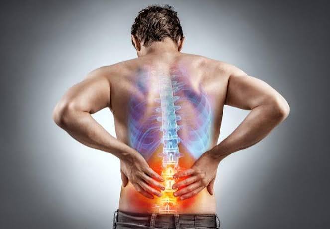
Kidney Stone Pain: What It Feels Like
Kidney stone pain — medically known as renal colic — is consistently described by patients who have experienced it as among the most severe pain a human being can feel. Studies comparing patient-rated pain intensity place renal colic alongside childbirth and post-operative pain in the top tier of acute pain experiences. Emergency physicians routinely see patients with renal colic in extremis: writhing, unable to find a comfortable position, pale, diaphoretic, and often vomiting — not because they are weak, but because the pain of ureteral obstruction from a stone triggers a physiological response that overwhelms even high pain tolerance. Understanding what kidney stone pain actually feels like — its character, location, progression, and the factors that make it better or worse — helps patients recognize it, communicate it accurately to medical providers, and seek the right level of care at the right time.
Not all kidney stone pain is the same. The experience depends on where in the urinary tract the stone is located, how large it is relative to the caliber of the structure it is obstructing, whether infection is coexisting with the obstruction, and the patient’s individual nervous system characteristics. Some patients pass small stones with nothing more than a brief ache; others require emergency intervention for stones that would ordinarily be expected to pass spontaneously. This variability in the pain experience is itself clinically informative — and understanding the different patterns of kidney stone pain helps distinguish it from the many other conditions it can mimic.
The Onset of Renal Colic: Abrupt and Escalating
One of the most characteristic features of kidney stone pain is its onset. Unlike the gradual onset of musculoskeletal back pain that worsens over hours or days, or the slowly escalating discomfort of a kidney infection building over a day or two, renal colic typically begins abruptly and escalates rapidly to maximum intensity within 20 to 30 minutes of onset. A patient may be completely comfortable at 9 PM and be calling for an ambulance by 9:30 PM — not because of any dramatic triggering event, but because the stone has shifted into a position of ureteral obstruction and the ureteral musculature has begun its spastic response.
The mechanism of the abrupt onset is straightforward: when a stone creates an acute obstruction in the ureter, pressure rises rapidly in the collecting system above it (the ureter, renal pelvis, and calices), stimulating pressure-sensitive nerves in the renal capsule and ureteral wall. Ureteral smooth muscle begins rhythmic peristaltic contractions — the normal mechanism for moving urine down the ureter — but these contractions cannot push the stone past its point of obstruction. Each contraction generates a wave of increased intra-ureteral pressure and stretching of ureteral muscle fibers — producing the characteristic crescendo pain of a colic episode.
Colicky Quality: Waves of Severe Pain
“Colicky” pain — pain that waxes and wanes in waves — is the defining characteristic of renal colic. Each wave of severe pain corresponds to a bout of ureteral peristalsis attempting (and failing) to expel the stone; between waves, the pain typically subsides from excruciating to severe, or in some cases to moderate. A full colic episode typically consists of pain that escalates sharply over a few minutes to a peak (often described as 9 or 10 out of 10 on a pain scale), sustains at that level for 20 to 60 minutes, then partially subsides before returning. Episodes typically recur every one to four hours, though the inter-episode pattern is highly variable.
It is important to note that “colicky” does not mean the pain is mild or intermittent in a comfortable way. Between colic peaks, patients remain in significant pain — simply at a level that is no longer absolutely unbearable. The partial relief between peaks can mislead patients into thinking the episode is resolving, only to be followed by another severe wave. Unlike bowel colic (the cramping of intestinal obstruction or gastroenteritis), which often does resolve substantially between episodes, renal colic between peaks remains significant — typically a 4 to 6 out of 10 even during partial remission for most patients with an actively obstructing stone.
Location: Where Does Kidney Stone Pain Occur?
The location of kidney stone pain is anatomically predictable and shifts as the stone moves down the ureter — one of the most useful features for distinguishing renal colic from other causes of acute pain. Understanding the pain location pattern also helps patients communicate what they are experiencing to emergency responders and clinicians.
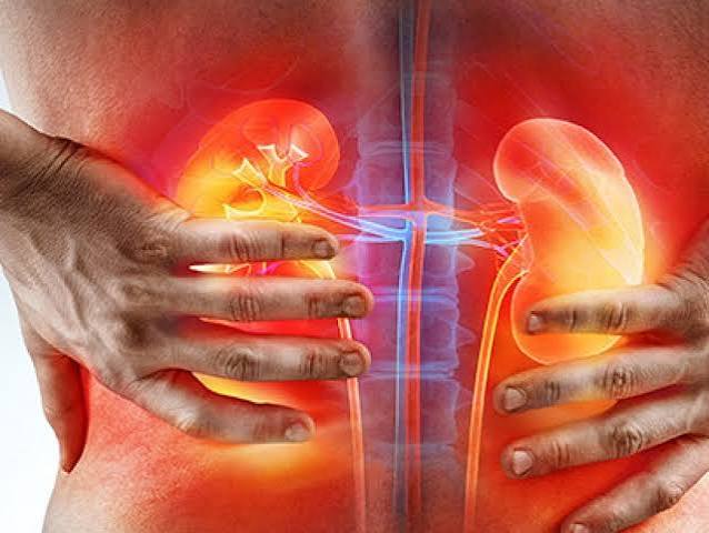
Flank and costovertebral angle: When a stone is in the ureter near the kidney (upper ureter or ureteropelvic junction), pain is most prominent in the flank — the area between the lowest rib and the iliac crest — and at the costovertebral angle (the angle formed by the spine and the lowest rib on the affected side). This location corresponds to where the kidney sits, and the pain reflects both renal capsular distension from rising pressure in the obstructed collecting system and ureteral spasm near the kidney. Costovertebral angle tenderness — elicited when a clinician taps on this area — is a reliable examination finding in upper ureteral obstruction.
Mid-flank to iliac fossa: As the stone moves to the mid-ureter (the segment crossing the iliac vessels), pain shifts from the flank toward the front of the lower abdomen, the iliac fossa (the area just above the hip on the affected side). This location can closely mimic appendicitis (right side) or ovarian pathology (right or left side in women), leading to diagnostic confusion. CT scanning is often the definitive test in this scenario — it simultaneously evaluates for appendicitis, ovarian pathology, and ureteral stone.
Groin, genitalia, and inner thigh: When the stone reaches the distal ureter near its insertion into the bladder, pain radiates into the groin, labia or scrotum, and inner thigh — following the path of the genitofemoral nerve, which is compressed by the inflamed ureteral wall. This distal radiation pattern is the classic “renal colic to the groin” that many patients report and that is highly specific for a distal ureteral stone. At this stage, irritative lower urinary tract symptoms — urgency, frequency, and sometimes burning with urination — typically join the pain picture as the stone irritates the bladder trigone.
The Restlessness of Renal Colic: Unable to Find a Comfortable Position
One of the most clinically distinctive features of renal colic — and one that emergency physicians use to distinguish it from other causes of acute pain — is the patient’s inability to find a comfortable position. Patients with peritonitis (inflammation of the abdominal lining, as from appendicitis or bowel perforation) characteristically lie very still because any movement worsens their pain. Patients with renal colic do the opposite: they pace, shift, writhe, curl, and change position repeatedly — not because movement helps (it generally does not reduce the pain significantly), but because the uncontrollable restlessness is a feature of the severe visceral pain and the autonomic response it triggers.
This restlessness is driven partly by the pain itself and partly by the nausea and autonomic activation (sweating, pallor, tachycardia) that accompany severe renal colic. Patients often describe a desperate urge to move, change position, or apply pressure to the flank — behaviors that represent the body’s attempt to relieve pain it cannot escape. For emergency providers, a patient who is writhing in a hospital gurney and cannot remain still is almost certainly in severe pain from a visceral source — kidney stone, biliary colic, or bowel obstruction — rather than a musculoskeletal or functional pain syndrome.
Associated Symptoms: What Accompanies the Pain
Nausea and vomiting accompany renal colic in approximately 50 to 80% of patients and can be as incapacitating as the pain itself. They arise from vagal activation triggered by the severe visceral pain and from the anatomical proximity of the renal nerves to the celiac plexus, which supplies the gastrointestinal tract. In some patients, vomiting is severe enough to prevent oral fluid and medication intake, making intravenous antiemetics and analgesics necessary. Vomiting during a kidney stone episode also worsens dehydration, increasing urine concentration and potentially promoting stone growth.
Hematuria (blood in the urine) is present in approximately 85% of patients with an actively symptomatic stone and ranges from microscopic (visible only on urinalysis) to grossly visible (pink, red, or brown urine). Many patients are alarmed by visible hematuria, but it is a predictable consequence of the stone abrading the ureteral mucosa and does not in itself indicate a serious complication. The absence of hematuria does not rule out a stone — approximately 15% of confirmed ureteral stones present without hematuria, particularly when the obstruction is complete and urine is not flowing past the stone.
Low-grade fever can occur in uncomplicated renal colic as part of the systemic inflammatory response to severe pain and ureteral inflammation — distinguishing this from the high fever (above 38.5°C) of infected obstruction requires clinical judgment. High fever combined with stone pain is a urological emergency requiring immediate drainage; low-grade temperature elevation (below 38°C) in an otherwise stable patient without rigors or hemodynamic instability is less alarming but warrants close monitoring and urology consultation.
Pain Management: What Helps During a Kidney Stone Episode
The primary goals of pain management during acute renal colic are reducing ureteral spasm (which drives the peak intensity of colic episodes), controlling associated nausea, and maintaining hydration. NSAIDs (non-steroidal anti-inflammatory drugs) such as ibuprofen, naproxen, and the injectable form ketorolac are the preferred analgesics for renal colic and have been shown in multiple randomized controlled trials to be as effective as opioids for most patients — and in some studies, superior — for controlling the pain of ureteral obstruction. Their mechanism is particularly well-suited to renal colic: NSAIDs reduce prostaglandin-mediated ureteral smooth muscle spasm and renal blood flow-driven increases in intra-pelvic pressure, directly addressing the two mechanisms driving the pain rather than merely blunting pain perception.
For patients who cannot tolerate NSAIDs (those with kidney disease, peptic ulcer disease, or NSAID allergy) or whose pain is not adequately controlled with NSAIDs alone, opioids (morphine, hydromorphone, or oxycodone) are effective second-line analgesics. Antiemetics — ondansetron (Zofran), promethazine, or metoclopramide — are essential when nausea is prominent and should be administered alongside analgesics rather than waiting until vomiting is established. Alpha-blockers (tamsulosin) reduce ureteral smooth muscle tone and have been shown to increase spontaneous stone passage rates for distal ureteral stones and to reduce the frequency and severity of colic episodes during conservative management — they are typically prescribed for all patients undergoing watchful waiting for a stone in the ureter.
For a clinical overview of kidney stone causes, types, and the full spectrum of treatment from conservative to surgical, see the kidney stones overview on Horizon Health Guide. Early warning signs before pain develops are covered in the early signs of kidney stones article on this site. For patients with recurrent stone episodes, the recurrent urinary conditions guide at Horizon Health Guide provides prevention context. External references include the NIDDK kidney stone patient guide, the StatPearls nephrolithiasis clinical review, and the American Urological Association kidney stone guidelines.
How Long Does Kidney Stone Pain Last?
The duration of kidney stone pain depends primarily on stone size, stone location, and whether the stone passes spontaneously or requires intervention. Small stones (less than 5 mm) in the ureter pass spontaneously approximately 68% of the time within four weeks — most within one to two weeks of the onset of symptoms. During this period, acute colic episodes may recur whenever the stone shifts position or triggers a wave of ureteral peristalsis, but many patients also have pain-free intervals of hours to days between episodes as the stone lodges in a position of partial rather than complete obstruction.
Stones between 5 and 10 mm pass spontaneously approximately 47% of the time, with average passage times of 12 to 22 days. Stones larger than 10 mm are unlikely to pass without intervention and are typically treated with ureteroscopy, shock wave lithotripsy, or percutaneous nephrolithotomy depending on size, location, and composition. Once a stone has either passed through the bladder into the ureter (felt as a sudden cessation of the renal colic, sometimes replaced by brief urethral discomfort) or been treated with a procedure, pain typically resolves rapidly — within hours for spontaneous passage, within one to two days after ureteroscopy.
Persistent pain after presumed stone passage, or pain that is atypical (constant rather than colicky, bilateral, or accompanied by fever), warrants follow-up imaging to confirm stone clearance and rule out complications such as residual stone fragments, ureteral injury, or secondary infection. Patients should not assume that the resolution of acute pain means the stone has passed — some stones lodge in the ureter without causing severe pain in the short term but continue to obstruct silently, risking progressive kidney damage. A follow-up CT scan or renal ultrasound within four to six weeks of the acute episode confirms stone clearance and baseline kidney function.
Sources: NIDDK — Kidney Stones · StatPearls — Nephrolithiasis · AUA Kidney Stone Guidelines
How Kidney Stone Pain Differs From Other Causes of Back and Abdominal Pain
Because kidney stone pain can occur in the flank, abdomen, groin, and genitalia — and because it is frequently accompanied by nausea, vomiting, and diaphoresis — it overlaps clinically with a number of other serious conditions. The ability to distinguish renal colic from its mimics is important both for patients deciding whether to seek emergency care and for clinicians selecting diagnostic tests.
Musculoskeletal back pain is the most common condition confused with kidney stone pain. The distinguishing features favor kidney stone in the following ways: renal colic is not reproducible by palpation of the paraspinal muscles or movement of the spine (musculoskeletal pain typically worsens with spinal flexion, extension, or rotation and is tenderness on palpation); renal colic is associated with hematuria and urinary symptoms; renal colic does not improve with a specific position (rest and lying down do not relieve it the way they relieve muscular pain); and renal colic does not follow the dermatomal distribution of nerve root compression. A patient who cannot recall any injury or physical activity that preceded the onset of flank pain, and whose pain is accompanied by urinary symptoms or hematuria, has renal colic until proven otherwise.
Appendicitis is confused with right-sided renal colic when the stone is in the mid-ureter crossing the iliac vessels. The distinguishing features of appendicitis include: pain that begins periumbilically and migrates to the right lower quadrant (McBurney’s point) rather than starting in the flank; pain that worsens with movement (especially the psoas sign and obturator sign on examination); fever typically present from early in the course; nausea proportional to but usually less extreme than in renal colic; and elevated white blood cell count. CT scan with contrast is the definitive study that distinguishes between these two diagnoses in most emergency settings.
Ovarian pathology — ovarian cyst rupture, ovarian torsion, or ectopic pregnancy — can produce left or right lower abdominal pain with nausea and vomiting that closely mimics distal ureteral stone pain in women. Ovarian torsion is a gynecologic emergency with sudden-onset severe unilateral lower abdominal pain that may include low-grade fever and vomiting; its distinction from renal colic usually requires pelvic ultrasound and measurement of urinary or serum beta-hCG to evaluate for ectopic pregnancy. Hematuria is absent in pure ovarian pathology but present in most kidney stone episodes.
Aortic aneurysm is the most dangerous condition confused with kidney stone pain — an expanding or ruptured abdominal aortic aneurysm can produce severe flank pain with hemodynamic instability that superficially resembles renal colic. The key clinical red flags that distinguish aneurysm from stone: sudden onset of the most severe pain the patient has ever experienced; pain that is tearing or ripping in quality rather than colicky; hemodynamic instability (low blood pressure, rapid heart rate, pallor, and diaphoresis out of proportion to the pain intensity); a pulsatile abdominal mass on examination; and risk factors including older age, male sex, smoking history, and known atherosclerosis. Any middle-aged or older patient with severe flank or back pain and hemodynamic instability requires immediate vascular evaluation before a kidney stone diagnosis is assumed.
The Psychological Experience of Renal Colic: What Patients Describe
Beyond the clinical description of pain character, location, and intensity, the subjective experience of renal colic has qualities that patients frequently describe and that deserve acknowledgment. The suddenness of onset — going from completely comfortable to 9 out of 10 pain within minutes — is profoundly disorienting. Many patients report experiencing fear alongside the pain: fear that something catastrophic is happening, fear that the pain will not stop, and a sense of helplessness against a pain that does not respond to any voluntary action. This psychological component is not trivial — it contributes to the distress of the episode and to the health-seeking behavior that drives patients to emergency departments even when they have some home analgesic supply.
Patients who have experienced multiple kidney stone episodes describe an evolution in their psychological response over time. First-time stone formers are typically terrified and are convinced something catastrophic is occurring — a reasonable assumption given the severity of the pain. Experienced stone formers, by contrast, may recognize the familiar pattern of onset and radiation, begin home analgesics and oral hydration, and seek care in a more organized manner. Some experienced patients develop what stone specialists call a “stone kit” — a home supply of oral NSAIDs or prescribed opioids, antiemetics, a urine strainer, and clear written instructions from their urologist about when to manage at home versus when to go to the emergency department.
This preparation does not minimize the severity of the pain — it reflects the reality that patients who understand their condition are better equipped to manage it effectively. For anyone experiencing kidney stone pain for the first time, the most important message is this: the pain of renal colic, while among the worst pain a human experiences, is not life-threatening in the absence of the red flags described in this article (fever, bilateral pain, anuria, hemodynamic instability). It is survivable. It will end. And with proper evaluation and follow-up, the factors that caused it can often be identified and modified to reduce the probability of it recurring.
A note on hydration during stone passage: drinking large volumes of water — sometimes suggested as a way to “flush the stone out” — should be approached with caution during active renal colic with complete obstruction. If the stone is fully blocking the ureter, the kidney above it cannot drain, and aggressive fluid loading increases renal pelvic pressure and may worsen pain without facilitating stone passage. Moderate, consistent fluid intake (sipping rather than gulping large volumes) is more appropriate during an acute colic episode; the high-volume hydration advice applies to the prevention of new stones rather than to the management of active obstruction. Once the stone has passed or been treated and the collecting system is draining normally, resuming high fluid intake helps clear the urinary tract and reduces crystallization risk.
I’ve had three kidney stone episodes and I’ve also broken my collarbone, had an appendectomy, and gone through a root canal without anesthesia. Nothing has come close to the kidney stone. The worst part was the first time — I was convinced I was dying because the pain escalated so fast and I had no idea what was happening. The description in this article — especially the restlessness and the inability to find any position that helped — is exactly right. I literally ran laps around my apartment at 3 AM because I couldn’t sit still. Paramedics were confused when I answered the door walking around instead of collapsed.
As an emergency medicine physician, this article accurately captures what we see at the bedside. The section on distinguishing renal colic from appendicitis and aortic aneurysm is clinically important — the aortic aneurysm mimicry is the one that keeps EM doctors up at night. An older male with sudden severe back or flank pain gets a vascular evaluation until proven otherwise. The detail about restlessness versus the stillness of peritonitis is a teaching pearl we use in medical education. Well done.
Thank you for that clinical perspective, Dr. Yamamoto — the aortic aneurysm mimicry point is one we wanted to include precisely because it is the life-threatening condition most likely to be missed in a patient presenting with what appears to be typical renal colic. The age/sex/risk factor profile often helps, but the key teaching point you raise — hemodynamic instability is not a feature of uncomplicated renal colic — is essential. A patient who looks shocked with severe flank pain warrants vascular imaging before a stone is assumed. We appreciate you sharing your teaching perspective, and we hope this article serves as a useful patient-education resource for your ED patients who want to understand what happened to them.