
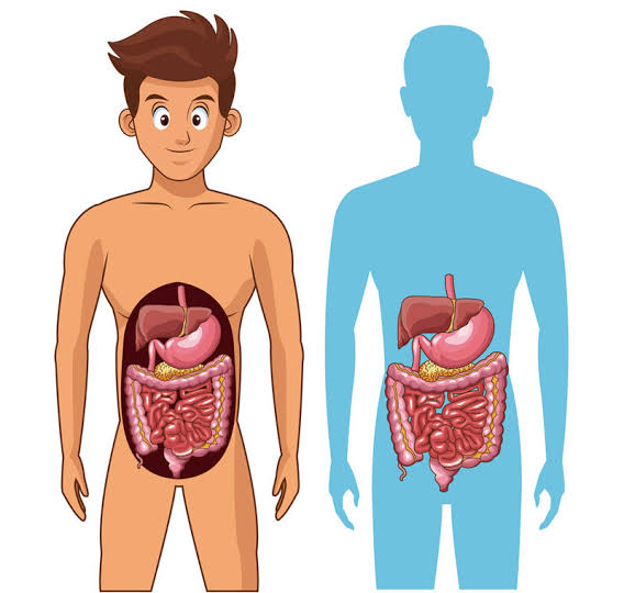
The digestive system is a continuous, coordinated tube running approximately 9 meters from the mouth to the anus, supported by accessory organs — the liver, gallbladder, and pancreas — that are essential to its function but do not form part of the main alimentary canal. Its job is deceptively simple to state: break down the food you eat into molecules small enough to absorb into the bloodstream, use those molecules to power the body and maintain its structure, and expel what cannot be used. The actual execution of that task involves an extraordinarily complex series of mechanical, chemical, neural, and hormonal processes that operate in precise sequence every time you eat. Understanding how the digestive system works at each stage clarifies why disruption at any point — from inadequate chewing to small intestinal bacterial overgrowth to impaired bile production — produces such specific and often puzzling symptoms.
The Mouth: Where Digestion Begins
Digestion does not begin in the stomach — it begins in the mouth, and the thoroughness of oral digestion directly affects everything that follows. Chewing (mastication) breaks food into smaller particles, increasing the surface area available to digestive enzymes. Saliva — approximately 1–1.5 liters produced daily — performs three simultaneous functions: it lubricates food to facilitate swallowing, it contains salivary amylase (also called ptyalin), which begins the chemical breakdown of starches into shorter carbohydrate chains, and it contains lingual lipase, which initiates fat digestion.
Salivary amylase remains active until it contacts stomach acid, meaning carbohydrate digestion begins in the mouth and continues briefly in the upper stomach before acid inactivates the enzyme. This is why swallowing unchewed food impairs digestion — the first enzymatic step is bypassed, and the burden on subsequent digestive processes increases. Digestion efficiency depends in part on the physical preparation of food at this stage: the smaller the particle size reaching the stomach, the more completely and quickly the stomach and small intestine can process it.
The Esophagus: Transport and Valve Function
The esophagus is a muscular tube approximately 25–30 centimeters long that connects the throat (pharynx) to the stomach. It performs no chemical digestion — its sole function is mechanical transport. Food moves through the esophagus via peristalsis: coordinated, rhythmic muscle contractions that move the food bolus downward in a wave. Peristalsis is involuntary and occurs even if you swallow in a position that opposes gravity — one reason people rarely have difficulty swallowing while lying down in the absence of disease.
At the lower end of the esophagus sits the lower esophageal sphincter (LES) — a ring of muscle that remains closed except during swallowing and belching. The LES serves as a valve preventing stomach acid from refluxing into the esophagus. When the LES is functioning correctly, it opens just long enough for food to pass and then closes tightly again. When LES tone is reduced — by certain foods (fatty meals, caffeine, chocolate, alcohol), medications, hiatal hernia, or other factors — stomach acid escapes upward, causing the burning sensation of heartburn and, if chronic, the esophageal lining damage of gastroesophageal reflux disease (GERD). This mechanism is discussed in detail in the article on GERD: a simple guide for adults.
The Stomach: Mechanical Churning and Chemical Breakdown
The stomach is a J-shaped muscular bag that can expand from its resting volume of approximately 75 mL to hold over 1 liter after a large meal. Its walls contain three layers of smooth muscle that contract rhythmically, churning food and mixing it with gastric secretions to produce chyme — a semi-liquid, acidic mixture. This mechanical churning is as important as the chemical digestion: grinding food against the stomach’s muscular walls further reduces particle size and increases enzyme access.
The stomach’s chemical environment is highly acidic — pH 1.5 to 3.5 — due to hydrochloric acid secreted by parietal cells. This acid serves three functions: it activates pepsinogen into pepsin (the enzyme that begins protein digestion), it sterilizes ingested food and fluids (killing most bacteria before they reach the intestines), and it creates the chemical environment needed for intrinsic factor — a glycoprotein also secreted by parietal cells — to bind vitamin B12. This B12-intrinsic factor complex is required for B12 absorption in the terminal ileum. Conditions that destroy parietal cells (autoimmune atrophic gastritis) or long-term use of acid-suppressing medications (particularly proton pump inhibitors) can impair B12 absorption and produce vitamin B12 deficiency over time.
Gastric emptying — the rate at which chyme moves from the stomach into the small intestine — is tightly regulated. Solid food takes 2–5 hours to empty; liquids empty faster; high-fat meals slow emptying significantly because fat must be emulsified and processed more slowly. The pyloric sphincter, a valve at the stomach’s exit, releases small amounts of chyme into the duodenum at intervals, coordinating with the small intestine’s capacity to receive and process it. Delayed gastric emptying — gastroparesis — causes nausea, bloating, and early satiety because food remains in the stomach far longer than normal.
The Small Intestine: Nutrient Absorption
The small intestine — named for its diameter (2.5–3 cm), not its length (approximately 6 meters) — is where the vast majority of nutrient digestion and absorption occurs. Chyme entering from the stomach triggers the release of two key hormones: secretin (which stimulates the pancreas to release bicarbonate, neutralizing the acid in the chyme) and cholecystokinin (CCK), which stimulates the pancreas to release digestive enzymes and triggers the gallbladder to contract and release bile.
The pancreas contributes three categories of enzymes to the small intestine: proteases (trypsin, chymotrypsin, elastase) to complete protein digestion into amino acids; pancreatic amylase to finish carbohydrate digestion into monosaccharides; and pancreatic lipase (with its cofactor colipase) to break triglycerides into fatty acids and monoglycerides. The role of the liver and gallbladder through bile acid secretion is to emulsify dietary fat — without emulsification, pancreatic lipase cannot access fat molecules efficiently, and fat absorption is severely impaired. This is why gallbladder disease, bile duct obstruction, and exocrine pancreatic insufficiency each produce fatty, floating, foul-smelling stools (steatorrhea).
The inner lining of the small intestine is anatomically specialized for maximum absorption. The mucosa is folded into circular folds (plicae circulares), which are covered with villi — finger-like projections 0.5–1 mm tall. Each villus is covered with microvilli (the brush border), creating a combined absorptive surface area of approximately 30 square meters — the size of half a badminton court. This vast surface area ensures that nutrients passing through the small intestine have maximum contact time with absorptive cells (enterocytes).
Nutrient absorption mechanisms vary by molecule type:
- Monosaccharides (glucose, fructose, galactose) are absorbed via active transport and facilitated diffusion across enterocyte membranes into the portal blood.
- Amino acids and small peptides are absorbed via specific amino acid transporters, also into the portal blood.
- Fatty acids and monoglycerides are reassembled into triglycerides inside enterocytes, packaged into chylomicrons (lipoprotein particles), and released into the lymphatic system (not directly into blood), eventually entering blood circulation via the thoracic duct.
- Fat-soluble vitamins (A, D, E, K) are absorbed with fat — conditions impairing fat absorption also impair these vitamins.
- Water-soluble vitamins are absorbed by specific transporters; B12 requires the intrinsic factor complex and is absorbed only in the terminal ileum.
- Minerals — calcium, iron, zinc, magnesium — are absorbed in the duodenum and upper jejunum, with absorption rates regulated by body stores and hormonal signals.
Transit time through the small intestine is approximately 2–6 hours in healthy adults. The NIDDK digestive system overview notes that any condition shortening small intestinal transit time — diarrheal illness, IBS-D, rapid gastric emptying — reduces nutrient contact time and can impair absorption even when the absorptive surface itself is intact.
The Large Intestine: Water Absorption and Microbiome
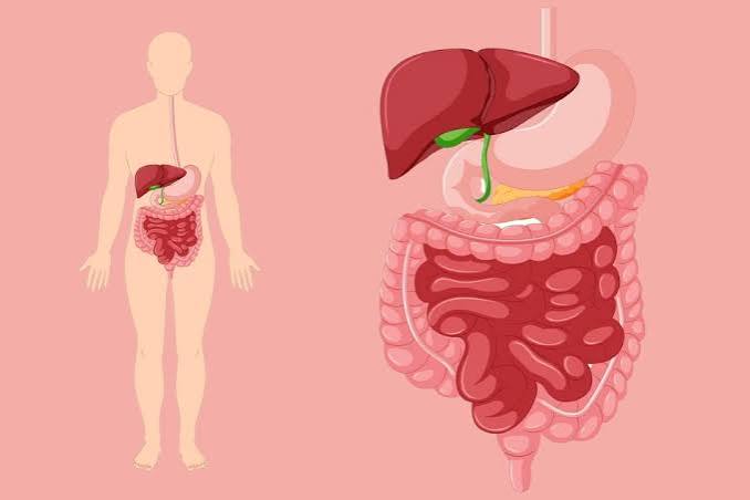
The large intestine (colon) is approximately 1.5 meters long and 6–7 cm in diameter. By the time chyme enters it from the small intestine, most nutrients have already been absorbed. The colon’s primary roles are to absorb water and electrolytes from the remaining liquid material, house the gut microbiome, and form, store, and ultimately expel solid stool.
Approximately 1–2 liters of fluid enter the colon daily from the small intestine; the colon reabsorbs most of it, producing the 100–200 mL of water in normal stool. When colonic absorption is impaired — by infection, inflammation, or laxative use — more water remains in the stool, causing diarrhea. When transit is too slow, the colon absorbs too much water, making stool hard and difficult to pass (constipation).
The colon houses the majority of the gut microbiome — approximately 100 trillion microorganisms, representing 1,000 or more species. Gut bacteria ferment the dietary fiber that escaped digestion in the small intestine, producing short-chain fatty acids (SCFAs): butyrate, propionate, and acetate. Butyrate is the primary energy source for colonocytes (the cells lining the colon) and has anti-inflammatory and anti-cancer properties in the colonic environment. Propionate and acetate enter the portal circulation and influence liver metabolism and systemic immunity. The role of this fermentation process in gut and systemic health is one of the most important and rapidly advancing areas of digestive research. More detail on this is available in the article on what is digestive health.
Colonic motility is driven by mass movements — large, coordinated contractions that propel contents toward the rectum. These occur approximately 3–4 times per day, typically stimulated by eating (the gastrocolic reflex — which is why the urge to defecate often occurs after meals). Stool consistency and transit time are the readouts of how well this entire process is working. The Bristol Stool Scale, which classifies stool appearance from type 1 (hard pellets) to type 7 (watery), serves as a practical proxy for colonic transit time: slow transit produces hard, dry stool (types 1–2); fast transit produces loose, watery stool (types 5–7).
The Liver, Gallbladder, and Pancreas: Essential Support
These three accessory organs are not part of the main digestive tube but are so integral to its function that failure of any one of them profoundly disrupts digestion.
The liver produces 600–800 mL of bile per day, synthesizes the proteins and enzymes needed for digestion and metabolism, processes all absorbed nutrients arriving from the gut via the portal vein, and detoxifies waste products and xenobiotics. The close relationship between liver and digestive function means that liver disease — fatty liver, hepatitis, cirrhosis — affects digestion directly, and gut health reciprocally affects the liver through the portal circulation. This bidirectional relationship is explored in the article on what is liver health.
The gallbladder concentrates bile (removing water to increase its potency) and stores it until fat consumption triggers CCK release, causing the gallbladder to contract and release bile into the duodenum. After cholecystectomy (gallbladder removal), bile drips continuously into the small intestine rather than being released in coordinated boluses. Most people adapt well to this change, but some experience loose stools and discomfort after fatty meals, particularly in the first months after surgery, as the body adjusts to continuous bile delivery.
The pancreas secretes both digestive enzymes (exocrine function) and hormones (endocrine function). Exocrine pancreatic insufficiency (EPI) — failure to produce adequate digestive enzymes — causes malabsorption that can closely mimic IBS symptoms: bloating, diarrhea, and steatorrhea. EPI is underdiagnosed and is seen in chronic pancreatitis, pancreatic cancer, and cystic fibrosis, but also occurs to a lesser degree in older adults and some people with celiac disease or Crohn’s disease. The pancreatic endocrine cells (islets of Langerhans) produce insulin and glucagon to regulate blood glucose — dysfunction of this system produces diabetes.
The Enteric Nervous System: The Gut’s Own Brain
The digestive system has its own nervous system — the enteric nervous system (ENS) — embedded in the walls of the GI tract. It contains approximately 500 million neurons, more than the spinal cord, arranged in two networks (Auerbach’s myenteric plexus and Meissner’s submucosal plexus) that run the full length of the digestive tract. The ENS can coordinate peristalsis, secretion, and blood flow entirely independently of the brain, which is why the gut continues to function even when the vagus nerve — the main connection to the central nervous system — is severed.
Communication between the ENS and the central nervous system flows in both directions via the gut-brain axis, primarily through the vagus nerve, enteroendocrine cells (which sense gut content and release over 20 gut hormones), and the immune system. This bidirectional pathway explains why psychological stress reliably produces GI symptoms — increased gut motility, altered secretion, heightened visceral pain sensitivity — in people prone to IBS and functional dyspepsia. The ENS also contains approximately 90% of the body’s serotonin, which regulates peristalsis and modulates pain signaling in the gut as well as mood and cognition at the systemic level.
How Long Does Digestion Take?
Total gut transit time — from eating to elimination — is typically 24 to 72 hours in healthy adults, though the range is wide. Colonic transit time (10–59 hours) accounts for most of the variability. Women on average have slower colonic transit than men, which contributes to higher rates of constipation in the female population. Diet — particularly fiber content — is the most consistent modifiable influence on colonic transit time. The American College of Gastroenterology patient resources on GI health include practical guidance on what affects transit and when slow or fast transit warrants evaluation.
What Disrupts Digestive Function
Disruption can occur at any stage of digestion, and identifying where in the system a problem originates helps explain both the symptom pattern and the appropriate response.
Upper GI disruption (mouth, esophagus, stomach) produces symptoms like heartburn, regurgitation, nausea, vomiting, early satiety, and difficulty swallowing. Common causes include GERD, gastritis, peptic ulcer disease, gastroparesis, and hiatal hernia.
Small intestinal disruption produces malabsorption — symptoms may include diarrhea, fatty stools, unexplained weight loss, nutrient deficiencies, and bloating. Causes include celiac disease, Crohn’s disease affecting the small intestine, small intestinal bacterial overgrowth (SIBO), exocrine pancreatic insufficiency, and bile acid malabsorption.
Large intestinal disruption typically produces altered bowel habits — constipation, diarrhea, or alternating patterns — along with bloating, cramping, and changes in stool character. Causes include IBS, ulcerative colitis, diverticular disease, colorectal cancer, and microscopic colitis.
Accessory organ disruption (liver, gallbladder, pancreas) produces fat malabsorption symptoms plus organ-specific signs: jaundice and altered urine/stool color in liver and bile duct disease, right upper quadrant pain after fatty meals in gallbladder disease, and steatorrhea with weight loss in pancreatic insufficiency.
The full scope of common digestive conditions and their symptoms is covered in the companion article on common digestive problems in adults.
Frequently Asked Questions
How does the digestive system break down food?
Through a combination of mechanical and chemical processes. Mechanical digestion — chewing, stomach churning, intestinal peristalsis — physically reduces food particle size. Chemical digestion uses enzymes (salivary amylase, pepsin, pancreatic enzymes), acid (stomach hydrochloric acid), and bile (from the liver and gallbladder) to break food into molecules small enough to absorb. The process begins in the mouth with salivary enzymes and is not complete until the small intestine finishes enzymatic digestion.
Where does most nutrient absorption happen?
The small intestine — particularly the duodenum and jejunum (the first two sections). The specialized mucosal surface with villi and microvilli gives the small intestine an absorptive area of approximately 30 square meters. The large intestine absorbs primarily water and electrolytes, not the macronutrients and micronutrients that fuel the body.
What does the liver do in digestion?
The liver produces bile, which is essential for fat digestion and fat-soluble vitamin absorption. It also processes every nutrient absorbed from the gut — arriving via the portal vein — before that blood reaches general circulation. This first-pass processing regulates blood glucose, detoxifies absorbed compounds, and synthesizes proteins needed throughout the body. Liver dysfunction therefore affects not just bile production but the processing of everything the gut absorbs.
How long does it take to digest food?
Total transit time from eating to elimination is typically 24–72 hours. The stomach empties solid food in 2–5 hours; the small intestine takes 2–6 hours; the large intestine adds 10–59 hours of variability depending on individual physiology, fiber intake, hydration, activity level, and stress.
Why do I get the urge to defecate after eating?
This is the gastrocolic reflex — a normal neural response to stomach distension that triggers mass movements in the colon. It is most pronounced after the first meal of the day. The reflex explains why breakfast commonly triggers a bowel movement and why increasing meal regularity can help people with constipation establish a more predictable bowel pattern.
What is the gut microbiome’s role in digestion?
Gut bacteria ferment dietary fiber that the human digestive system cannot process on its own, producing short-chain fatty acids that nourish colonic cells, regulate inflammation, and influence metabolism systemically. The gut microbiome also influences intestinal motility, gut barrier integrity, immune function, and neurotransmitter production. Without gut bacteria, colonic function — and many downstream systemic health effects — would be substantially impaired.
Sources: National Institute of Diabetes and Digestive and Kidney Diseases (NIDDK), Your Digestive System and How It Works; American College of Gastroenterology (ACG), GI Health and Disease; Johnson LR (ed.), Gastrointestinal Physiology, 8th ed., Elsevier; Furness JB, The Enteric Nervous System, Blackwell (2006); Cummings JH et al., Gut 1978 (transit time reference values).
How to Support Each Stage of Digestion
Understanding how the digestive system works at each stage translates directly into practical habits that support it. Because the process is sequential — each stage preparing food for the next — the most effective approach addresses multiple points in the chain rather than targeting a single organ or symptom.
Support oral digestion by chewing thoroughly. Eating quickly and swallowing large food particles burdens the stomach and small intestine with work that should have been done in the mouth. Chewing each bite 15–20 times — or until food is soft and uniform — increases salivary enzyme contact, reduces particle size, and measurably reduces bloating and post-meal discomfort in people prone to functional GI symptoms. Eating slowly also gives stretch receptors in the stomach time to signal satiety to the brain (a process that takes 15–20 minutes), which helps prevent overeating.
Protect gastric acid levels. Adequate stomach acid is essential for protein digestion, B12 absorption, and sterilization of ingested food. Long-term acid suppression with PPIs should be reviewed periodically — these medications are appropriate for active peptic ulcer disease and moderate-to-severe GERD, but many patients continue them indefinitely for mild symptoms that could be managed with lifestyle changes and H2 blockers. The AGA patient guidance on GI health addresses when acid suppression is appropriate and when deprescribing should be considered.
Support small intestinal absorption with dietary fiber and variety. A diverse, fiber-rich diet maintains the integrity of the small intestinal lining, supports the gut microbiome composition that modulates immune function at the gut wall, and provides the prebiotics needed to sustain beneficial bacteria in the colon. The synergy between what enters the small intestine and what reaches the colon intact determines both absorptive function and downstream microbiome health — which is why dietary quality is the single most consistent predictor of long-term digestive health across population studies.
The section on small intestinal bacterial overgrowth (SIBO) touched on something I spent three years trying to diagnose. I had all the classic symptoms — bloating that made me look five months pregnant by evening, loose stools, fatigue, and eventually significant nutrient deficiencies — but every standard test came back normal. It wasn’t until I requested a hydrogen breath test from a gastroenterologist who specialized in motility disorders that SIBO was confirmed. The treatment was a course of rifaximin, and the improvement in symptoms was dramatic within the first two weeks. What this article explains about the small intestine’s transit time and bacterial environment really puts into context why SIBO develops — slow transit or disrupted motility allows bacteria to migrate upward from the colon where they belong and colonize the small intestine, where they ferment food before it can be properly absorbed.
Rachel, the SIBO journey you describe — years of normal standard tests before the underlying motility issue is identified — is unfortunately typical. Standard upper endoscopy and colonoscopy do not visualize the small intestine, and even imaging studies miss functional problems like impaired motility. The gastrocolic reflex disruption and retrograde bacterial migration you’re describing are well-established mechanisms in SIBO pathogenesis, and the success of rifaximin in your case is consistent with the trial data — it is a non-absorbable antibiotic that acts locally in the gut without significant systemic effects, which is why it is the preferred treatment when SIBO is confirmed.
I teach biology and I use the digestive system as one of my favorite examples of organ system integration — this article is an excellent patient-accessible explanation that I’ll share with students and their parents. The detail about the enteric nervous system containing 500 million neurons — more than the spinal cord — is the kind of fact that reframes people’s understanding of the gut. Most students think of digestion as something the stomach does. Understanding that the entire GI tract has its own nervous system, produces its own neurotransmitters, and maintains its own regulatory circuitry independent of the brain completely changes the picture of what the gut actually is.