
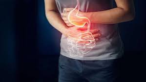
Upper abdominal pain — pain above the navel — is one of the most common presenting complaints in primary care and emergency medicine, and one of the most diagnostically important. The upper abdomen contains a concentration of digestive and hepatobiliary organs: the stomach and duodenum, liver, gallbladder and bile ducts, pancreas, spleen, and the transverse colon. Pain anywhere in this region can originate from any of these structures, and identifying the correct source determines whether the condition is managed conservatively, with medication, or requires urgent intervention. Location within the upper abdomen — epigastric (central), right upper quadrant, or left upper quadrant — provides the most useful initial diagnostic framework. Timing, character of the pain, associated symptoms, and whether the pain is meal-related, positional, or constant refine the differential further. This article covers the full range of digestive and liver causes of upper abdominal pain, with particular attention to the patterns that distinguish them. For the post-meal subset of upper abdominal pain, see stomach pain after eating.
Epigastric (Central) Upper Abdominal Pain
The epigastric region — the central upper abdomen between the sternum and the navel — is where stomach, duodenal, lower esophageal, and pancreatic pain most commonly presents. It is also the region most closely associated with GERD-related burning, making it one of the most frequently symptomatic areas in the upper abdomen.
Functional dyspepsia is the most common cause of chronic epigastric discomfort in adults, affecting approximately 10 to 20 percent of the adult population. It is defined by the Rome IV criteria as bothersome upper abdominal symptoms (fullness, early satiety, burning, nausea) without a structural explanation on endoscopy. The discomfort is characteristically post-meal and may be accompanied by early satiety, bloating, and nausea. Peptic ulcer disease produces epigastric pain with a characteristic timing pattern: gastric ulcers worsen with food (acid stimulated by eating contacts the ulcer), while duodenal ulcers peak 1 to 3 hours after eating when gastric acid is no longer buffered by the meal and often wake the patient at night. H. pylori infection is the most common cause, followed by NSAID use. H. pylori testing by breath test or stool antigen is the appropriate first step. Gastritis from H. pylori, NSAIDs, alcohol, or autoimmune disease produces diffuse epigastric discomfort, burning, nausea, and early satiety. Autoimmune gastritis destroys parietal cells, causing progressive achlorhydria and pernicious anemia. GERD and reflux esophagitis produce burning epigastric and retrosternal pain that is characteristically post-meal and worsened by lying down. For a detailed GERD discussion, see GERD: a simple guide for adults. Acute pancreatitis produces severe, constant epigastric pain radiating to the back — classically described as boring or band-like — that begins 30 to 60 minutes after a large fatty meal or alcohol ingestion and is accompanied by nausea, vomiting, and fever. Serum lipase is elevated more than three times the upper limit of normal. Acute pancreatitis requires hospitalization. The most common causes are gallstones (obstructing the ampulla of Vater) and alcohol use. Chronic pancreatitis produces recurrent or constant epigastric and left upper quadrant pain that is worsened by eating and may improve leaning forward. It is associated with fat malabsorption (steatorrhea — oily, floating, foul-smelling stools), weight loss, and diabetes from progressive exocrine and endocrine insufficiency.
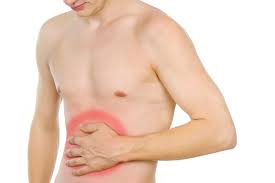
Right Upper Quadrant Pain — Biliary and Liver Causes
The right upper quadrant contains the liver, gallbladder, bile ducts, and the hepatic flexure of the colon. Pain in this region most commonly arises from the biliary system or liver and has a characteristic relationship to fat intake (through cholecystokinin-mediated gallbladder contraction) and to liver function abnormalities.
Biliary colic produces episodic severe RUQ pain lasting 30 minutes to 6 hours after fatty or large meals, accompanied by nausea and vomiting. It resolves completely between episodes. Ultrasound identifies gallstones with high sensitivity. Laparoscopic cholecystectomy is the definitive treatment. Acute cholecystitis occurs when a gallstone causes sustained cystic duct obstruction — pain lasting more than 6 hours, fever, and positive Murphy’s sign distinguish it from biliary colic. It requires hospitalization and cholecystectomy. Choledocholithiasis — a stone in the common bile duct — produces RUQ pain with elevated bilirubin, alkaline phosphatase, and GGT, indicating biliary obstruction. MRCP or endoscopic ultrasound confirms the diagnosis; ERCP with stone extraction is the treatment. Ascending cholangitis is a biliary emergency. Charcot’s triad — fever, jaundice, and RUQ pain — indicates infection ascending from an obstructed bile duct and requires immediate broad-spectrum antibiotics and urgent biliary drainage. Reynolds’ pentad adds confusion and hypotension to the triad, indicating septic shock. Hepatitis from viral infection (A, B, C, E), alcohol, drug-induced liver injury (DILI), or autoimmune disease produces RUQ aching or fullness from hepatic inflammation and capsular stretch, accompanied by elevated transaminases (ALT, AST) and sometimes jaundice. The pain is typically constant, dull, and non-episodic — unlike biliary colic. Congestive heart failure causes hepatic congestion from elevated right-sided pressures, producing RUQ fullness and tenderness from hepatomegaly, sometimes with jaundice from hepatocellular dysfunction. For a detailed discussion of right upper quadrant and liver causes, see right upper abdominal pain and liver health. The NIDDK digestive diseases library provides patient-level guides for each of these conditions.
Left Upper Quadrant Pain
Left upper quadrant pain is less common than epigastric or RUQ pain and has a narrower differential — but includes several serious causes that should be recognized.
Pancreatitis frequently radiates from the epigastrium into the LUQ, particularly when the body and tail of the pancreas are primarily involved. The pain pattern, severity, and associated features are the same as described above. Splenic causes of LUQ pain include splenic infarct (most commonly in patients with hematologic malignancy, atrial fibrillation, or sickle cell disease), splenomegaly from any cause (which produces LUQ fullness and discomfort from the enlarged spleen pressing on surrounding structures), and spontaneous splenic rupture — an emergency seen most commonly with infectious mononucleosis (EBV). Splenic rupture presents with sudden severe LUQ pain, left shoulder pain (Kehr’s sign — referred pain from diaphragmatic irritation by blood), and hemodynamic instability. Any patient with recent EBV infection who develops sudden LUQ pain should be evaluated for splenic rupture. Gastric causes of LUQ pain include fundal ulcer, gastric volvulus (twisting of the stomach — a rare emergency producing sudden severe pain, retching, and inability to pass a nasogastric tube), and, less commonly, gastric cancer when involving the gastric fundus. Left renal and ureteral causes — left renal colic and left pyelonephritis — produce left flank pain that may radiate anteriorly into the LUQ and are distinguished by their association with hematuria, costovertebral angle tenderness, and characteristic CT findings.
Liver-Specific Upper Abdominal Pain
The liver itself has no pain fibers in its parenchyma — pain from liver conditions is produced by stretching Glisson’s capsule (the fibrous covering of the liver), by peritoneal inflammation adjacent to the liver, or by referred pain through diaphragmatic irritation via the phrenic nerve (which produces right shoulder tip pain). Understanding this anatomy explains why liver diseases are often asymptomatic until they cause significant capsular stretch or complication.
Hepatitis (viral, alcoholic, or drug-induced) produces RUQ aching from hepatic inflammation causing capsular stretch as the liver swells. The pain is constant and dull rather than sharp or episodic. Jaundice, dark urine, pale stools, and fatigue accompany the pain in significant hepatitis. Transaminase levels (ALT, AST) are markedly elevated — often in the thousands in acute viral hepatitis. Nonalcoholic fatty liver disease (NAFLD) and its more advanced form MASH (metabolic dysfunction-associated steatohepatitis) produce RUQ fullness and vague discomfort in some patients — mainly from hepatomegaly — but are frequently entirely asymptomatic until late fibrosis or cirrhosis develops. Many patients with NAFLD are diagnosed incidentally on imaging done for other reasons. Liver abscess — pyogenic (bacterial) or amebic — produces constant RUQ pain, fever, and weight loss. Pyogenic liver abscess is most commonly from biliary sources (cholangitis, cholecystitis) or hematogenous spread. Amebic liver abscess from Entamoeba histolytica is more common in travelers and immigrants from endemic areas. Both require drainage and antimicrobial therapy. Budd-Chiari syndrome — hepatic vein thrombosis — produces acute or subacute RUQ pain, hepatomegaly, and ascites from outflow obstruction. It is associated with hypercoagulable states (polycythemia vera, factor V Leiden, pregnancy, oral contraceptives, paroxysmal nocturnal hemoglobinuria). Doppler ultrasound of hepatic veins confirms the diagnosis. Hepatocellular carcinoma (HCC) typically arises in the setting of cirrhosis and produces RUQ pain from rapid tumor growth stretching Glisson’s capsule or from invasion of adjacent structures. Any patient with known cirrhosis who develops new RUQ pain warrants imaging to screen for HCC. The ACG liver disease patient resource covers the spectrum of liver conditions that produce upper abdominal pain.
Red Flags and Emergencies
Several causes of upper abdominal pain represent medical or surgical emergencies requiring urgent evaluation. Recognizing these patterns allows the appropriate triage — emergency department vs. urgent clinic vs. elective evaluation.
Acute cholangitis (fever + jaundice + RUQ pain) requires emergency admission, IV antibiotics, and urgent biliary drainage. Delay increases mortality from sepsis. Acute pancreatitis with severe pain, vomiting, and markedly elevated lipase requires emergency evaluation for hospitalization, IV fluids, monitoring for complications (necrosis, pseudocyst), and identification of the precipitating cause (gallstone requiring ERCP, alcohol cessation). Abdominal aortic aneurysm (AAA) expansion or rupture presents with sudden severe epigastric or back pain with a pulsatile epigastric mass in an at-risk patient (older male, smoker, hypertension). It is a vascular emergency with high mortality; immediate CT angiography and surgical consultation are required. Acute liver failure — sudden onset of jaundice, coagulopathy, and encephalopathy — is a multi-organ emergency requiring immediate transfer to a liver transplant center. Causes include acetaminophen overdose, drug-induced liver injury, acute viral hepatitis, and Wilson’s disease. Splenic rupture — sudden severe LUQ pain with hemodynamic instability — requires immediate surgical evaluation. For a framework on when upper abdominal pain specifically requires same-day evaluation, see the discussion in common digestive problems in adults.
Evaluating Upper Abdominal Pain
The initial evaluation of upper abdominal pain begins with history and physical examination — the location, timing, character, associated symptoms, and triggers of the pain guide test selection. Indiscriminate testing without a clinical framework delays diagnosis and increases costs without improving accuracy.
Laboratory evaluation includes complete blood count (CBC), comprehensive metabolic panel with liver enzymes (ALT, AST, ALP, GGT, bilirubin), lipase and amylase, H. pylori testing (breath test or stool antigen), C-reactive protein, and thyroid function. An elevated lipase suggests pancreatitis; elevated transaminases suggest hepatic inflammation; elevated ALP and bilirubin suggest biliary obstruction; anemia suggests chronic GI blood loss or malabsorption. Right upper quadrant ultrasound is the first-line imaging for RUQ pain — it identifies gallstones, biliary duct dilation (indicating obstruction), liver size and echotexture, and free fluid (ascites). It is fast, widely available, and involves no radiation. CT abdomen and pelvis with IV contrast is used for severe acute upper abdominal pain requiring detailed anatomical assessment — pancreatitis staging, liver abscess, AAA, splenic pathology, and mesenteric ischemia. CT is also the initial imaging for presentations where the diagnosis is uncertain and urgent evaluation is needed. MRCP (Magnetic resonance cholangiopancreatography) provides detailed non-invasive imaging of the bile ducts and pancreatic duct — used for suspected choledocholithiasis, primary sclerosing cholangitis, and pancreatic ductal pathology. Upper endoscopy is indicated for epigastric pain with alarm features, suspected peptic ulcer disease, treatment failure, or when mucosal biopsies are needed. The Mayo Clinic’s abdominal pain causes guide provides a patient-accessible overview of the upper abdominal differential.
Frequently Asked Questions
What causes burning pain in the upper middle abdomen?
Burning epigastric pain most commonly reflects GERD (acid reflux irritating the lower esophageal mucosa), gastritis (gastric mucosal inflammation), or peptic ulcer disease. Less commonly it reflects functional dyspepsia with epigastric pain syndrome. The burning character — as opposed to dull aching or cramping — and its relationship to meals, position, and antacids help narrow the cause. GERD-related burning is most prominent after meals and when lying down; peptic ulcer burning has the characteristic timing pattern tied to the ulcer’s location.
Can liver problems cause upper abdominal pain?
Yes — the liver produces pain by stretching its fibrous capsule (Glisson’s capsule) when the organ swells from inflammation, congestion, or rapidly growing masses. Hepatitis (viral, alcoholic, or drug-induced) typically produces a constant, dull RUQ ache accompanied by elevated liver enzymes and sometimes jaundice. Fatty liver disease (NAFLD) can produce RUQ fullness in some patients but is often asymptomatic. Acute liver conditions — liver abscess, Budd-Chiari syndrome, acute liver failure — produce more prominent pain with associated systemic features. The location is typically the right upper quadrant, sometimes with right shoulder radiation from phrenic nerve irritation.
How do I know if upper abdominal pain is from the gallbladder?
Gallbladder pain (biliary colic) has a distinctive pattern: episodic, severe, located in the right upper quadrant or epigastrium, triggered by fatty or large meals, lasting 30 minutes to 6 hours, accompanied by nausea, and resolving completely between episodes. This pattern — particularly the episodic nature and fatty-meal trigger — is one of the more specific patterns in upper abdominal pain. However, not all gallbladder pain has the classic presentation; some patients have biliary dyskinesia without visible stones, or stones that cause atypical symptoms. RUQ ultrasound is the appropriate first test when biliary colic is suspected.
What does pancreatic pain feel like?
Pancreatic pain is characteristically epigastric, constant and severe, and radiates to the back (often described as a band or boring sensation encircling the upper abdomen). It is typically worsened by eating and may improve when the patient leans forward. In acute pancreatitis, the pain begins relatively rapidly after a triggering event (large fatty meal or alcohol ingestion) and is accompanied by nausea, vomiting, and often fever. In chronic pancreatitis, the pain is recurrent and may be accompanied by symptoms of pancreatic exocrine insufficiency — oily, floating stools, weight loss, and eventually diabetes.
When does upper abdominal pain need emergency evaluation?
Upper abdominal pain requires emergency evaluation when: pain is severe, sudden, and rapidly worsening; pain is accompanied by fever and jaundice (suggesting cholangitis or severe cholecystitis); pain radiates to the back with nausea and elevated lipase (pancreatitis); a pulsatile mass is felt in the epigastrium in a high-risk patient (AAA); there is evidence of GI bleeding (hematemesis, melena); signs of acute liver failure are present (jaundice, confusion, coagulopathy); or there is hemodynamic instability. These presentations require same-day emergency assessment, not watchful waiting.
Sources: National Institute of Diabetes and Digestive and Kidney Diseases (NIDDK); American College of Gastroenterology (ACG); Rome IV Functional GI Disorders Criteria; American Gastroenterological Association (AGA); Mayo Clinic; American Liver Foundation.
Upper Abdominal Pain and Liver Disease — A Closer Look
Liver disease is a particularly important cause of upper abdominal symptoms to understand, both because the liver is so often asymptomatic until advanced disease develops and because the symptoms it does produce — RUQ fullness, fatigue, jaundice — overlap with biliary and other causes. Understanding when upper abdominal pain suggests liver disease versus biliary disease versus GI disease helps patients communicate more precisely with their clinicians and reduces diagnostic delay.
Fatty liver disease and upper abdominal discomfort: Nonalcoholic fatty liver disease (NAFLD) — now increasingly referred to as metabolic dysfunction-associated steatotic liver disease (MASLD) — is the most prevalent liver condition in Western countries, affecting approximately 25 to 30 percent of adults globally. Most patients with NAFLD are completely asymptomatic. A minority report vague RUQ fullness, discomfort after large meals, or fatigue — symptoms that correlate with the degree of hepatomegaly rather than with the histologic severity of steatosis or inflammation. The important clinical point is that asymptomatic NAFLD can progress silently through steatohepatitis, fibrosis, and cirrhosis without ever producing warning pain — which is why periodic monitoring of liver enzymes and liver ultrasound is appropriate for at-risk patients, even in the absence of symptoms.
Alcoholic liver disease: The spectrum ranges from alcoholic fatty liver (often asymptomatic) to alcoholic hepatitis (presenting with RUQ pain, fever, jaundice, and elevated ALT/AST with a characteristic AST:ALT ratio above 2:1) to alcoholic cirrhosis (presenting with the complications of portal hypertension — ascites, variceal bleeding, encephalopathy). Alcoholic hepatitis can be severe enough to cause acute-on-chronic liver failure and carries significant short-term mortality. Any patient with a history of significant alcohol use who presents with jaundice and RUQ pain requires urgent evaluation.
Drug-induced liver injury (DILI): DILI is one of the most underrecognized causes of upper abdominal pain and abnormal liver tests in adults. Hepatotoxic medications include acetaminophen at high doses (the most common cause of acute liver failure in Western countries), statins (usually causing asymptomatic transaminase elevation rather than pain), herbal and dietary supplements (the fastest-growing category of DILI — including kava, green tea extract, pyrrolizidine alkaloids), antibiotics (amoxicillin-clavulanate being the most common prescription antibiotic cause), and dozens of other commonly prescribed medications. Any new upper abdominal pain or jaundice should prompt a review of all prescription medications, over-the-counter medications, and herbal supplements — including products started in the preceding 3 to 12 months. Many patients do not volunteer supplement use unless specifically asked.
Liver cysts and benign hepatic masses: Simple liver cysts are common incidental findings on imaging — they are almost always asymptomatic and require no treatment. Large cysts (over 5 to 7 cm) occasionally produce RUQ fullness or discomfort from mass effect. Hepatic hemangiomas — the most common benign liver tumor — are similarly almost always asymptomatic and require no treatment except for very large lesions. Both are often found incidentally on ultrasound or CT ordered for other reasons; the key clinical task is distinguishing benign from malignant hepatic lesions, which requires characterization by MRI or dedicated contrast-enhanced CT in most cases.
When liver causes produce referred pain: The diaphragm sits immediately above the liver, and peritoneal or capsular irritation from liver conditions can produce referred pain to the right shoulder tip or right trapezius ridge through the phrenic nerve (C3-C5 dermatome). Right shoulder pain associated with RUQ disease — without any shoulder-specific injury or history — is a classical referred pain pattern. It is most commonly seen in biliary colic, acute cholecystitis, hepatic abscess, and, rarely, liver tumor or Budd-Chiari syndrome. Patients who present with right shoulder pain and do not have an orthopedic explanation should have RUQ ultrasound as part of their evaluation if abdominal pathology is plausible.
Upper abdominal pain that is new, persistent, or accompanied by fever, jaundice, or weight loss deserves a systematic evaluation rather than indefinite empiric treatment. The majority of causes are identifiable with a focused history, targeted laboratory tests, and ultrasound — and the majority respond well to treatment once correctly diagnosed. Recognizing the anatomical patterns outlined in this guide is the starting point for getting to the right evaluation efficiently.
The section on drug-induced liver injury was really important for me personally. I’ve been taking herbal supplements for about a year and recently noticed my liver enzymes were elevated at a routine blood test. My doctor just said to monitor it. This article made me realize I should specifically mention the supplements and ask about DILI — I hadn’t connected the two at all.
Heather, that’s exactly the right question to raise with your doctor. Herbal and dietary supplements are consistently underestimated as a source of liver injury — many patients don’t think of them as ‘medications’ and don’t mention them unless specifically asked, and many clinicians don’t ask. Providing your complete supplement list, including product names and how long you’ve been taking them, gives your doctor the information needed to evaluate DILI properly. Stopping any supplement that temporally correlates with the enzyme elevation, with monitoring to confirm improvement, is often both diagnostic and therapeutic.
I appreciated the section on referred pain to the right shoulder. I had months of intermittent right shoulder pain that three orthopedic evaluations found nothing for. It finally turned out to be biliary colic — gallstones found on ultrasound. If I’d read this article earlier I might have asked for an abdominal ultrasound a lot sooner.