
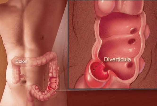
Diverticulosis and diverticulitis are two closely related but distinct conditions affecting the colon — and understanding the difference between them matters. Diverticulosis is the presence of small outpouching sacs (diverticula) in the colon wall. It is extremely common, mostly harmless, and affects approximately 50–60% of people over 70 in Western populations. Diverticulitis is what happens when one or more of those pouches becomes inflamed or infected — causing left lower abdominal pain, fever, and tenderness. Most people with diverticulosis will never develop diverticulitis, but for those who do, the condition ranges from mild and self-limiting to a surgical emergency.
What Is Diverticulosis?
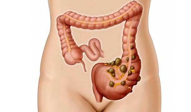
Diverticulosis is the presence of diverticula — small, outpouching sacs — in the wall of the colon. These pouches form when the inner lining of the colon herniates through weakened points in the muscular wall, typically the sites where blood vessels penetrate to supply the mucosal lining.
Diverticulosis is extremely common in Western populations and becomes increasingly prevalent with age:
- Approximately 10% of people under 40 have diverticula
- Approximately 50–60% of people over 70 have diverticula
- In Western countries, diverticula form predominantly in the sigmoid colon — the left-sided, S-shaped segment — because it has the highest intraluminal pressure and the narrowest diameter
- In Asian populations, diverticula are more commonly right-sided
The vast majority of people with diverticulosis are completely asymptomatic and will remain so throughout their lives. Diverticula discovered incidentally during colonoscopy or CT scan require no specific treatment beyond dietary advice.
Symptomatic Uncomplicated Diverticular Disease (SUDD): A subset of people with diverticulosis develop persistent left lower quadrant pain and altered bowel habit in the absence of confirmed acute diverticulitis. The mechanism may involve low-grade mucosal inflammation, altered gut microbiome, and visceral hypersensitivity — in some respects resembling IBS. Management includes high-fibre diet and mesalazine, which has some evidence in SUDD for reducing recurrence.
What Is Diverticulitis?
Diverticulitis is the inflammation and infection of one or more diverticula. It develops when a faecalith (hardened piece of stool) obstructs the narrow neck of a diverticulum, causing bacterial overgrowth, local inflammation, and — in more severe cases — microperforation or macroperforation of the diverticular wall.
Approximately 5–25% of people with diverticulosis develop diverticulitis at some point in their lives. The classic presentation includes:
- Left lower quadrant (LLQ) pain: constant, not colicky, localised to the left side of the lower abdomen over the sigmoid colon
- Fever: typically low-grade in uncomplicated cases; high fever suggests abscess or perforation
- Tenderness: localised LLQ tenderness on palpation; guarding and rigidity suggest peritonitis
- Nausea: common; vomiting in more severe cases
- Constipation or altered bowel habit: the inflamed segment may reduce transit
Right-sided diverticulitis: In Asian populations where right-sided diverticula are more common, diverticulitis presents with right lower quadrant pain and fever — closely mimicking appendicitis. This diagnostic challenge means right-sided diverticulitis is often only confirmed at CT or at surgery.
How Diverticula Form
The formation of diverticula is driven by two interacting factors: increased intraluminal pressure within the colon, and localised weakness in the colonic muscle wall.
The role of low-fibre diet: A low-fibre diet produces small-volume, hard stools that require more forceful muscular contractions to propel through the colon. The sigmoid colon generates the highest intraluminal pressures during these contractions. Over decades of these pressure spikes, the mucosa herniates through the anatomically weakest points in the muscular wall — the sites where arterioles penetrate from the outer wall to the inner mucosa.
This mechanism explains the epidemiology: diverticulosis is far more prevalent in Western, low-fibre diet populations than in rural sub-Saharan Africa, where traditional high-fibre plant-based diets produce bulky stools with lower intraluminal pressure.
Connective tissue changes: Age-related changes in collagen structure — reduced cross-linking, altered elastin — reduce the tensile strength of the colonic wall and increase susceptibility to herniation. This explains why diverticulosis becomes near-universal in people reaching their 80s in Western populations.
Diagnosing Diverticulitis
CT scan with intravenous contrast is the gold standard for diagnosing and staging acute diverticulitis. CT confirms the diagnosis, identifies the extent of inflammation, and detects complications (abscess, perforation, fistula). Sensitivity for complicated diverticulitis is approximately 90%. CT also excludes alternative diagnoses — colonic cancer, ovarian pathology, or appendicitis — that can closely mimic diverticulitis clinically.
Blood tests: CRP is often elevated above 150 mg/L in acute diverticulitis, which helps distinguish it from mild uncomplicated diverticular discomfort. Elevated white cell count and ESR are additional markers of active infection. Falling CRP is the most reliable indicator of treatment response.
Ultrasound: Less sensitive than CT but avoids radiation. It may be the initial imaging choice in young women (to first exclude gynaecological causes), in pregnancy, or when CT is not immediately available.
Colonoscopy timing: Colonoscopy is not performed during an acute episode — the risk of perforation through the inflamed colonic wall is significant. It is performed 6–8 weeks after clinical resolution to:
- Confirm diverticula presence and distribution
- Exclude colonic cancer — a colonic tumour can present with a pericolonic inflammatory mass indistinguishable from diverticulitis on CT
- Exclude Crohn’s colitis, which can cause similar LLQ inflammation (see our guide to inflammatory bowel disease)
How Severe Is Diverticulitis? The Hinchey Classification
The Hinchey classification is the most widely used system for staging acute diverticulitis severity:
- Uncomplicated diverticulitis (~75% of cases): inflammation limited to the pericolic fat and colon wall without abscess, perforation, or fistula — managed conservatively in most cases
- Hinchey Stage I — Pericolic abscess: a small, contained abscess immediately adjacent to the inflamed diverticulum — often treated with IV antibiotics alone
- Hinchey Stage II — Pelvic abscess: the abscess has tracked into the pelvis — typically requiring CT-guided percutaneous drainage plus IV antibiotics
- Hinchey Stage III — Generalised purulent peritonitis: perforation with pus spreading throughout the peritoneal cavity — a surgical emergency requiring laparoscopic washout or Hartmann’s procedure
- Hinchey Stage IV — Generalised faecal peritonitis: free perforation with faecal contamination of the peritoneum — the most dangerous presentation; high mortality; requires emergency Hartmann’s procedure
Treatment for Acute Diverticulitis
Uncomplicated mild diverticulitis: The management of mild uncomplicated diverticulitis has been revised following several randomised controlled trials — DIABOLO, AVOD, and DIVER — that showed no significant difference in outcomes between antibiotic treatment and observation without antibiotics in otherwise healthy patients with CT-confirmed uncomplicated diverticulitis.
Current guidance: antibiotics are not universally required for mild uncomplicated diverticulitis in immunocompetent patients without significant fever or systemic upset. Management consists of:
- Clear liquid diet for 2–3 days, advancing to low-residue solid food as symptoms improve
- Analgesia with paracetamol (NSAIDs should be avoided — they worsen intestinal perforation risk)
- Oral antibiotics remain recommended for: fever above 38°C; CRP above 100 mg/L; immunocompromised patients; significant comorbidity; or failure to improve within 48–72 hours
Moderate-severe uncomplicated diverticulitis: Hospital admission; IV antibiotics (co-amoxiclav or piperacillin-tazobactam); nil by mouth advancing to clear fluids; IV fluids; monitoring of CRP response.
Abscess (Hinchey I-II): Pericolic abscesses smaller than 3–4cm may resolve with IV antibiotics alone. Larger abscesses typically require CT-guided percutaneous drainage plus antibiotics.
Perforation with peritonitis (Hinchey III-IV): Emergency surgery. Hartmann’s procedure — resection of the sigmoid colon with end colostomy — is the standard emergency operation. Hartmann’s reversal (restoring bowel continuity) can be performed 3–6 months later if the patient is fit, but only approximately 50% of patients ultimately achieve successful reversal.
Diverticular Complications
Fistula: When diverticulitis causes transmural erosion into an adjacent organ, a fistula forms. The most common is a colovesical fistula (between the colon and bladder) — causing pneumaturia (gas in the urine, producing bubbles during urination), faecaluria (stool particles in the urine), and recurrent polymicrobial urinary tract infections resistant to standard single-agent antibiotics. Any person with recurrent UTIs caused by gut organisms should have a colovesical fistula considered. Surgical repair is required.
Obstruction: Recurrent diverticulitis causes pericolic fibrosis and scarring that can progressively narrow the sigmoid colonic lumen, presenting as increasing constipation, bloating, and colicky abdominal pain. Surgical sigmoid resection resolves the obstruction.
Diverticular haemorrhage: This is a separate entity from diverticulitis — and the most common cause of significant acute lower GI bleeding in adults. Haemorrhage occurs when an artery adjacent to the dome of a diverticulum ruptures — a process not driven by inflammation. Presentation: sudden-onset painless large-volume fresh rectal bleeding, often without any prior LLQ pain. Bleeding stops spontaneously in approximately 80% of cases. Persistent bleeding requires colonoscopy with endoscopic haemostasis, or angiography with embolisation.
Surgery and Elective Management
Elective laparoscopic sigmoid colectomy is the definitive treatment for diverticular disease. Indications for elective surgery include:
- Recurrent symptomatic diverticulitis (typically 2+ episodes) with significant impact on quality of life
- Failure to exclude colonic cancer after imaging and colonoscopy
- Colovesical or other fistula requiring repair
- Persisting SUDD symptoms after adequate conservative management
Laparoscopic sigmoid colectomy in elective settings carries a lower complication rate than emergency surgery and is associated with shorter hospital stays and faster recovery. It reduces but does not entirely eliminate the risk of further diverticulitis — diverticula may be present proximal to the resected segment.
For people managing ongoing colon health, including diverticular disease risk reduction, our guide to colon health after age 50 covers screening, dietary strategies, and lifestyle modifications in detail.
Prevention and Diet
High-fibre diet: The most evidence-supported lifestyle modification for reducing diverticulosis progression and diverticulitis risk. Dietary fibre increases stool bulk, reduces transit time, and lowers intraluminal colonic pressure. Target: 25–35g of dietary fibre per day (most Western adults consume 15–18g). Good sources: vegetables (especially legumes), whole grains (oats, wholemeal bread), fruit, nuts, and seeds.
Seeds and nuts — the myth: For decades, patients with diverticulosis were routinely advised to avoid seeds, nuts, popcorn, and similar foods. Multiple large prospective cohort studies — including the Health Professionals Follow-Up Study — have shown no increased risk of diverticulitis from these foods. In fact, higher nut consumption was associated with a lower diverticulitis risk in some analyses. Patients with diverticulosis can eat seeds and nuts freely.
Physical activity: Physically active people have significantly lower rates of diverticulitis than sedentary individuals across large prospective cohort studies. Even moderate activity (30 minutes of brisk walking most days) is associated with measurable risk reduction.
NSAIDs: Non-steroidal anti-inflammatory drugs (ibuprofen, naproxen) are strongly associated with both diverticulitis onset and diverticular complications, particularly perforation. People with known diverticulosis should use paracetamol as their first-choice analgesic. For people also managing conditions like ulcerative colitis where NSAID avoidance is also critical, this consistent guidance reinforces the importance of paracetamol as the safer alternative.
Obesity and smoking both independently increase diverticulitis risk. Obesity is associated with increased intra-abdominal pressure and a pro-inflammatory state. Smoking impairs mucosal healing and increases infection risk.
Diverticulitis in Special Populations
While diverticulitis in older adults follows the typical pattern described above, certain populations face distinct challenges in both presentation and management.
Immunocompromised patients: People receiving immunosuppressive therapy (organ transplant recipients, patients on long-term corticosteroids, those receiving chemotherapy or biologic treatments for conditions such as inflammatory bowel disease, rheumatoid arthritis, or psoriasis) are at significantly higher risk of complicated diverticulitis and present with attenuated symptoms. Because the immune response is suppressed, fever and elevated CRP may be minimal even when significant perforation or abscess is present. A lower threshold for CT imaging and a lower threshold for hospitalisation and IV antibiotics is warranted in immunocompromised patients with suspected diverticulitis — the clinical picture reliably underestimates the pathological severity.
Young adults (under 50): Diverticulitis in younger adults is less common but tends to present with a more aggressive clinical course when it does occur. Historically, guidelines recommended earlier recourse to elective sigmoid colectomy in young patients after a first episode, on the assumption that the cumulative lifetime risk of recurrence justified early definitive surgery. This view has been revised — current guidance is to individualise the decision based on symptom burden, episode severity, and patient preference rather than using age as a primary criterion. However, diverticulitis in a young adult without obvious risk factors (low-fibre diet, obesity, sedentary lifestyle) should prompt consideration of Crohn’s colitis as an alternative diagnosis, since Crohn’s can cause a pericolonic inflammatory mass and left-sided colonic thickening indistinguishable from diverticulitis on CT.
Pregnancy: Diverticulitis in pregnancy is rare (diverticulosis affects a much younger age group than typical diverticulitis, but can occur in the third trimester when an enlarged uterus compresses the sigmoid colon). Diagnosis is challenging because: LLQ pain in pregnancy has a broad differential (round ligament pain, ovarian torsion, preterm labour); radiation exposure from CT must be carefully weighed; and the gravid uterus can displace the sigmoid colon, altering typical pain localisation. MRI is the preferred imaging modality when available. Conservative management with IV antibiotics is the primary approach; surgery carries significant obstetric risk and is reserved for life-threatening complications.
People taking anticoagulants or antiplatelet agents: Diverticular haemorrhage is more likely to be severe and less likely to stop spontaneously in people taking warfarin, direct oral anticoagulants (DOACs), or dual antiplatelet therapy. If diverticular bleeding occurs in a patient on anticoagulation, the bleeding and anticoagulation management must be coordinated carefully — typically involving gastroenterology, haematology, and the prescribing team (for example, the cardiologist prescribing antiplatelet therapy post-coronary stent).
Monitoring and Long-term Follow-up After Diverticulitis
After an episode of acute diverticulitis resolves — whether managed conservatively or surgically — long-term follow-up has several goals: confirming the diagnosis, excluding malignancy, monitoring for recurrence, and optimising prevention.
Post-acute colonoscopy: As noted in the diagnosis section, colonoscopy 6–8 weeks after resolution is standard practice. The most important purpose is excluding colonic cancer — a colonic carcinoma can cause a pericolonic inflammatory mass that is radiologically identical to diverticulitis on CT, and a malignancy missed because it was attributed to diverticulitis carries serious consequences. Studies suggest that approximately 1–2% of patients diagnosed with diverticulitis on CT are subsequently found to have colonic cancer on colonoscopy; in patients over 50 with a first episode of diverticulitis, this figure is the primary driver for routine post-acute colonoscopy.
Recurrence surveillance: After a first episode, approximately 30–40% of people have a second episode of diverticulitis. Risk factors for recurrence include: obesity (adipose tissue produces pro-inflammatory cytokines that promote colonic inflammation); NSAID use; smoking; low dietary fibre; immunosuppression; and residual narrowing or pericolic fibrosis at the site of prior diverticulitis. Addressing modifiable risk factors — achieving a healthy weight, stopping NSAIDs, stopping smoking, increasing dietary fibre — forms the cornerstone of post-episode care. There is no evidence that routine interval CT imaging or colonoscopy surveillance (beyond the initial post-acute colonoscopy) is beneficial in asymptomatic patients who have had uncomplicated diverticulitis.
Surgery timing: For patients who have had multiple episodes and are being considered for elective sigmoid colectomy, the optimal timing is during a quiescent (asymptomatic) interval — not during an acute episode (when the operation is technically more difficult, complication rates are higher, and primary anastomosis is less safe) and not too soon after an acute episode (when pericolic inflammation and oedema increase the difficulty and risks of resection). Most surgeons prefer an interval of at least 6–12 weeks between the most recent episode and elective sigmoid colectomy to allow inflammation to fully resolve.
Mesalazine (5-ASA) in SUDD: In patients with Symptomatic Uncomplicated Diverticular Disease — persistent LLQ discomfort, bloating, and altered bowel habit without confirmed acute diverticulitis — mesalazine has been studied in the PREVENT trials as a maintenance therapy to reduce pain and recurrence. Evidence is mixed; some trials showed benefit in reducing symptomatic recurrence while others showed no significant difference from placebo. Mesalazine is generally well-tolerated and is used in some centres for symptomatic diverticular disease pending further evidence. It is the same medication used in the management of ulcerative colitis, though the doses and mechanisms may differ.
Frequently Asked Questions
What is the difference between diverticulosis and diverticulitis?
Diverticulosis is the presence of diverticula — small outpouching sacs — in the colon wall. It is very common (affecting around 50% of people over 70 in Western populations) and usually causes no symptoms. Diverticulitis is what happens when one or more of those diverticula becomes inflamed or infected — causing left lower abdominal pain, fever, and tenderness. Most people with diverticulosis never develop diverticulitis. Diverticulosis is a structural finding; diverticulitis is an acute medical condition requiring assessment and treatment.
What does diverticulitis feel like?
The hallmark symptom is constant (not colicky) pain in the left lower abdomen, often described as a deep ache or pressure. Unlike IBS or constipation, the pain in diverticulitis is typically constant rather than coming in waves. It is usually accompanied by fever, tenderness when the lower left abdomen is pressed, nausea, and sometimes constipation or reduced bowel movements. Severe diverticulitis with abscess or perforation causes more intense pain, higher fever, and may produce signs of peritonitis — rigid, board-like abdomen.
What foods should I avoid with diverticulitis?
During an acute episode, a clear liquid diet for the first 2–3 days, advancing to low-residue diet as symptoms improve. Seeds and nuts are not specifically restricted — the old advice to avoid them has been shown to be unsupported by evidence. Once recovered, a high-fibre diet (25–35g/day) is recommended for long-term prevention. Red meat and processed meat consumption is associated with higher diverticulitis risk and is worth moderating. NSAIDs should be avoided — use paracetamol for pain relief instead.
Can diverticulitis be cured?
A single episode of uncomplicated diverticulitis typically resolves completely with conservative management. However, diverticulosis — the underlying diverticula — persists, and approximately 30–40% of people have a recurrent episode of diverticulitis. Surgical sigmoid colectomy removes the diverticula-bearing segment and is considered curative for that segment, but diverticula in the remaining colon can still cause future symptoms. The most accurate framing is that diverticulitis can be successfully treated and risk can be reduced with dietary changes, but existing diverticula cannot be reversed.
Does diverticulitis always need antibiotics?
No — this is one of the most important changes in recent diverticulitis management. Randomised controlled trials (DIABOLO, AVOD, and DIVER) have demonstrated that, in otherwise healthy people with CT-confirmed uncomplicated diverticulitis, antibiotic treatment does not significantly reduce complications, hospital readmissions, or the need for surgery compared with observation without antibiotics. Antibiotics are still recommended when there is significant fever, markedly elevated CRP, signs of systemic infection, or in immunocompromised patients — but routine prescribing for all diverticulitis cases is no longer supported by evidence.
When does diverticulitis require surgery?
Emergency surgery is required for: free perforation with generalised peritonitis (Hinchey III/IV); large abscesses failing CT-guided drainage; and bowel obstruction not resolving with conservative management. Elective surgery (sigmoid colectomy) is indicated for: recurrent symptomatic episodes significantly affecting quality of life; fistula formation (particularly colovesical); persistent inability to exclude cancer; and SUDD with chronic symptoms unresponsive to medical management.
Can diverticulosis go away?
No — once diverticula have formed, they do not disappear. The structural change to the colonic wall is permanent. However, the risk of developing diverticulitis from existing diverticula can be reduced through dietary and lifestyle changes, and many people with diverticulosis live their entire lives without ever developing symptoms or complications. The focus of management is preventing new diverticula formation, reducing pressure in the colon, and minimising risk factors for diverticulitis.
Medical disclaimer: This article is for educational purposes only and does not constitute medical advice. People experiencing persistent left lower abdominal pain, fever, blood in the stool, or symptoms that may represent diverticulitis should seek prompt medical assessment.
References:
1. Stollman N et al. AGA Institute Guideline on the Management of Acute Diverticulitis. Gastroenterology. 2015;149(7):1944–9.
2. Tursi A et al. Diverticulosis and diverticular disease of the colon. Lancet. 2020;396(10257):1025–37.
3. Peery AF et al. Burden and cost of gastrointestinal, liver, and pancreatic diseases in the US. Gastroenterology. 2019;156(1):254–72.
4. Böhm SK. Risk factors for diverticulosis, diverticulitis, diverticular perforation, and bleeding. Viszeralmedizin. 2015;31(2):84–94.
The section on the antibiotic controversy was genuinely eye-opening. I had diverticulitis twice in the past three years — both times managed with a course of antibiotics at home — and had always assumed antibiotics were simply mandatory. Reading about the DIABOLO and AVOD trials is the first time I’ve encountered this evidence presented clearly. My GP mentioned at my last visit that there was ‘some debate’ about antibiotics but didn’t elaborate. I’m going to bring this up at my next appointment and ask specifically whether watchful waiting would be appropriate for me if I have another mild episode.
Robert — you are asking exactly the right question, and it is a conversation very much worth having with your GP. The DIABOLO trial (Netherlands, published in Annals of Surgery), the AVOD trial (Scandinavian), and the DIVER trial (Spanish) all looked at this question prospectively and found no statistically significant difference in complication rates, recurrence rates, or need for surgery between patients treated with antibiotics and patients treated with observation and analgesia alone for mild uncomplicated diverticulitis confirmed on CT. The key phrase is ‘mild uncomplicated’ — these findings apply specifically to patients who are not systemically unwell, do not have significant fever or very elevated CRP, and are immunocompetent. If you meet those criteria during a future episode, there is now good evidence that a trial of observation with clear liquids, paracetamol, and close monitoring for 48–72 hours is a reasonable and guideline-supported approach. Your GP may be comfortable managing this; some GPs prefer to involve gastroenterology for the initial shared decision around this approach. Sandra — you are right to flag the NSAID issue, and this is an important one. Ibuprofen, naproxen, and other NSAIDs inhibit prostaglandin synthesis in the colonic mucosa — prostaglandins play a key role in maintaining the protective mucosal lining and supporting healing. In people with diverticulosis, NSAID use is independently associated with a significantly increased risk of diverticulitis onset, and is particularly associated with complicated diverticulitis (perforation, abscess) rather than just uncomplicated episodes. For regular back pain in someone with known diverticulosis, paracetamol at adequate doses (up to 1g four times daily) is the first-line alternative, and it would be reasonable to discuss this specifically with your GP — both for your diverticulosis and your back pain management. Some people with back pain benefit from physiotherapy-directed exercise programmes that reduce the need for regular analgesia altogether.
I was told to avoid nuts and seeds about four years ago when diverticulosis was found on my colonoscopy. I’ve been meticulously avoiding them ever since — including avoiding foods I genuinely enjoy. Finding out this advice was never evidence-based and has been debunked by large prospective studies is frustrating but also a huge relief. I wish someone had told me sooner. I’m also going to ask about the connection between NSAIDs and diverticulitis — I take ibuprofen fairly regularly for back pain and had no idea this could be increasing my risk.