
Clicking or popping joints — the sounds and sensations produced when a joint moves through its range — are among the most common phenomena that prompt people to ask whether something is wrong. The answer depends almost entirely on context. A knee that clicks loudly but is completely painless is almost certainly not a sign of pathology; a knee that clicks with pain and swelling requires urgent assessment. Understanding the mechanism behind different joint sounds, which joints are most commonly affected, and which associated features change the clinical significance allows an informed judgement about whether a sound needs medical attention.
This article covers the physiology and causes of joint clicking and popping, the most frequently affected joints and their specific causes, the clinical features that distinguish benign from pathological crepitus, and a clear guide to when assessment is needed. The terminology matters: crepitus refers to a broader category of sounds and sensations — including grinding, crackling, and grating — that arise from joint surfaces, while cracking specifically refers to the high-amplitude, often single-event sound of gas bubble cavitation. Both are addressed here.
Why Do Joints Click and Pop?
Several distinct mechanisms produce the sounds and sensations that people describe as clicking, popping, cracking, or crepitus. They are not all the same phenomenon, and they do not all carry the same significance:
- Gas bubble cavitation (knuckle cracking): When a joint is distracted (pulled apart) rapidly, the decrease in intra-articular pressure causes dissolved gases — primarily carbon dioxide — to form a bubble in the synovial fluid. The collapse or formation of this bubble produces the characteristic loud crack. The joint capsule must be re-pressurised before the crack can occur again — usually 15–30 minutes. This is the mechanism behind knuckle cracking, toe cracking, and many similar joint manipulations.
- Tendon snapping: A tendon moving over a bony prominence can snap audibly as it flicks across the surface. This produces a repeatable click at a specific point in the range of motion. Common examples include iliotibial band snap over the greater trochanter (coxa saltans — snapping hip), the peroneal tendons snapping over the lateral malleolus, and the long head of biceps tendon in the shoulder.
- Articular surface irregularity (crepitus): When cartilage is worn, eroded, or irregular — as in osteoarthritis — the opposing joint surfaces produce a crackling, grinding, or crunching sensation as they move across each other. This crepitus is different in character from cavitation cracking: it tends to be more sustained, lower in amplitude, repetitive, and often felt rather than heard. It is pathological when associated with pain and stiffness.
- Meniscal movement (knee): The medial and lateral menisci of the knee can click as the femoral condyle moves over a damaged or displaced meniscal segment. A mechanical click that occurs at a specific flexion angle, is associated with catching or locking, and is reproducible on examination is a strong indicator of a meniscal tear.
- Ligamentous or capsular tissue movement: Lax ligaments and loose intra-articular tissue can produce clicking sounds as they intermittently tighten or snap under load. This is common in hypermobile individuals.
Knuckle Cracking: Does It Cause Arthritis?
The belief that knuckle cracking causes arthritis is widespread and persistent — and is not supported by the evidence. The most definitive individual study in the literature is Donald Unger’s self-experiment: a physician who cracked the knuckles of one hand every day for sixty years and left the other hand as a control, finding no difference in arthritis development. Larger systematic reviews have similarly found no significant association between habitual knuckle cracking and the development of hand osteoarthritis. Knuckle cracking may be associated with slight soft tissue swelling of the fingers and a mild reduction in grip strength in habitual crackers, but neither finding is clinically significant. The sound is cavitation, not bone-on-bone contact, and the mechanism does not damage articular cartilage.
The Knee: Most Commonly Noticed Joint
The knee is the joint that most frequently prompts concern about clicking and popping. The range of conditions that cause knee sounds is wide:
Patellofemoral Crepitus
Crepitus felt under or around the kneecap (patella) during squatting, stair climbing, or rising from a chair is extremely common in adults. It arises from mild roughening of the cartilage on the undersurface of the patella or the femoral groove (trochlea) over which it moves. In the absence of anterior knee pain, swelling, or functional limitation, patellofemoral crepitus is a benign finding. When associated with peripatellar aching, particularly after prolonged sitting (the “theatre sign” or “cinema sign”) or going down stairs, the diagnosis of patellofemoral pain syndrome (PFPS) or early patellofemoral OA is suggested.
Meniscal Clicking
A click that occurs at a reproducible point during knee flexion or extension, particularly if associated with catching, locking, giving way, or joint line tenderness, is the hallmark of a meniscal tear. The medial meniscus is most commonly involved. McMurray’s test — flexion and rotation of the knee with valgus or varus stress — and Thessaly’s test (standing on one leg and rotating the knee) are used to reproduce the click and confirm meniscal involvement. MRI is the investigation of choice for confirming a tear and planning surgical management. Knee and leg pain that worsens with specific movement patterns frequently reflects meniscal or cartilage pathology.
Loose Bodies
Fragments of cartilage or bone floating within the joint — loose bodies — can cause intermittent locking, catching, and clicking as they become temporarily trapped between the articular surfaces. They are most common in OA, osteochondritis dissecans (most common in adolescent males after repetitive sport), and synovial chondromatosis (a rare benign condition where the synovium metaplastically produces cartilaginous nodules). The key clinical feature distinguishing loose bodies from other causes is intermittent locking — the joint suddenly becoming unable to fully extend or flex — which then resolves when the loose body moves back out of the articular surface.
The Hip: Snapping Hip Syndrome
Snapping hip syndrome (coxa saltans) is a common condition, particularly in young active adults and dancers, characterised by an audible or palpable snap around the hip during movement. There are two main anatomical variants:
- External (iliotibial band snapping, most common): The posterior edge of the iliotibial band (or gluteus maximus) snapping over the greater trochanter during hip flexion and extension. The snap is felt or heard on the lateral hip and is reproducible with hip circumduction. In most cases it is painless; when associated with pain and bursal inflammation, it is called greater trochanteric pain syndrome.
- Internal (iliopsoas tendon snapping): The iliopsoas tendon snapping over the iliopectineal eminence or femoral head during hip flexion-extension. The snap is felt in the groin and may be associated with iliopsoas bursitis. It is particularly common in dancers performing hip flexion movements.
An intra-articular cause — labral tear or loose body within the hip joint — produces a less clearly reproducible click, often accompanied by groin pain that worsens with hip loading and rotation. An increasing loss of hip range of motion alongside clicking suggests labral pathology or developing hip OA and warrants assessment.
The Shoulder: Multiple Sources of Clicking
The shoulder produces clicking sounds from several potential sources that are anatomically distinct and have different implications:
- Acromioclavicular joint crepitus: The AC joint — where the clavicle meets the acromion — is a common site of OA and produces palpable crepitus over the superior shoulder. It is worsened by cross-body movements and direct pressure. Isolated AC joint OA rarely requires surgical intervention; physiotherapy and corticosteroid injection are effective in most cases.
- Glenohumeral crepitus: Crackling within the ball-and-socket joint itself suggests chondral damage, OA, or inflammatory arthritis. It is best appreciated by placing a hand over the shoulder while the patient actively rotates the arm.
- Scapulothoracic crepitus: The scapula moving over the thoracic rib cage can produce a grinding sensation when scapular movement mechanics are disrupted — through muscle imbalance, a subscapular bursitis, or a bony lesion on the anterior scapular surface. A snapping scapula is audible and palpable posteriorly.
- Biceps tendon clicking: The long head of the biceps tendon running in the bicipital groove of the humerus can click when the tendon is thickened, damaged, or the groove is irregular. A sudden painful click and visible deformity (Popeye deformity — distal migration of the biceps belly) indicates long head biceps tendon rupture, most common in older men with pre-existing rotator cuff disease.
The Neck and Spine
Clicking and grinding in the neck on movement is extremely common and increases with age as cervical disc dehydration and facet joint OA develop — affecting the majority of adults over 50. In isolation, cervical crepitus without associated arm pain, numbness, or weakness is almost always benign. The same applies to thoracic and lumbar spinal crepitus. However, manipulation of the cervical spine — including self-manipulation or therapeutic manipulation — carries a small but real risk of vertebral artery dissection, and people who routinely forcefully rotate or crack their own necks should be aware of this. The safe alternative is mobilisation within comfort, avoiding high-velocity thrust movements.
Osteoarthritis and Joint Crepitus
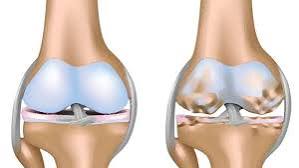
Osteoarthritis is the most common cause of pathological joint crepitus in adults over 50. The articular cartilage that provides frictionless joint movement is gradually eroded over time; as the cartilage thins and the underlying bone becomes exposed, the joint produces a sustained crackling, grinding, or grating sensation during movement. OA crepitus is typically accompanied by:
- Morning stiffness lasting less than 30 minutes (longer stiffness suggests inflammatory arthritis)
- Pain that is worse with activity and improved by rest (the opposite pattern to inflammatory arthritis)
- Bony enlargement and joint-line tenderness on examination
- Reduced range of motion
- Plain X-ray showing joint space narrowing, subchondral sclerosis, osteophytes, and subchondral cysts
The knees, hips, and hands (particularly the first carpometacarpal joint at the base of the thumb and the DIP joints — Heberden’s nodes) are the most commonly affected sites. Hip and spine OA can also contribute to leg and back pain alongside crepitus. Management is stepwise: weight management, physiotherapy, and appropriately chosen analgesia for mild to moderate OA; total joint replacement for severe disease that has failed conservative management.
Inflammatory Arthritis and Clicking Joints
Inflammatory arthritis — including rheumatoid arthritis, psoriatic arthritis, and reactive arthritis — produces joint symptoms that are distinct from OA in several ways. Morning stiffness lasting more than 60 minutes is the cardinal distinguishing feature. Swelling is soft and boggy (synovitis) rather than hard and bony. Symptoms improve with activity rather than worsening. Joints are warm and tender to pressure. Crepitus in inflammatory arthritis reflects pannus (inflamed synovial tissue) and damaged articular surfaces. Any joint that is persistently hot, swollen, and stiff for more than six weeks, particularly in a younger adult, warrants blood tests (CRP, ESR, rheumatoid factor, anti-CCP antibodies) and referral to rheumatology. Early disease-modifying treatment dramatically improves long-term joint outcomes in inflammatory arthritis.
Hypermobility and Joint Clicking
Joint hypermobility — ligamentous laxity producing a greater than usual range of motion — is extremely common and is found on the spectrum from normal variation through benign joint hypermobility syndrome (BJHS) to hereditary connective tissue disorders including Ehlers-Danlos syndrome. Hypermobile joints click more frequently because lax ligaments allow greater joint translation and more opportunities for tendons and ligaments to snap across bony prominences. In benign hypermobility, clicking is a normal feature of joint movement and is not in itself harmful. However, hypermobile individuals are at higher risk of joint subluxation, proprioceptive deficit, and musculoskeletal pain, and benefit from physiotherapy programmes specifically targeting joint stabilisation and proprioceptive retraining.
- Benign: Painless click; no swelling; full range of movement; no change over time; no locking or giving way
- Needs assessment: Click with pain; swelling (especially if warm); catching or locking; giving way; progressive worsening; limited range of motion
When to See a Doctor About Joint Clicking
The majority of joint sounds need no investigation. The presence of any of the following features changes the clinical significance and warrants assessment:
- Pain with or after clicking — pain transforms a physiological sound into a symptom that needs explanation
- Swelling — any persistent joint swelling, particularly if warm or red, requires assessment to exclude an effusion, infection, or inflammatory arthritis
- Locking or catching — the joint suddenly unable to complete its range; strongly suggests a structural cause (meniscal tear, loose body)
- Giving way — the joint buckling or collapsing under load; may indicate ligament damage or muscle weakness
- Progressive worsening — clicking that is getting louder, more frequent, or more uncomfortable over weeks to months
- New onset after injury — a click that appeared after a specific trauma (fall, sport injury, twist) may indicate structural damage
- Associated systemic features — fever, fatigue, weight loss, rash, or morning stiffness lasting over an hour alongside joint symptoms warrants urgent blood tests and rheumatology referral
- A joint that is hot, red, and severely swollen — exclude septic arthritis (joint infection), which is a surgical emergency
- A joint that has locked and will not unlock
- Joint symptoms after significant trauma with suspected fracture
- Any large joint swollen for the first time without a clear cause
Investigation of Symptomatic Joint Clicking
When clicking joints require investigation, the pathway follows clinical assessment. Plain X-ray identifies joint space narrowing (OA), loose bodies (osteochondral fragments), calcification (chondrocalcinosis — calcium pyrophosphate deposition), and structural bone changes. MRI is the gold standard for soft tissue structures: menisci, cartilage, labra (hip and shoulder), tendons, and ligament integrity. Ultrasound is particularly useful for tendons and dynamic assessment — it can visualise a snapping tendon in real time. Blood tests (FBC, CRP, ESR, uric acid, rheumatoid factor, anti-CCP, ANA) are indicated when inflammatory or systemic disease is in the differential. Joint aspiration is performed when there is a significant effusion — the fluid analysis distinguishes inflammatory (high white cell count), septic (very high white cell count, organisms on gram stain and culture), crystal-induced (gout, pseudogout — crystals under polarised microscopy), and traumatic (haemarthrosis) causes.
Key Resources
Frequently Asked Questions
Is it bad if my joints crack a lot?
Frequent joint cracking is not harmful in itself if the cracking is painless and the joint moves normally. The sound of a crack is caused by gas bubble formation in the synovial fluid — a physical phenomenon, not damage. Research has not found that habitual joint cracking accelerates arthritis development. The key question is always whether the cracking is associated with pain, swelling, or reduced movement — if none of these features is present, frequent cracking is a benign finding. If any of these features accompanies the cracking, clinical assessment is warranted.
Why does my knee click when I bend it?
Knee clicking during flexion has several possible causes. In a young person with a painless click that has always been present, tendon or ligament snapping over a bony prominence is the most likely cause. In someone with anterior knee pain that is worse going down stairs or after prolonged sitting, patellofemoral crepitus from cartilage irregularity under the kneecap is common. A click at a specific flexion angle associated with catching, locking, or giving way — especially after a twisting injury — suggests a meniscal tear. Sustained crepitus throughout the range of motion in an older adult, with progressive pain and stiffness, points toward OA.
Does joint cracking cause arthritis?
No — the evidence does not support a causal relationship between habitual joint cracking and arthritis. Multiple studies, including a long-running self-experiment, have found no significant difference in arthritis rates between habitual crackers and non-crackers. The mechanism of joint cracking — gas bubble cavitation in synovial fluid — does not damage articular cartilage. Arthritis develops through a combination of mechanical wear, inflammatory processes, and genetic factors that are separate from joint cracking behaviour.
What causes clicking in the shoulder?
Shoulder clicking arises from several possible sources. A superficial click over the top of the shoulder joint typically involves the acromioclavicular joint and often indicates AC joint OA. Clicking deep within the shoulder with internal or external rotation may arise from the glenohumeral joint itself — from chondral damage, a labral tear (the fibrocartilaginous rim around the socket), or loose bodies. A palpable snap over the anterior shoulder during arm movement may reflect the long head of biceps tendon. Clicking or clunking from the posterior shoulder blade area during arm movement is scapulothoracic in origin. Each pattern has a different examination finding and investigation pathway.
What is snapping hip syndrome?
Snapping hip syndrome (coxa saltans) is a condition in which a tendon — most commonly the iliotibial band or the iliopsoas tendon — snaps over a bony prominence during hip movement, producing an audible or palpable snap. External snapping (IT band over the greater trochanter) is felt on the lateral hip and is common in young active adults and dancers. Internal snapping (iliopsoas over the eminence or femoral head) is felt in the groin. Both are usually painless; when associated with pain, bursal inflammation is likely and physiotherapy is the primary treatment. Intra-articular causes of hip clicking — labral tear, loose body — produce less reproducible symptoms and are usually accompanied by groin pain with loading.
When does clicking in a joint mean something is wrong?
Clicking indicates something is wrong when it is accompanied by pain, swelling, catching or locking, giving way, or progressive worsening. A click alone — without any of these features — is almost always benign. The most important red flags are: a joint that is simultaneously hot, red, and swollen (exclude infection and inflammatory arthritis); a joint that locks (strong indicator of a mechanical structural problem such as a meniscal tear or loose body); and a click that appeared after a specific injury. In older adults, sustained crepitus with progressive pain and stiffness in a large joint almost always warrants investigation for OA.
Can physiotherapy help joint clicking?
Physiotherapy is a first-line treatment for most causes of symptomatic joint clicking. Snapping hip syndrome, patellofemoral crepitus, AC joint OA, scapulothoracic crepitus, and hypermobility-related clicking all respond well to targeted physiotherapy programmes. The mechanisms include: strengthening the muscles that stabilise the joint (reducing abnormal translation and snapping), correcting muscle imbalances that alter tracking mechanics, and proprioceptive retraining to improve neuromuscular control. For structural causes such as meniscal tears, loose bodies, and full-thickness labral tears, physiotherapy may manage symptoms but surgical intervention is often needed for definitive treatment.
References
- Unger DL. Does knuckle cracking lead to arthritis of the fingers? Arthritis Rheum. 1998;41(5):949–950.
- Castellanos J, Axelrod D. Effect of habitual knuckle cracking on hand function. Ann Rheum Dis. 1990;49(5):308–309.
- Polousky JD. Juvenile osteochondritis dissecans. Sports Med Arthrosc Rev. 2011;19(1):56–63.
- Sanders TL, et al. Incidence of snapping hip syndrome in US military personnel. Orthop J Sports Med. 2016;4(7).
- Brignardello-Petersen R, et al. Knee arthroscopy versus conservative management in OA. BMJ. 2017;357:j1982.
- NHS. Osteoarthritis. nhs.uk. Updated 2023.
- Nijs J, et al. Diagnosis and management of hypermobility. Phys Ther. 2017;97(12):1240–1251.
- Birch NC, et al. Crepitus in chondromalacia patellae. J Bone Joint Surg Br. 1991;73(4):655–658.
I have cracked my knuckles my whole life and I’ve been told by so many people it causes arthritis. I’m glad this article cites Donald Unger’s self-experiment — I had no idea someone had actually studied it that rigorously. The explanation of cavitation and gas bubble collapse is also very clear. I do have some mild OA in my left thumb DIP joint at 54, but given I only cracked my right hand knuckles for decades, I’m fairly confident causality runs the other way. The article correctly notes that the cracking sound itself is not a sign of joint damage.
Chloe, the Unger self-experiment is genuinely one of the more entertaining pieces of clinical research — the dedication required to crack only the right-hand knuckles every day for sixty years and then compare hand radiographs is impressive. The conclusion is consistent with the body of evidence: habitual knuckle cracking does not cause OA. The thumb DIP joint OA you mention is strongly genetically determined — the risk is substantially increased by family history and female sex, with the knuckle cracking having negligible contribution to that trajectory. Stephen, the presence of a meniscal tear on MRI in an active person requires careful interpretation — meniscal tears are common incidental findings on MRI in adults over 45 and may predate symptoms. The key question is whether the tear is mechanically symptomatic: clicking or catching with pain that is reproduced by McMurray’s test or joint-line palpation suggests mechanical significance, while diffuse aching after loading suggests more of a degenerative/inflammatory response. Your physiotherapy approach of load management is appropriate for a degenerative radial tear.
My knee has been clicking for about a year. It’s painless most of the time but after a longer run it becomes a dull ache that lasts a day. My GP referred me for physiotherapy which has helped with the aching but the clicking persists. I had an MRI which showed a small radial tear of the lateral meniscus. The physiotherapist said the tear is unlikely to be causing the clicking itself, but the loading from running is aggravating the perimeniscal soft tissue. I found the section on meniscal clicking versus patellofemoral crepitus very useful for understanding what’s likely happening.