
Gout is the most common inflammatory arthritis in adults, affecting approximately 2.5% of the UK population — around 1.7 million people. It is caused by the deposition of monosodium urate (MSU) crystals in joints and soft tissues, resulting from sustained elevated serum uric acid levels (hyperuricaemia). Gout produces acutely painful attacks of joint inflammation that can be almost incapacitating in their severity, yet it remains both underdiagnosed and undertreated — many patients experience repeated attacks for years before receiving urate-lowering therapy that could prevent attacks entirely.
This guide covers gout comprehensively: the uric acid physiology behind it, what causes hyperuricaemia, how an acute gout attack develops, how gout is diagnosed, what the long-term consequences of untreated gout are, and how modern treatment achieves complete prevention of attacks for most patients who receive it.
What Is Uric Acid and Why Does It Accumulate?
Uric acid is the end product of purine metabolism in humans. Purines are nitrogen-containing compounds found in the nucleotides of DNA and RNA and are therefore present in all cells. They are obtained both from the breakdown of the body’s own cells — endogenous production, approximately two-thirds of daily uric acid production — and from dietary sources rich in purines (approximately one-third). Unlike most other mammals, humans lack the enzyme uricase, which converts uric acid to the more soluble compound allantoin. As a result, uric acid is the final product of purine catabolism in humans and must be excreted primarily by the kidneys (approximately 70%) and the gut (approximately 30%).
Normal serum uric acid levels are below 360 µmol/L (6 mg/dL) for women and below 420 µmol/L (7 mg/dL) for men. Hyperuricaemia — serum uric acid above these thresholds — does not cause symptoms in the majority of people. Approximately 90% of individuals with hyperuricaemia never develop gout. However, hyperuricaemia is a necessary precondition for gout: without sustained elevated serum urate, MSU crystals do not form in the joints, and gout does not occur.
Hyperuricaemia arises from three mechanisms:
- Underexcretion (approximately 90% of cases): The kidneys fail to excrete sufficient uric acid, allowing it to accumulate in the serum. This is the most common cause and is influenced by genetic variants in renal urate transporters (URAT1, ABCG2), kidney disease, and medications such as diuretics.
- Overproduction (approximately 10% of cases): Accelerated cell turnover (haematological malignancies, psoriasis, chemotherapy) or rare inborn errors of purine metabolism (Lesch-Nyhan syndrome) increase the purine load beyond what the kidneys can excrete.
- Combined overproduction and underexcretion: Often seen with high purine diet and alcohol combined with mild renal impairment.
Causes and Risk Factors for Gout
Gout is strongly influenced by a combination of genetic predisposition, dietary habits, comorbidities, and medications. Understanding the modifiable risk factors is central to both prevention and treatment.
Diet and Alcohol
Dietary purines significantly influence serum urate levels. High-purine foods that raise serum urate include red meat (particularly organ meats such as liver, kidney, and sweetbreads), shellfish (mussels, scallops, lobster), anchovies, sardines, and herring. Plant-based purines — found in legumes, mushrooms, and asparagus — raise serum urate considerably less than animal-derived purines and are not associated with the same gout risk in prospective cohort studies.
Alcohol raises serum urate through multiple mechanisms: ethanol metabolism increases purine production via accelerated ATP degradation; alcohol promotes dehydration and lactic acidosis, which reduce renal urate excretion; and beer contains significant quantities of guanosine, a direct purine precursor. Beer and spirits are most strongly associated with gout risk in epidemiological data. Wine is less strongly associated, though still a relevant contributor at high consumption. Fructose-sweetened beverages — particularly soft drinks containing high-fructose corn syrup — raise serum urate because fructose metabolism generates AMP, a purine substrate catabolised to uric acid. Consuming two or more sugar-sweetened drinks per day approximately doubles gout risk.
Several foods are associated with reduced gout risk. Low-fat dairy products have a mildly uricosuric effect through the protein casein and are associated with lower serum urate and reduced gout incidence in prospective cohort studies. Coffee (both caffeinated and decaffeinated) is associated with a modest reduction in serum urate and gout risk. Staying well hydrated (2–3 litres daily) dilutes serum urate and supports renal excretion.
Comorbidities
Gout clusters strongly with several common conditions that impair urate excretion or increase production:
- Chronic kidney disease: The strongest comorbid driver of hyperuricaemia — reduced glomerular filtration reduces renal urate clearance. The relationship is bidirectional: hyperuricaemia may also contribute to CKD progression through tubular crystal deposition and endothelial inflammation.
- Obesity: Independently associated with both increased uric acid production and reduced renal excretion. Weight loss of 5–10% produces a clinically meaningful reduction in serum urate.
- Type 2 diabetes and insulin resistance: Insulin resistance promotes renal urate retention via upregulation of the URAT1 transporter. Metabolic syndrome — obesity, hypertension, dyslipidaemia, insulin resistance — is present in the majority of gout patients.
- Hypertension: Both directly through reduced renal blood flow and indirectly through diuretic use. Losartan (an ARB antihypertensive) has a mild uricosuric effect and is the preferred antihypertensive in gout patients.
- Heart failure: Reduced cardiac output and associated diuretic use substantially raise serum urate.
Medications That Raise Uric Acid
Several commonly prescribed medications raise serum urate and can precipitate gout in susceptible patients:
- Diuretics: Both thiazide (bendroflumethiazide) and loop diuretics (furosemide) significantly reduce renal urate excretion — the most common drug-induced cause of gout. These are often essential in heart failure and should not be stopped for gout; instead, urate-lowering therapy addresses the elevated urate.
- Low-dose aspirin: At antiplatelet doses (75–300 mg), aspirin reduces renal urate excretion. It should not be stopped for gout management given its cardiovascular benefit.
- Ciclosporin: A potent inhibitor of renal urate excretion — gout occurs in approximately 80% of ciclosporin-treated renal transplant patients.
- Pyrazinamide and ethambutol: Antituberculous agents that reduce renal urate excretion.
How an Acute Gout Attack Develops
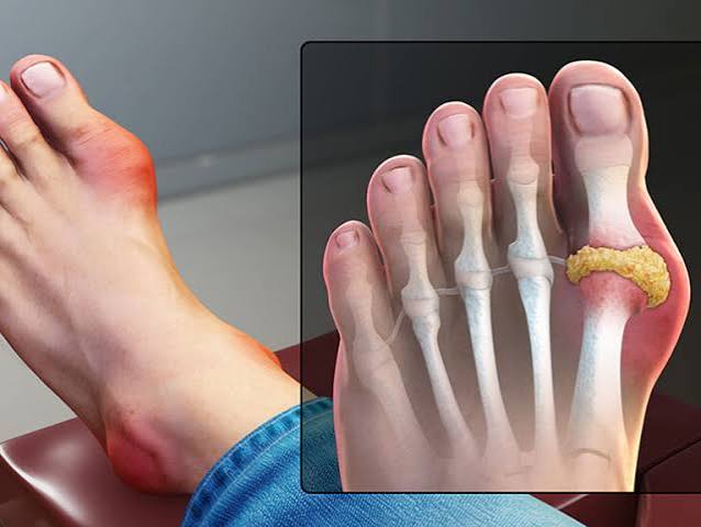
The transition from asymptomatic hyperuricaemia to acute gout occurs when serum urate reaches a sustained level at which MSU crystals precipitate in the relatively avascular, lower-temperature environment of joint cartilage and synovium. Crystals form gradually over months to years, depositing particularly in the fibrocartilage of the first metatarsophalangeal joint (MTP1), ankles, knees, wrists, and bursae. During this prolonged period of crystal deposition, patients are typically asymptomatic.
The acute attack is triggered when crystals shed from the cartilaginous matrix into the joint space — typically after a rapid change in serum urate. This can be caused by an abrupt rise (from alcohol, a purine-rich meal, or dehydration) or, paradoxically, an abrupt fall — which is why initiating allopurinol can trigger an attack in the first months of treatment. In the joint fluid, MSU crystals are recognised by macrophages and neutrophils through the NLRP3 inflammasome, triggering production of IL-1β, TNF-α, and IL-8. IL-1β drives a massive neutrophil influx — synovial fluid during an acute attack contains 10,000–100,000 cells per microlitre. Neutrophils attempt to phagocytose the crystals but cannot digest them, leading to cell death and further release of inflammatory mediators in a self-amplifying inflammatory cycle.
The acute attack typically peaks within 12–24 hours of onset and resolves spontaneously over 5–14 days as anti-inflammatory mechanisms — including TGF-β and IL-1 receptor antagonist — gradually suppress the response. Between attacks, patients are symptom-free: this is the intercritical period. Crystals continue to accumulate during this period, and without urate-lowering therapy, attacks become more frequent and prolonged over years.
Symptoms of an Acute Gout Attack
Gout attacks are typically monoarticular — affecting a single joint — especially in early disease. The first metatarsophalangeal joint (the base of the big toe) is affected in approximately 70% of first attacks, giving rise to the term podagra. Other commonly affected joints include the ankle, midfoot, knee, wrist, and olecranon bursa. Hip and spinal gout are uncommon.
The attack typically begins at night or in the early morning, often waking the patient from sleep. Pain builds from mild discomfort to excruciating within hours. The joint becomes hot, erythematous, and exquisitely tender — patients frequently describe the inability to tolerate the weight of bedclothes over the affected joint. Even light touch is intensely painful (allodynia). The joint shows visible swelling with tense, shiny, erythematous overlying skin. In severe attacks, systemic features — low-grade fever and malaise — accompany the joint inflammation, occasionally causing confusion with septic arthritis at initial presentation.
Without treatment, the acute attack resolves over 5–14 days. Subsequent attacks in untreated patients tend to be more frequent, more prolonged, and affect more joints. Polyarticular gout — affecting multiple joints simultaneously — becomes more common as the crystal burden accumulates over years of untreated hyperuricaemia.
Tophaceous Gout
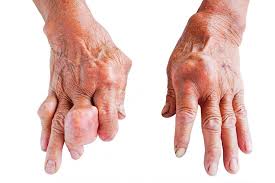
With sustained hyperuricaemia over many years, MSU crystals accumulate into palpable deposits called tophi — chalky, white nodular deposits visible or palpable under the skin at characteristic sites: the helix of the ear, olecranon bursae, Achilles tendon, extensor surfaces of the fingers, and around the first MTP joint. Tophi are the hallmark of chronic tophaceous gout, developing when the total body urate crystal burden accumulates without adequate treatment over years to decades.
Tophaceous deposits are destructive: they erode into adjacent bone, producing the characteristic “punched-out” or “rat-bite” erosions with overhanging edges seen on X-ray. They damage joint cartilage, cause progressive joint deformity, and compress adjacent structures — tophi in the carpal tunnel produce median nerve compression indistinguishable from conventional carpal tunnel syndrome. Tophaceous deposits in the renal collecting ducts can cause urate nephropathy and contribute to CKD progression. Modern urate-lowering therapy dissolves tophi over months to years when serum urate is maintained well below 300 µmol/L — complete resolution of tophi is achievable in most patients with sustained, consistent treatment.
Diagnosing Gout
Gout diagnosis is clinical in the majority of cases, particularly when the presentation is classic (acute podagra in a middle-aged or older man with hyperuricaemia). Several investigations support or confirm the diagnosis:
- Serum uric acid: Should be measured in all suspected cases. A level above 360 µmol/L supports the diagnosis. Importantly, serum urate during an acute attack may be falsely normal or low — the inflammatory response redistributes urate — so a normal result during an attack does not exclude gout. Measurement should be repeated 4–6 weeks after the attack resolves.
- Synovial fluid analysis: The gold standard for gout diagnosis. MSU crystals are identified under polarised light microscopy as needle-shaped, negatively birefringent crystals (yellow when aligned parallel to the compensator axis). Synovial fluid analysis also excludes septic arthritis — which requires urgent joint washout and intravenous antibiotics and must not be missed.
- Dual-energy CT (DECT): A non-invasive modality that identifies urate crystal deposits based on their X-ray attenuation. Highly specific for gout; increasingly used when synovial aspiration is impractical or the diagnosis remains uncertain.
- Musculoskeletal ultrasound: Detects the “double contour” sign — urate crystal deposition on the surface of articular cartilage — which is specific for gout. Also identifies tophi and synovitis in clinically uncertain presentations.
- Plain X-rays: Normal in early gout; show characteristic “punched-out” erosions with sclerotic margins and overhanging edges in established tophaceous gout. Useful for baseline assessment and monitoring erosive progression.
The 2015 ACR/EULAR gout classification criteria assign points based on joint symptoms, synovial fluid findings, serum urate level, and imaging features. A score of 8 or more classifies gout with 92% sensitivity and 89% specificity — useful in clinically uncertain cases.
Treating an Acute Gout Attack
The goals of acute treatment are rapid pain control and resolution of the inflammatory episode. An important principle: urate-lowering therapy should not be started during an acute attack — initiating allopurinol or febuxostat during the attack can prolong or worsen it by rapidly changing serum urate and mobilising crystal deposits. ULT is started 2–4 weeks after complete attack resolution.
- NSAIDs: First-line in patients without contraindications. Naproxen, indomethacin, and etoricoxib are used at full anti-inflammatory doses for 5–7 days. Contraindicated in severe renal impairment, active peptic ulcer disease, and significant anticoagulation.
- Colchicine: Highly effective when started within 24–36 hours of onset. The modern low-dose regimen (500 µg two to three times daily for 3–5 days) has equivalent efficacy to the older high-dose regimen with considerably fewer gastrointestinal side effects. Particularly useful when NSAIDs are contraindicated. Should be avoided in severe renal or hepatic impairment and with strong CYP3A4 inhibitors (clarithromycin, ciclosporin).
- Corticosteroids: Oral prednisolone (30–35 mg daily for 5 days) or intra-articular corticosteroid injection are effective alternatives when both NSAIDs and colchicine are contraindicated, for example in severe CKD. Repeated systemic corticosteroid courses carry risks of adrenal suppression, hyperglycaemia, and bone loss.
Urate-Lowering Therapy: Long-Term Prevention
Urate-lowering therapy (ULT) is the cornerstone of long-term gout management and the only intervention that addresses the underlying pathology — elevated serum urate and crystal deposition — rather than just the symptoms. NICE guidelines recommend ULT for patients with two or more attacks per year, tophaceous gout, gout with CKD stage 2 or above, urate nephropathy or urolithiasis, or gout with radiographic joint damage. The serum urate target is below 360 µmol/L for most patients and below 300 µmol/L in tophaceous gout to accelerate tophus dissolution.
Allopurinol
Allopurinol is the first-line ULT for most patients. It is a xanthine oxidase inhibitor — it blocks the enzyme that converts hypoxanthine and xanthine to uric acid, substantially reducing uric acid production. It is started at a low dose (100 mg daily, or 50 mg in CKD) and increased in 100 mg increments every 4 weeks until the serum urate target is achieved. Most patients require 300–600 mg daily; the maximum dose is 900 mg.
The most serious adverse effect is allopurinol hypersensitivity syndrome (DRESS — drug reaction with eosinophilia and systemic symptoms), occurring in approximately 1 in 1,000 patients and carrying a mortality of around 25% without prompt discontinuation and specialist management. The HLA-B*58:01 allele strongly predicts this reaction and is substantially more prevalent in Han Chinese, Thai, Korean, and Vietnamese populations — genetic screening before allopurinol initiation is recommended in individuals from these ethnic groups.
Febuxostat
Febuxostat is a non-purine xanthine oxidase inhibitor used when allopurinol is not tolerated or fails to achieve the serum urate target. It does not require dose reduction in mild-to-moderate renal impairment. Febuxostat 80 mg achieves the serum urate target in approximately 40% of patients; at 120 mg, approximately 55%. The CARES trial (2018, NEJM) raised concern about increased cardiovascular mortality compared with allopurinol in patients with established cardiovascular disease. However, the subsequent FAST trial (Lancet, 2020) found febuxostat non-inferior to allopurinol for cardiovascular outcomes in gout patients with established CV disease, somewhat tempering the initial concern. NICE currently recommends reserving febuxostat for allopurinol intolerance or failure, with caution in ischaemic heart disease and heart failure.
Prophylaxis During ULT Initiation
A critical aspect of gout management that is frequently overlooked: approximately 50% of patients experience acute attacks during the first 3–6 months of ULT without prophylaxis. This is the most common reason patients discontinue their allopurinol — they conclude, understandably, that the drug is making them worse. The mechanism is that rapidly lowering serum urate destabilises existing crystal deposits and triggers their shedding into the joint space. The crystals were already there; the allopurinol simply initiates the mobilisation process that precedes long-term crystal dissolution.
Low-dose colchicine (500 µg once or twice daily) for the first 3–6 months of ULT prevents most of these flares. Low-dose naproxen (250 mg twice daily) with a proton pump inhibitor is an alternative when colchicine is not tolerated. The prophylactic course continues until serum urate has been at target for at least 3–6 months and no attack has occurred for at least 6 months.
Lifestyle Changes to Prevent Gout
Lifestyle modification reduces serum urate, lowers attack frequency, and improves the cardiometabolic risk profile of gout patients — who have elevated baseline cardiovascular risk. In most patients with established gout, lifestyle changes work best as an adjunct to urate-lowering therapy rather than a replacement for it.
- Reduce alcohol: Beer and spirits are most harmful; wine less so but still relevant at high consumption. Aim for no more than 14 units per week with at least two alcohol-free days; during periods of frequent attacks, complete abstinence is advisable.
- Reduce high-purine animal foods: Limit red meat, organ meats, and shellfish. Lean white meat (chicken, turkey) is a lower-purine substitute. Plant-based purines in legumes and vegetables do not need to be restricted.
- Eliminate fructose-sweetened drinks: Replace sugary soft drinks with water, low-fat milk, or coffee — all of which have neutral or beneficial effects on serum urate.
- Stay well hydrated: A daily fluid intake of 2–3 litres supports renal urate excretion. Extra hydration during hot weather and exercise is important, as dehydration concentrates serum urate and precipitates attacks.
- Achieve and maintain a healthy weight: Weight loss of 5–10% produces a clinically meaningful reduction in serum urate. Avoid crash dieting or prolonged fasting, which causes ketosis and reduces renal urate excretion, potentially triggering an attack.
- Eat low-fat dairy: Low-fat milk, yoghurt, and cheese are associated with reduced serum urate and lower gout risk in prospective studies.
- Antihypertensive choice matters: If hypertension requires drug treatment, losartan is the preferred agent in gout patients because it has a mild uricosuric effect — unlike thiazide and loop diuretics, which worsen hyperuricaemia.
Comparing gout with osteoarthritis and rheumatoid arthritis helps clarify why gout — despite feeling like a joint problem — is metabolic at its root and responds to a completely different treatment approach. Understanding the spectrum of inflammatory arthritis places gout alongside psoriatic arthritis and reactive arthritis as crystal-mediated and immune-mediated conditions. Psoriatic arthritis and gout can coexist — psoriasis is itself a cause of hyperuricaemia through increased cell turnover, and patients with psoriatic arthritis have a higher-than-average gout risk. Distinguishing gout from rheumatoid arthritis is clinically important: both cause joint swelling and tenderness, but the treatment pathways are entirely different.
Key Resources
Frequently Asked Questions
What causes gout?
Gout is caused by monosodium urate (MSU) crystal deposition in joints and soft tissues, resulting from sustained elevated serum uric acid (hyperuricaemia). Uric acid is the end product of purine metabolism. Hyperuricaemia arises most commonly from reduced renal urate excretion (90% of cases), influenced by CKD, diuretic use, and genetic variants in renal urate transporters. Dietary purines (red meat, shellfish, alcohol) and fructose-sweetened drinks contribute. Without sustained elevated urate, crystals do not form and gout does not occur.
What does a gout attack feel like?
A gout attack typically starts at night or early morning with rapidly escalating pain in a single joint — most commonly the base of the big toe (podagra). The joint becomes exquisitely tender, hot, red, and swollen — many patients describe the weight of bedclothes as unbearable. The pain peaks within 12–24 hours and resolves over 5–14 days without treatment. With appropriate treatment (NSAIDs, colchicine, or corticosteroids started within 24–36 hours of onset), most attacks resolve within 3–7 days.
How long does a gout attack last?
With treatment started within 24–36 hours of onset, most gout attacks resolve within 3–7 days. Without treatment, attacks typically resolve spontaneously over 5–14 days. After resolution, there is an intercritical period — usually weeks to months in early gout — during which the joint is asymptomatic, though urate crystals remain deposited. Without urate-lowering therapy, attacks become more frequent and prolonged over time.
Does gout go away on its own?
Individual acute attacks resolve spontaneously without treatment over 5–14 days, though with significant pain. However, the underlying cause — hyperuricaemia and crystal deposition — does not resolve without urate-lowering therapy. Without ULT, attacks become more frequent and eventually polyarticular, tophi develop, and joint and renal damage accrue. Allopurinol, taken consistently to maintain serum urate below 360 µmol/L, prevents attacks in the vast majority of patients and gradually dissolves existing crystal deposits.
Can gout be cured?
Gout can be put into complete remission — no attacks, no tophus progression, gradual crystal dissolution — with consistent urate-lowering therapy. However, permanent remission after stopping ULT is unusual, since the underlying hyperuricaemia is typically genetic and metabolic and recurs when medication is stopped. Most patients with recurrent gout require lifelong ULT. Patients who achieve major lifestyle change (significant weight loss, stopping a causative medication such as a diuretic) may sustain low serum urate without drugs, but this is the exception rather than the rule.
What foods should I avoid with gout?
The foods most strongly associated with gout attacks are: beer and spirits; red meat and organ meats (liver, kidney, sweetbreads); shellfish (mussels, scallops, lobster); anchovies and sardines; and fructose-sweetened soft drinks. Plant-based purines in legumes, mushrooms, and asparagus are less risky and can generally remain in the diet. Low-fat dairy and coffee are associated with reduced gout risk and can be consumed freely. Staying well hydrated (2–3 litres daily) is important for renal urate excretion.
When should I start allopurinol?
Allopurinol should be started 2–4 weeks after a gout attack has fully resolved — not during the attack, as doing so can prolong or worsen the inflammation. NICE recommends ULT for patients with two or more attacks per year, tophaceous gout, gout with kidney disease, or radiographic joint damage. Start at 100 mg daily (50 mg in CKD) and increase by 100 mg every 4 weeks until serum urate falls below 360 µmol/L. Always co-prescribe prophylactic colchicine (500 µg once or twice daily) for the first 3–6 months to prevent attack flares during dose titration — without it, approximately half of patients experience attacks and discontinue their allopurinol.
References
- Dalbeth N, et al. Gout. Lancet. 2016;388(10055):2039–2052.
- NICE CG56. Gout: diagnosis and management. 2012 (updated 2022).
- Richette P, et al. 2016 updated EULAR recommendations for gout management. Ann Rheum Dis. 2017;76(1):29–42.
- FitzGerald JD, et al. 2020 ACR guideline for gout management. Arthritis Care Res. 2020;72(6):744–760.
- Perez-Ruiz F, et al. Gout. BMJ. 2023;381:e070748.
- Neogi T, et al. 2015 gout classification criteria. Arthritis Rheumatol. 2015;67(10):2557–2568.
- MacDonald TM, et al. Febuxostat vs allopurinol — cardiovascular outcomes (FAST trial). Lancet. 2020;396(10264):1745–1757.
- NHS. Gout. nhs.uk. Updated 2023.
I had my first gout attack at 52 — in my right big toe, in the middle of the night. I had no idea what was happening and genuinely thought I had broken something. Within three hours the pain was so severe I could not bear the weight of the sheet on my foot. My GP diagnosed it at the next morning appointment from examination alone, confirmed with a blood test showing serum uric acid of 540. I was put on allopurinol after my second attack six months later and have had no further attacks in four years. The section on starting allopurinol gradually and using colchicine to prevent flares in the first months was something nobody explained to me when I started — I had two attacks in the first month before my GP added the colchicine prophylaxis and they stopped completely.
Martin, the experience you describe — attacks in the first month of allopurinol — is extremely common and is the most frequent reason patients discontinue ULT. They reasonably conclude the medication is causing more harm than good, when in fact the mechanism is that allopurinol rapidly lowers serum urate, which destabilises existing crystal deposits and triggers their shedding into the joint space. The crystals were already there; the allopurinol simply initiated the mobilisation process. Colchicine prophylaxis at 500 µg once or twice daily for 3–6 months is now standard in NICE and EULAR guidelines specifically to prevent this, and it is worth making sure any GP initiating allopurinol is familiar with co-prescribing it. With sustained allopurinol and serum urate consistently below 360 µmol/L, existing crystal deposits continue to dissolve over months to years, which is why attack frequency continues to fall the longer treatment is maintained. Patricia, your situation reflects the intersection of gout, CKD, and cardiovascular disease that makes management genuinely complex. In CKD stage 3, allopurinol doses above 100–200 mg carry an increased risk of allopurinol hypersensitivity and are used cautiously, which can make reaching the serum urate target of 360 µmol/L difficult. The FAST trial (Lancet, 2020) did find febuxostat non-inferior to allopurinol for cardiovascular outcomes in patients with gout and established cardiovascular disease — this has somewhat reduced the earlier CARES concern, and your rheumatologist may factor this into any reassessment of febuxostat as an option for you.
I have had gout for eight years alongside stage 3 chronic kidney disease, which makes management more complicated than for most patients. My nephrologist is cautious about allopurinol dosing because of the kidney function, so I am on 100 mg daily and my uric acid is still borderline at 370. The section on febuxostat was interesting — my rheumatologist mentioned it as an option but was concerned about the cardiovascular data as I have had a previous heart attack. I have been managing primarily through diet — I stopped beer entirely five years ago and reduced red meat — which has reduced my attack frequency from four or five per year to one or two, but has not eliminated them. The combination of CKD and cardiovascular disease makes this a genuinely difficult condition to treat optimally.