The skin is the largest organ of the human body and, in a very real sense, one of its most communicative. Changes in the skin can reflect what is happening locally — primary cancers arising from the skin cells themselves — or what is happening systemically, in organs that the eye cannot see.
Skin changes are responsible for detecting one of the most successfully screened cancers (melanoma, when caught early), but also for providing some of the earliest and most specific signals of internal malignancies that would otherwise go undetected for months or years. Recognizing which skin changes are warning signs — and which are benign — is knowledge that genuinely saves lives.
Melanoma: The Most Dangerous Skin Cancer
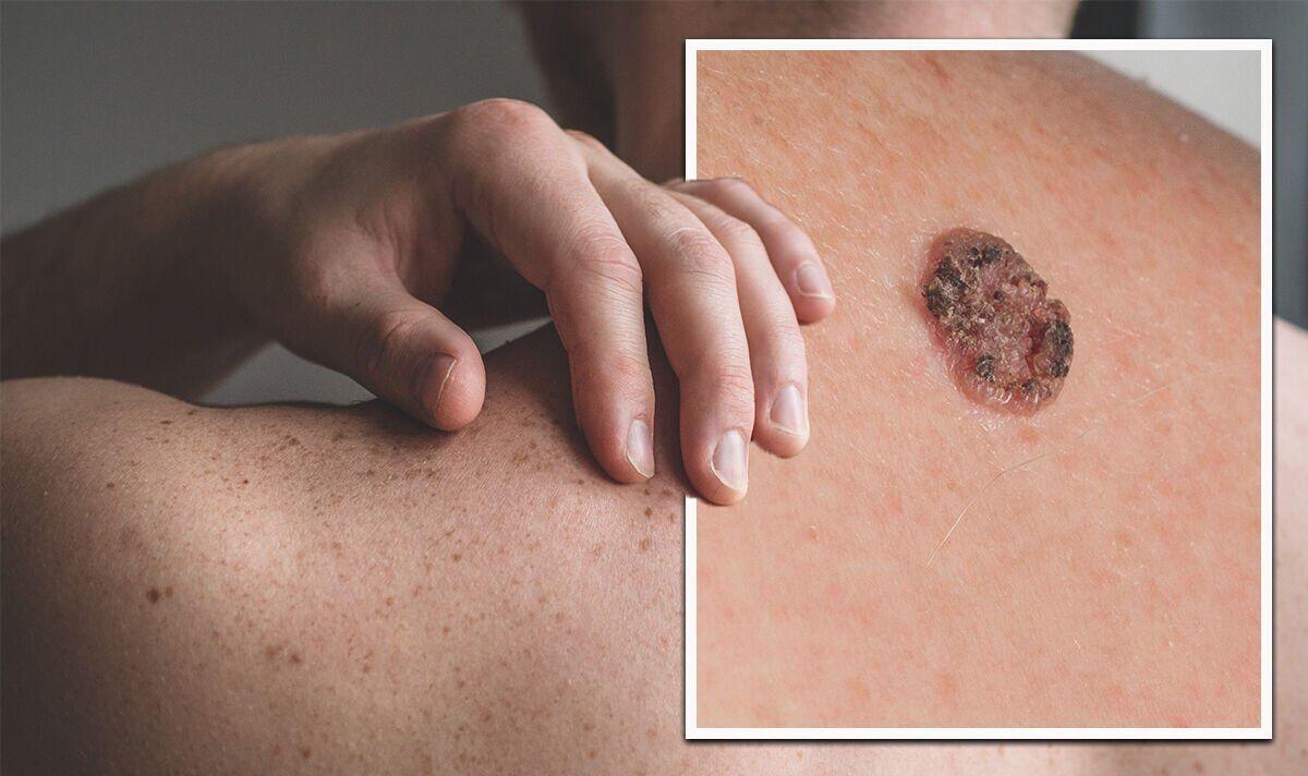
Melanoma is not the most common skin cancer — that distinction belongs to basal cell carcinoma by a wide margin. But melanoma causes approximately 8,000 deaths annually in the United States from roughly 100,000 new cases. Its lethality comes from its capacity to metastasize early and widely — to lymph nodes, the liver, lungs, brain, and bone.
What makes melanoma both uniquely dangerous and uniquely catchable is that it arises on the visible surface of the skin, where it can be seen for months to years before becoming deep enough to spread. The challenge is recognizing it.
| Criterion | What It Means | Benign vs. Concerning |
|---|---|---|
| A | Asymmetry | Benign: both halves match. Concerning: one half does not match the other |
| B | Border | Benign: smooth, round, well-defined. Concerning: irregular, ragged, notched, blurred |
| C | Color | Benign: single uniform shade of brown. Concerning: multiple shades (tan, dark brown, black, red, white, blue) within one lesion |
| D | Diameter | Melanomas are often >6 mm at diagnosis but can be smaller — diameter alone is NOT the deciding factor |
| E | Evolution — THE MOST IMPORTANT | Any mole that is CHANGING — growing, darkening, lightening, itching, bleeding, crusting — requires evaluation regardless of other criteria |
Basal Cell and Squamous Cell Carcinoma
Basal cell carcinoma (BCC) is the most common cancer in humans — approximately 4 million cases per year in the United States. It rarely metastasizes, but its danger lies in local invasion: a neglected BCC can grow through the dermis into underlying fat, muscle, cartilage, and bone. Classic appearance: a pearly or waxy nodule with telangiectasias (tiny dilated blood vessels) visible on the surface, raised rolled borders, and a central area that may ulcerate. Found on sun-exposed areas — face, ears, scalp, neck.
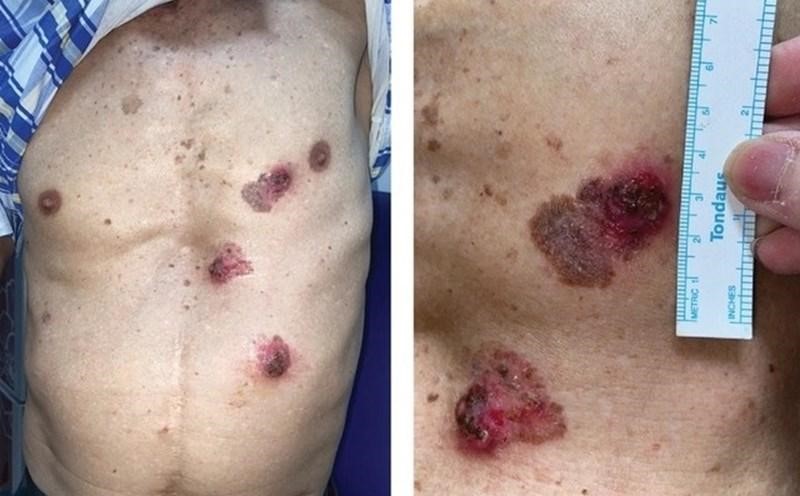
Squamous cell carcinoma (SCC) is the second most common skin cancer and kills approximately 15,000 Americans annually — sometimes exceeding melanoma deaths. SCC develops from actinic keratoses (AKs) — rough, scaly patches on chronically sun-damaged skin. Classic appearance: a scaly, rough red patch that doesn’t resolve; a hardened nodule with a wart-like or crusted surface; an ulcerating lesion with raised, firm edges. The common feature: it does not heal. SCC metastasizes in approximately 3–5% of cases overall, with higher rates on the lip, ear, and in immunocompromised patients.
- Any mole that is asymmetric, has irregular borders, multiple colors, or exceeds 6 mm
- Any mole that is changing in size, shape, color, or developing new symptoms
- A mole or skin lesion that is bleeding, itching persistently, or crusting
- A rapidly growing flesh-colored or violaceous nodule in an older adult (Merkel cell)
- Any skin wound, sore, or ulcer not healing within 4–6 weeks
- New moles appearing after age 40
Paraneoplastic Skin Changes: The Skin as a Window to Internal Cancer
Some of the most clinically important skin changes are not cancers of the skin itself but manifestations of cancer growing elsewhere in the body. Through mechanisms involving tumor-released hormones, growth factors, and immune activation, the skin displays visible signs that an internal malignancy is present. These paraneoplastic skin findings can predate the cancer diagnosis by months.
| Skin Finding | Appearance | Associated Cancer |
|---|---|---|
| Malignant acanthosis nigricans | Sudden-onset velvety hyperpigmented thickened skin in skin folds (axillae, neck, groin) — in a non-obese adult | Gastric cancer (most common); lung, uterine, ovarian |
| Dermatomyositis | Heliotrope rash (violaceous periorbital); Gottron’s papules (over knuckles); proximal muscle weakness | Lung, ovarian, GI, breast, cervical (15–30% of adult cases) |
| Jaundice (yellow skin/sclera) | Yellowing of skin and whites of eyes; dark urine; pale stools | Pancreatic cancer (head), cholangiocarcinoma, liver mets |
| Leser-Trélat sign | Sudden explosion of multiple seborrheic keratoses (waxy “barnacle” skin growths), often with AN | Gastric cancer; lung, breast, lymphoma |
| Necrolytic migratory erythema | Blistering, crusting, erythematous migrating rash on face, groin, extremities | Glucagonoma (pancreatic alpha-cell tumor) — near-pathognomonic |
| Erythema gyratum repens | Rapidly advancing concentric wood-grain skin pattern across large body surface areas | Lung cancer (most common) — nearly pathognomonic of malignancy |
Jaundice: Yellow as an Alarm Color
Jaundice — the yellowing of the skin and sclera (whites of the eyes) from elevated bilirubin — is among the most visually recognizable signs of serious illness. The most cancer-relevant type is obstructive jaundice — bile cannot flow from the liver to the small intestine because of a mechanical blockage.
Pancreatic cancer (head of pancreas) is the most important cancer cause of painless obstructive jaundice. As the tumor grows, it compresses the common bile duct that passes through the pancreatic head. The classic presentation: painless progressive jaundice, dark urine (bilirubinuria), pale stools (from absent bile), and severe pruritus (itching) — in an older adult, often with weight loss and back pain. Painless progressive jaundice in an older adult is pancreatic cancer until proven otherwise. This requires urgent CT abdomen/pelvis and biliary imaging (MRCP or ERCP).
Any new diagnosis of dermatomyositis in an adult mandates a comprehensive malignancy workup — CT chest/abdomen/pelvis, gynecologic evaluation in women, and age-appropriate cancer screening. The skin findings (heliotrope rash, Gottron’s papules) can precede the cancer diagnosis by months. Failing to screen is a recognized medical error.
The Non-Healing Wound: A Biopsy Mandatory
Any wound, sore, or ulceration that has not healed within four to six weeks should be biopsied. This applies to:
- A sore on the lip, tongue, or mouth that does not heal — oral SCC or minor salivary gland carcinoma
- An ulceration on the lower leg in a patient with chronic venous disease — Marjolin’s ulcer (SCC arising in a chronic wound)
- A healing wound from surgery or trauma that breaks down again and persists
- Any skin lesion in a field of prior radiation — radiation-induced SCC or sarcoma
Chronic inflammation is a carcinogenic stimulus. Wounds that fail to heal deserve biopsy before they are given years of topical wound care that covers a growing cancer.
Frequently Asked Questions
References
- NCCN Clinical Practice Guidelines: Melanoma; Squamous Cell Carcinoma of the Skin; Basal Cell Carcinoma; Merkel Cell Carcinoma. 2024.
- Siegel RL, et al. Cancer Statistics 2023. CA Cancer J Clin. 2023.
- Shih BB, et al. Paraneoplastic dermatoses and their association with malignancy. Br J Dermatol. 2016.
- Thiers BH, Sahn RE, Callen JP. Cutaneous manifestations of internal malignancy. CA Cancer J Clin. 2009.
- Gaudy-Marqueste C, et al. Eruptive seborrheic keratoses as a diagnostic clue to cancer. Eur J Dermatol. 2009.
- Rogers HW, et al. Incidence of Nonmelanoma Skin Cancer in the US. JAMA Dermatol. 2015.