When melanoma is caught at Stage I — thin, localized to the skin’s surface — the 5-year survival rate exceeds 98%. When it is caught at Stage IV, after spreading to the lungs, liver, or brain, that number historically hovered around 15%. Even with today’s remarkable immunotherapy advances, metastatic melanoma remains a formidable disease.
The difference between those two outcomes is almost entirely about one thing: how early a warning sign is recognized and acted upon.
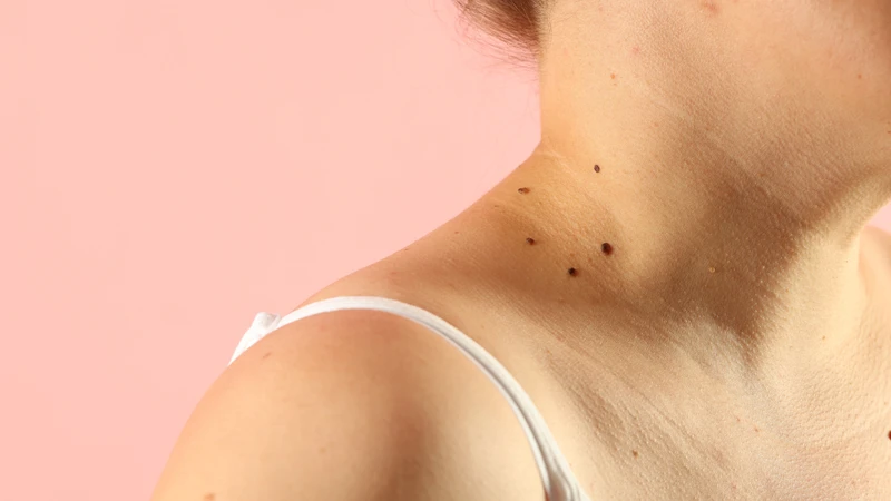
Melanoma starts in melanocytes, the pigment-producing cells in your skin. Most melanomas look like a mole, arise from a mole, or are easily mistaken for one. Understanding what a normal mole looks like — and recognizing exactly what changes demand attention — is a practical, potentially life-saving skill.
What Is a Mole — and How Does It Become Cancer?
A mole (medical term: nevus) forms when melanocytes cluster together rather than distributing individually throughout the basal layer of the skin. Most adults have between 10 and 40 moles. The vast majority will never cause any problem.
Moles typically appear during childhood and early adulthood. A new mole appearing after age 40 is uncommon — and worth noting, because melanocytes have had decades to accumulate UV-induced DNA damage by that point.
Malignant transformation happens when a melanocyte accumulates enough critical DNA mutations to begin dividing without normal controls. The most common molecular driver is a mutation in the BRAF gene — specifically the V600E substitution — which occurs in approximately 50% of all melanomas. UV radiation causes characteristic DNA damage (“UV signature mutations”) that initiates this process.
The ABCDE Warning Signs: What Each Criterion Actually Means
Developed in 1985 by dermatologist Ronald Friedman and colleagues, the ABCDE framework remains the most widely used guide for self-examination. Understanding each criterion deeply — including its limitations — makes you far more effective at using it.
| Letter | Criterion | What to Look For |
|---|---|---|
| A | Asymmetry | One half does not mirror the other — draw a line through the middle; the two halves are unequal in size, shape, or color |
| B | Border | Edges are ragged, notched, scalloped, or blurred rather than smooth and well-defined |
| C | Color | Multiple shades within the same mole — brown, black, red (inflammation), white (regression), blue-gray (deep pigment) |
| D | Diameter | Larger than 6 mm (pencil eraser size) — though many early melanomas are smaller, and not all large moles are cancerous |
| E | Evolving ⚠️ | Any change in size, shape, or color — or any new symptom (itching, bleeding, tenderness, crusting) — is the most important criterion |
The Ugly Duckling Sign: Catching What ABCDE Misses
On any given person, most moles resemble each other — a personal “pattern” of roughly similar size, shape, and color. A mole that clearly does not fit this pattern — the outlier, the one that stands apart — warrants evaluation regardless of whether ABCDE criteria are formally met.
This sign catches nodular melanoma (dark, round, dome-shaped — growing rapidly but not triggering ABCDE concern), patients with multiple atypical nevi (where the ugly duckling is the one that looks unlike all the others), and amelanotic melanoma (a pink or red spot surrounded by otherwise pigmented moles).
When examining your skin: step back and look at your moles as a group. Does one look like it belongs to a different person’s body? That is the one to have evaluated.
The 5 Types of Melanoma — Why Each One Matters
| Type | Frequency | Key Features |
|---|---|---|
| Superficial spreading | ~70% | Most common; grows horizontally first; ABCDE features usually visible; on trunk (men) or legs (women) |
| Nodular | ~15–20% | Most dangerous; grows vertically from the start; dome-shaped dark nodule; rapid growth; ABCDE often negative |
| Lentigo maligna | ~5–10% | Elderly patients; sun-damaged skin of face/neck/ears; flat tan-brown patch; often mistaken for age spot |
| Acral lentiginous | ~5% overall | Palms, soles, under nails; NOT UV-related; most common type in dark-skinned populations; frequently diagnosed late |
| Amelanotic | ~5% | No pigment — appears pink, red, or skin-colored; most commonly misdiagnosed; highest rate of delayed diagnosis |
Warning Signs That Fall Outside ABCDE
- Bleeding without trauma — spontaneous bleeding from a mole is a significant red flag
- Persistent itch or tenderness — a stable benign mole is asymptomatic; recurring symptoms demand evaluation
- Ulceration — skin breaking down over the lesion indicates deep invasion
- Satellite lesions — small separate pigmented spots near a primary lesion; indicates in-transit metastasis
- New mole after age 40 — uncommon and merits professional evaluation
- Dark streak under a nail — especially if new, widening, or spreading onto surrounding skin (Hutchinson sign)
Who Is at Highest Risk?
Skin type and personal history: Fitzpatrick Type I–II skin (very pale, burns easily, red or blonde hair, blue or green eyes) carries the highest baseline risk. A personal history of melanoma confers a 5× increased risk of a second primary. First-degree relatives with melanoma: 2–3× increased risk. FAMMM syndrome (CDKN2A mutation): lifetime risk 17–76%.
Mole burden: More than 50 acquired nevi increases risk. Atypical (dysplastic) nevi carry ~10× higher risk per lesion. Giant congenital nevi (>20 cm) carry 5–10% lifetime melanoma risk. Immunosuppression raises risk 2–8×.
Tanning beds: IARC Group 1 human carcinogen — the same category as asbestos and tobacco. Use before age 35 increases melanoma risk by 59%. There is no safe dose.
Why Every Month of Delay Matters: Breslow Thickness
Breslow thickness — the depth of the tumor from the skin’s surface to the deepest melanoma cell — is the single most important prognostic measurement. It determines surgical margins, need for sentinel lymph node biopsy, adjuvant therapy eligibility, and 5-year survival.
| Breslow Thickness | 5-Year Survival | Clinical Implication |
|---|---|---|
| ≤1.0 mm | >98% | Wide local excision; near-certain cure |
| 1.01–2.0 mm | 87–93% | Excision + sentinel lymph node biopsy |
| 2.01–4.0 mm | 75–82% | Surgery + staging + likely adjuvant therapy |
| >4.0 mm | 60–71% | Extensive surgery + imaging + immunotherapy |
The cancer is biologically the same disease at every stage. The outcome is radically different based purely on how thick it is when removed. And thickness is determined by time.
What Happens When You See a Dermatologist
Dermoscopy: A dermatologist evaluates suspicious lesions with a dermoscope — a device using polarized light and 10× magnification. Dermoscopy improves diagnostic accuracy by 10–27% over naked-eye examination. Sensitivity in experienced hands exceeds 90–95%.
Biopsy: Excisional biopsy removes the entire lesion with a 1–2 mm margin. Critical fact: biopsy does not cause melanoma to spread — this persistent myth delays life-saving diagnoses. Shave biopsy should not be used for suspected melanoma as it may prevent accurate Breslow measurement.
If positive: Breslow ≥0.8 mm triggers sentinel lymph node biopsy. A positive result qualifies patients for adjuvant immunotherapy — pembrolizumab (KEYNOTE-054: 75.4% 5-year recurrence-free survival) or nivolumab — dramatically improving Stage III outcomes.
How to Do a Monthly Skin Self-Examination
What you need: A well-lit room, a full-length mirror, a handheld mirror, and ideally a partner to check your back and scalp.
- Face, ears (front and back), neck
- Scalp — part hair in sections with a comb or hairdryer
- Chest and abdomen
- Both arms — all surfaces, armpits, between fingers
- Back and buttocks — handheld mirror + full-length mirror
- Thighs, lower legs, behind the knees
- Feet — soles, between toes, under each toenail
- Fingernails and toenails — check for dark streaks
Document your baseline. Photograph moles you are monitoring. Consistent smartphone photos from the same angle let you track genuine change month to month without uncertainty.
Frequently Asked Questions
Can a mole stable for 20 years suddenly turn into cancer?
Yes. Melanocytes accumulate UV-induced DNA damage over decades. A mole that was benign for many years can undergo additional critical mutations at any point. The E (evolving) criterion applies to long-standing moles, not only new ones.
Is a bleeding mole always cancer?
Not always — but spontaneous bleeding without obvious trauma is a meaningful warning sign. Moles can bleed from friction or scratching without malignancy. What is concerning is unexplained, recurring, or unprovoked bleeding.
How quickly does melanoma spread?
It varies enormously by subtype. Lentigo maligna may remain in situ for 5–15 years. Nodular melanoma can progress from thin to deeply invasive in weeks. There is no safe window to “watch and wait” — you cannot know which timeline applies to your lesion.
Can melanoma occur where the sun never reaches?
Absolutely. Acral lentiginous melanoma (palms, soles, under nails) is not UV-related. Melanoma also occurs in the eye, in mucosal surfaces (sinuses, rectum, genitals), and anywhere melanocytes exist.
What is a dysplastic nevus — does it mean I have cancer?
A dysplastic nevus shows cellular irregularity under the microscope — it is not cancer, but it confers increased risk. Multiple dysplastic nevi with family history defines FAMMM syndrome. A report saying “moderately dysplastic” is a reason for closer monitoring, not a cancer diagnosis.
I have a dark mark under my toenail — should I be worried?
Dark nail marks are most often subungual hematomas from minor forgotten trauma. However, if the mark appeared without injury, is widening, has irregular edges, or pigment is spreading onto the surrounding skin (Hutchinson sign), have it evaluated. The distinction is clinically important and usually straightforward for a dermatologist.
⚠️ Medical Disclaimer: This article is for educational purposes only and does not constitute medical advice. Any mole or skin change that concerns you should be evaluated by a qualified dermatologist. Self-examination is a complement to — not a replacement for — regular professional skin examinations.
The Bottom Line
Today’s immunotherapy — ipilimumab combined with nivolumab — achieves a 5-year overall survival of 52% for Stage IV melanoma, against a historical baseline of approximately 15%. That is genuine, hard-won scientific progress.
But it is no substitute for what early detection achieves: a thin melanoma removed in a minor procedure under local anesthesia, with >98% long-term cure, at a cost of one appointment.
Check your skin once a month. Know the ABCDE criteria. Look for the ugly duckling. Pay attention to what is changing. And when something changes — see a dermatologist. Not eventually. Now.
Sources
- American Cancer Society. Cancer Facts & Figures 2024.
- Gershenwald JE, et al. Melanoma staging: AJCC 8th edition. CA Cancer J Clin. 2017.
- NCCN Guidelines: Cutaneous Melanoma. Version 2.2024.
- Eggermont AMM, et al. Adjuvant pembrolizumab in Stage III melanoma — 5-year analysis. NEJM. 2021.
- Larkin J, et al. Five-year survival with combined nivolumab and ipilimumab in advanced melanoma. NEJM. 2019.
- Boniol M, et al. Cutaneous melanoma attributable to sunbed use: systematic review and meta-analysis. BMJ. 2012.
- IARC. Solar and ultraviolet radiation. Monographs Volume 100D. 2012.