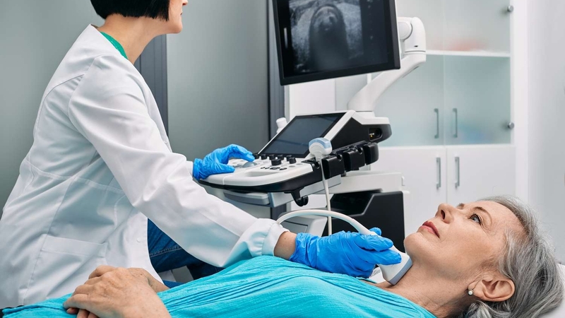
Ultrasound for cancer is one of the most versatile and accessible diagnostic tools in oncology — using sound waves instead of radiation to create real-time images of organs, masses, and blood flow. Because ultrasound involves no ionizing radiation, it is safe for all ages, safe during pregnancy, and safe to repeat as often as clinically needed. That combination of safety, speed (most scans take 5–30 minutes), widespread availability, and real-time biopsy guidance makes ultrasound for cancer a cornerstone of thyroid nodule evaluation, breast mass characterization, liver cancer surveillance, and percutaneous biopsy guidance across virtually every organ that can be imaged without bone or bowel gas in the way.
Ultrasound for cancer does not replace CT, MRI, or PET — it answers specific clinical questions its properties make it uniquely suited for: Is this thyroid nodule suspicious enough to biopsy? Is this breast lump a cyst or a solid mass? Is this liver lesion consistent with HCC? Is the needle in the right position? For a broader overview of how all imaging modalities fit together in oncology, see our guide to imaging tests for cancer.
How Ultrasound for Cancer Works
Ultrasound transmits high-frequency sound waves (2–18 MHz) from a handheld transducer into the body. Waves reflect off tissues based on their physical properties — density, stiffness, and the interface with neighboring structures. The transducer receives these reflected echoes and a computer converts them into real-time grayscale images.
The fundamental image principle:
- Fluid (cyst fluid, blood, ascites) = black (anechoic) — sound waves pass through without reflection
- Solid tissue = gray to white (echogenic) — higher density reflects more sound back
- Calcifications = bright white with a dark acoustic shadow behind them
Key ultrasound techniques in cancer:
- B-mode (grayscale): Standard real-time imaging — the primary mode for thyroid, breast, liver, and pelvic evaluation
- Doppler: Detects blood flow velocity and direction; tumors often develop abnormal neovascularity that appears as chaotic flow on color Doppler
- Elastography: Measures tissue stiffness — malignant lesions are typically stiffer than normal tissue or benign lesions; displayed as a color map
- Contrast-enhanced ultrasound (CEUS): Microbubble contrast agents injected IV — immediately characterizes vascular enhancement patterns at bedside, with no radiation
No radiation is produced at any stage. Unlike CT and PET, there is no cumulative dose concern — which is why ultrasound is the standard modality for ongoing surveillance in cirrhosis, where patients need imaging every 6 months indefinitely.
Thyroid Nodule Evaluation — ACR TI-RADS
Thyroid nodule evaluation is the most common cancer-related application of ultrasound. High-resolution thyroid ultrasound detects nodules in up to 68% of adults — yet only 5–15% are malignant. The clinical challenge is stratifying which nodules need biopsy and which can be safely monitored.
The ACR TI-RADS (Thyroid Imaging Reporting and Data System) scores thyroid nodules on five ultrasound features and places each nodule into one of five risk categories:
| TI-RADS | Description | Action |
|---|---|---|
| TR1 | Benign (score 0) | No biopsy, no follow-up needed |
| TR2 | Not suspicious (score 2) | No biopsy; follow-up if preferred |
| TR3 | Mildly suspicious (score 3) | FNA if ≥2.5 cm; follow-up US if ≥1.5 cm |
| TR4 | Moderately suspicious (score 4–6) | FNA if ≥1.5 cm; follow-up US if ≥1 cm |
| TR5 | Highly suspicious (score ≥7) | FNA if ≥1 cm; follow-up US if ≥0.5 cm |
Features that increase TI-RADS score:
- Solid composition (vs. cystic or spongiform)
- Hypoechogenicity — darker than surrounding thyroid tissue; very hypoechoic adds 3 points
- Irregular or lobulated margins
- Taller-than-wide orientation (AP diameter exceeds transverse diameter)
- Punctate echogenic foci (microcalcifications) — scores 3 points (the highest single feature); strongly associated with papillary thyroid cancer
If a thyroid nodule requires biopsy, this is typically done as an ultrasound-guided fine-needle aspiration (FNA) — a quick outpatient procedure. See our biopsy for cancer guide for what to expect during the procedure and how to interpret the Bethesda category results.
Breast Cancer — Ultrasound for Diagnosis and Dense Breast Screening
Diagnostic Breast Ultrasound
The most important thing ultrasound tells you about a breast mass is whether it is cystic (fluid-filled) or solid. Simple cysts — completely anechoic, with thin walls and posterior acoustic enhancement — are classified as BI-RADS 2 (benign) and require no further workup. Complicated cysts and all solid masses require full characterization.
BI-RADS ultrasound features associated with malignancy:
- Irregular shape (not round or oval)
- Taller-than-wide orientation — more AP height than width in a transverse image
- Spiculated or angulated margins (instead of smooth, circumscribed)
- Marked hypoechogenicity
- Posterior acoustic shadowing
- Calcifications visible within the mass on ultrasound
A BI-RADS 4 or 5 mass → ultrasound-guided core needle biopsy. Most breast biopsies today are done under ultrasound guidance — real-time visualization allows the radiologist to confirm needle position within the mass before firing the biopsy device. Ultrasound also evaluates axillary lymph nodes at the same visit — suspicious nodes (cortical thickening >3 mm, absent fatty hilum) can be sampled with FNA at the time of the breast evaluation.
Supplemental Ultrasound Screening in Dense Breasts
Women with heterogeneously dense (BI-RADS C) or extremely dense (BI-RADS D) breast tissue face two problems: mammography misses roughly 50% of cancers in dense breasts, and dense breast tissue is itself an independent breast cancer risk factor.
The ACRIN 6666 trial (JAMA 2012) — 2,659 high-risk women with dense breasts — found supplemental ultrasound detected an additional 4.2 cancers per 1,000 women compared to mammography alone. However, the false-positive recall rate was substantially higher, leading to nearly twice as many unnecessary biopsies.
Liver — Ultrasound for HCC Surveillance in Cirrhosis
All patients with liver cirrhosis — from any cause — face substantially elevated HCC risk. The AASLD and EASL recommend semiannual liver ultrasound (± AFP) for HCC surveillance in all cirrhotic patients.
What surveillance ultrasound looks for: a new hepatic lesion not present on prior scan, growth of a previously identified lesion, or a new vascular abnormality. Any new finding → contrast CT or MRI with hepatobiliary contrast for LI-RADS characterization.
Sensitivity limitations: Ultrasound sensitivity for HCC in cirrhosis ranges from 58–89% — with sensitivity substantially lower for lesions ≤2 cm in the nodular cirrhotic background. This limitation is unavoidable, which is why any abnormal finding on surveillance ultrasound prompts immediate cross-sectional imaging rather than watchful waiting.
CEUS for immediate bedside characterization: When a new lesion appears on grayscale ultrasound, CEUS can characterize it on the spot — arterial phase hyperenhancement (APHE) + marked washout = CEUS LI-RADS-5 = high probability HCC. CEUS is particularly useful in patients with poor renal function where CT contrast is risky. AFP monitoring alongside surveillance ultrasound is described in our guide to cancer blood tests.
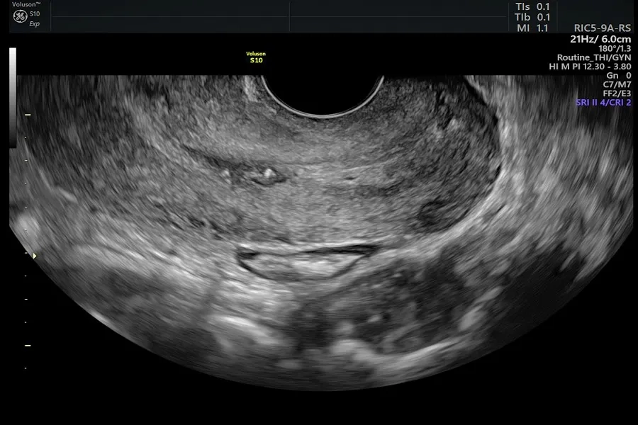
Ovarian and Pelvic Cancer — Transvaginal Ultrasound
Transvaginal ultrasound (TVUS) is the primary first-line imaging tool for evaluating adnexal masses and pelvic symptoms that may indicate ovarian cancer.
Features on TVUS associated with malignancy:
- Solid components within an otherwise cystic mass
- Papillary projections (internal wall excrescences)
- Thick irregular septations (>3 mm)
- Bilateral adnexal masses
- Ascites (free peritoneal fluid)
- Doppler showing low-resistance arterial flow (neovascularity)
The ACR O-RADS (Ovarian-Adnexal Reporting and Data System) 2022 standardizes adnexal mass reporting with a 1–5 risk classification, similar to TI-RADS and BI-RADS. Simple cysts in premenopausal women carry very low malignancy risk even up to 5–10 cm — annual follow-up is appropriate rather than immediate surgery.
Ultrasound-Guided Biopsy — Real-Time Needle Guidance
Ultrasound is the most widely used imaging modality for guiding percutaneous biopsy — providing real-time, portable needle visualization across multiple organ sites:
- Thyroid FNA: 25–27G needle; ultrasound guidance markedly improves diagnostic yield vs. palpation-guided sampling. Rapid on-site evaluation (ROSE) by cytopathologist during the procedure reduces inadequate samples.
- Breast core needle biopsy: 14G automated core needle; ultrasound provides real-time needle visualization within the mass. 3–6 cores typically obtained per lesion. Avoids open surgical biopsy for most breast lesions.
- Liver biopsy: 16–18G core needle; ultrasound confirms needle position in real time during the biopsy.
- Lymph node FNA: Direct sampling of suspicious cervical, axillary, or inguinal nodes under ultrasound guidance.
- Renal mass biopsy: For ambiguous renal masses where biopsy would determine management and potentially avoid nephrectomy for a benign lesion.
For a complete guide to biopsy types, what pathology does with the tissue, and how to interpret Bethesda/BI-RADS/other pathology categories, see our biopsy for cancer overview.
Limitations of Ultrasound for Cancer
- Operator-dependent: Image quality varies more between operators in ultrasound than in any other imaging modality — inter-observer variability is a real clinical limitation
- Cannot penetrate bone or bowel gas: The pancreas is the most common casualty — bowel gas frequently obscures it; CT is the preferred modality for pancreatic cancer
- Limited in obese patients: Sound waves attenuate with depth; image quality degrades significantly with thick abdominal walls
- Not useful for lung cancer: Air-filled lungs are essentially invisible to ultrasound
- No whole-body staging: Cannot stage most solid tumors — CT or PET-CT required for comprehensive staging
- HCC sensitivity: The nodular cirrhotic background limits sensitivity for small HCC — a critical limitation given that surveillance is most valuable for detecting early-stage disease
If you are experiencing symptoms that may indicate cancer but imaging has been inconclusive, our cancer symptoms checklist can help you organize what to discuss with your provider about next diagnostic steps.
Frequently Asked Questions
Can ultrasound detect cancer?
Yes, in specific locations. Ultrasound is effective for detecting thyroid nodules, breast masses, liver lesions (especially in cirrhosis surveillance), ovarian masses, and suspicious lymphadenopathy. It is not useful for lung cancer and has limited sensitivity for pancreatic cancer. According to the National Cancer Institute, ultrasound is particularly useful for distinguishing solid from fluid-filled masses and for guiding biopsies of suspicious lesions. Ultrasound detects suspicious findings — final cancer diagnosis always requires tissue biopsy.
What cancers does ultrasound detect best?
Ultrasound is most valuable for thyroid cancer evaluation (TI-RADS nodule scoring), breast cancer diagnosis and supplemental screening in dense breasts, hepatocellular carcinoma surveillance in cirrhosis, and ovarian cancer risk evaluation via transvaginal ultrasound. It is also the primary guidance tool for biopsies of the thyroid, breast, liver, and lymph nodes. For staging most solid tumors and evaluating lung cancer, CT or PET-CT is used. See the American Cancer Society ultrasound overview for additional cancer applications.
Is ultrasound good enough to rule out cancer?
Not for most situations. A normal thyroid ultrasound is reassuring for most nodule types but not conclusive for all. A negative liver ultrasound does not rule out small HCC in a cirrhotic patient — sensitivity for lesions <2 cm is 50–60%. A normal pelvic ultrasound in a woman with elevated CA-125 may not exclude ovarian cancer. Ultrasound typically raises or lowers suspicion rather than definitively ruling cancer in or out. Additional imaging (CT, MRI) or biopsy is often needed to reach a definitive answer.
What does cancer look like on an ultrasound?
Malignant lesions typically appear as irregular, hypoechoic (darker than surrounding tissue) solid masses with ill-defined, spiculated, or angulated margins. Thyroid cancers often show microcalcifications (bright punctate foci) and taller-than-wide shape. Breast cancers are often non-parallel with posterior acoustic shadowing. Liver HCC may show arterial hyperenhancement on CEUS. However, appearance alone is never diagnostic — many benign lesions look suspicious and some cancers appear benign. Biopsy is required for definitive diagnosis in virtually all cases.
Can I use ultrasound instead of mammography for breast cancer?
No — ultrasound is not recommended as a mammography replacement for average-risk women. Ultrasound misses calcification-only cancers (DCIS presenting as microcalcifications is invisible on ultrasound), and has a substantially higher false-positive rate. Ultrasound is used as a supplemental tool alongside mammography in women with dense breast tissue, or as a diagnostic tool when mammography finds something suspicious. High-risk women (>20% lifetime risk) should receive annual breast MRI in addition to mammography — not ultrasound as the primary supplemental modality.
How often should I have liver ultrasound if I have cirrhosis?
The AASLD and EASL recommend semiannual (every 6 months) liver ultrasound for all patients with cirrhosis — regardless of the cause of liver disease — for HCC surveillance. AFP testing alongside each ultrasound visit is recommended by most guidelines. If a new lesion is found, CT or MRI with contrast is the next step for LI-RADS characterization. If you are managing cirrhosis and concerned about liver cancer risk, ensuring this surveillance schedule is in your care plan is an important topic for your regular cancer checkup.
Sources & Further Reading
- NCI — Ultrasound for Cancer Fact Sheet
- ACR — TI-RADS Thyroid Nodule Scoring System
- American Cancer Society — Ultrasound for Cancer
- Tessler FN et al. — ACR TI-RADS White Paper, JACR 2017
- Berg WA et al. — ACRIN 6666 Trial (supplemental ultrasound in dense breasts), JAMA 2012
- Jacobs IJ et al. — UKCTOCS Trial (ovarian cancer screening), Lancet 2016
- AASLD Practice Guidance — HCC Surveillance 2023
- ACR O-RADS Ultrasound 2022
This article is for educational purposes only and does not constitute medical advice. Ultrasound recommendations and results should be discussed with your physician or radiologist.