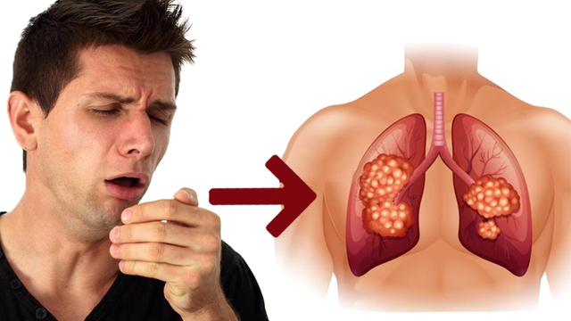
A persistent cough is the most common presenting symptom of lung cancer — present in 50 to 75% of patients at the time of diagnosis. Yet most persistent coughs are benign, caused by post-nasal drip, acid reflux, asthma, or the chronic bronchitis of long-term smoking. The challenge is knowing when a cough is just a cough, and when it may signal something that requires urgent investigation.
Lung cancer cough carries no single defining characteristic that sets it apart from benign causes — it cannot be identified by sound or feel alone. What distinguishes it is context: who has it, how long it has persisted, whether it has changed in character, and what accompanies it. Studies consistently show an average delay of three to six months between the onset of respiratory symptoms and a lung cancer diagnosis, much of it driven by patients attributing a new cough to smoking, a recent infection, or acid reflux — and waiting to see if it resolves.
This guide explains how lung cancer causes cough, what features are worrying versus reassuring, when blood in the sputum requires urgent evaluation, how doctors investigate a suspicious cough, and how cough is managed when lung cancer is the diagnosis.
How Lung Cancer Causes a Cough
Not all lung tumors cause cough equally. The lung’s parenchyma — the air-exchanging tissue — contains no cough receptors, so tumors growing there are silent. Cough only arises when the cancer reaches or affects structures with cough receptors: the bronchial and tracheal epithelium, the pleura, or adjacent structures.
Central / Endobronchial Tumor
Squamous cell carcinoma and small cell lung cancer typically arise near central airways. These tumors directly irritate bronchial mucosa, stimulating vagal cough receptors. Result: a persistent, often dry cough that may precede other symptoms by months.
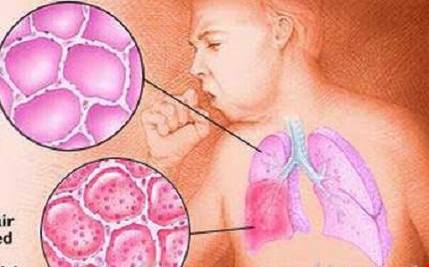
Post-Obstructive Pneumonia
A growing tumor partially or completely blocking a bronchus causes secretions to accumulate and become infected. Result: a productive, purulent cough clinically identical to pneumonia. The clue: recurrence in the same lobe, or failure to clear after antibiotics.
Pleural Effusion
Tumor invasion of the pleura causes fluid accumulation that compresses the lung and lower airways. This produces cough combined with dyspnea — often the patient’s most disabling symptom pair.
Laryngeal Nerve Involvement
The left recurrent laryngeal nerve loops under the aortic arch. Tumor or lymph node compression causes hoarseness — and laryngeal area irritation may also contribute to cough in patients with mediastinal involvement.
What Does a Lung Cancer Cough Look Like?
There is no single sound or sensation that identifies a lung cancer cough — it may be dry or productive, harsh or soft, continuous or paroxysmal. What distinguishes it is clinical context. The following features raise concern and warrant prompt evaluation:
Duration ≥3 Weeks (Smoker ≥40)
Any new or changed cough lasting 3 or more weeks in a current or former smoker aged 40 or older warrants chest imaging within 2 weeks per NICE NG12 and similar guidelines.
Change in Character
A long-term smoker who notices their chronic cough has become more frequent, more severe, or qualitatively different from baseline should not wait — this is the most important red flag in this population.
Progressive Worsening
Benign causes typically plateau or improve. A cough that continuously worsens over weeks to months with no other explanation warrants investigation.
Accompanying Hemoptysis
Any amount of blood in sputum in a smoker ≥40 requires urgent same-week evaluation. Do not wait to see if a single episode resolves on its own.
Accompanying Weight Loss
New cough + unexplained weight loss (>5% body weight) in a smoker is a high-yield pattern for urgent chest imaging in primary care.
Accompanying Hoarseness
New hoarseness lasting more than 3 weeks alongside cough suggests left recurrent laryngeal nerve compression — indicating at minimum stage III disease. Requires urgent imaging.
Unilateral Wheeze
A fixed, monophonic wheeze affecting only one side that does not respond to bronchodilators — unlike asthma — suggests partial airway obstruction by an endobronchial tumor.
Productive Cough Not Clearing with Antibiotics
Post-obstructive pneumonia from a tumor-obstructed bronchus will not fully clear with antibiotics alone. Persistent or recurrent consolidation in the same location requires CT chest.
Hemoptysis — When Lung Cancer Cough Produces Blood
Hemoptysis in a Smoker ≥40 Is a Red Flag — No Safe Amount to Ignore
Hemoptysis (coughing up blood) occurs in 20 to 30% of lung cancer patients at diagnosis. In a current or former smoker aged 40 or older, any amount of blood in sputum demands same-week evaluation — even a single episode of blood-streaked mucus.
Types: Blood-streaked sputum (most common; from mucosal tumor irritation) → frank hemoptysis (pure bright-red blood) → rust-colored sputum (older blood) → massive hemoptysis (>300–600ml/24h; ~5% of lung cancer hemoptysis cases; life-threatening emergency from tumor erosion into a large vessel).
What to do: Blood-streaked sputum → call your doctor for same-day or next-day evaluation. Frank hemoptysis → emergency department immediately. Massive hemoptysis → call emergency services; lie with the suspected bleeding side down to protect the other lung.
Not only lung cancer: Other causes include pulmonary tuberculosis, bronchiectasis, pulmonary embolism, lung abscess, anticoagulation therapy, and AVM. Full evaluation rules these out — but lung cancer must always be on the differential in any smoker ≥40.
Most Coughs Are NOT Lung Cancer — The Differential Diagnosis
The vast majority of persistent coughs — even in smokers — are benign. Understanding common benign causes helps calibrate evaluation while not missing warning signs.
Upper Airway Cough Syndrome (Post-Nasal Drip)
The most common cause of chronic cough. Mucus from sinuses/nasopharynx drains to the back of the throat. Sensation of mucus, nasal discharge, frequent throat-clearing. Often responds to antihistamine/decongestant treatment.
Asthma / Cough-Variant Asthma
Episodic cough; worse at night, with exercise or cold air. Responds to bronchodilators. In cough-variant asthma, cough is the dominant symptom without obvious wheeze. Spirometry and methacholine challenge are diagnostic.
GERD (Acid Reflux)
Acid reaching the upper esophagus/larynx stimulates cough reflex. Often occurs without heartburn (silent GERD). Worse after meals and lying flat. Empirical PPI trial for 4–8 weeks is both diagnostic and therapeutic.
ACE Inhibitor Cough
Approximately 5–15% of patients on ACE inhibitor medications develop a dry, persistent cough. Starts weeks to months after beginning the drug; resolves within 1–4 weeks of stopping. Switching to an ARB eliminates the cough.
Chronic Bronchitis / COPD
Productive cough in smokers is nearly universal in chronic bronchitis. The key warning sign is a change in the baseline cough — more frequent, different quality, or newly associated with weight loss or hemoptysis.
Post-Viral Cough
Persists 3–8 weeks or more after an acute respiratory infection. Benign; resolves spontaneously. Reassurance is appropriate if no worrying features accompany it, particularly in non-smokers under 40.
When to See a Doctor About a Cough
Seek Evaluation — Don’t Wait to See If It Resolves
- Smoker or ex-smoker aged 40+ with a persistent cough of 3 weeks or more → chest X-ray within 2 weeks
- Any hemoptysis in a smoker aged 40+ → same-week evaluation regardless of amount
- Cough + unexplained weight loss → prompt evaluation even if the cough has been brief
- Cough + new hoarseness lasting more than 3 weeks → imaging required
- Cough + unilateral fixed wheeze → endobronchial lesion must be excluded
- Productive cough failing antibiotics, or recurrent pneumonia in the same lobe → CT chest required
- Never-smoker aged 40+ with unexplained cough ≥8 weeks → evaluate (15% of lung cancers occur in never-smokers)
How Cough Leads to Lung Cancer Diagnosis
Chest X-ray: Standard first-line investigation. Misses approximately 25 to 40% of lung cancers. When abnormal, urgent CT is ordered. When normal but suspicion remains high (especially with hemoptysis), CT should still be obtained.
CT chest: Far more sensitive than chest X-ray. Identifies the primary tumor, lymph node involvement, and pleural effusion. For detail on what LDCT screening involves, see our guide on low dose CT lung cancer screening.
Bronchoscopy: For central tumors or suspected endobronchial lesions — particularly when hemoptysis is the presenting symptom. Allows direct visualization with biopsy, brushings, and lavage for cytology. Navigational bronchoscopy extends reach to peripheral lesions.
CT-guided biopsy: For peripheral lesions not accessible by bronchoscopy. Provides tissue for histology and molecular testing (EGFR, ALK, ROS1, PD-L1) that determines targeted therapy eligibility.
PET/CT: Used for staging once a histological diagnosis is confirmed. Identifies mediastinal lymph node involvement and distant metastases. Informs treatment planning.
Managing Cough When Lung Cancer Is Diagnosed
Cough management in lung cancer combines treating the underlying cancer (which often reduces cough substantially) with symptomatic relief when cancer-directed treatment is not immediately sufficient.
Cancer-Directed Treatment
Surgery, chemotherapy, targeted therapy, and radiation all reduce cough by reducing tumor volume. External beam radiation targeting an endobronchial tumor is particularly effective for palliating cough in non-surgical candidates.
Endobronchial Interventions
Laser ablation, cryotherapy, and airway stent placement can physically open obstructed airways — rapidly relieving cough from post-obstructive pneumonia and dyspnea from proximal airway compromise.
Opioid Antitussives
Most effective for refractory cancer-associated cough. Codeine 15–30mg every 4–6 hours, or low-dose oral morphine (5–10mg every 4 hours), acts centrally to suppress the cough reflex. Used in palliative and supportive care.
Other Medications
Gabapentin (modulates cough sensitivity), systemic corticosteroids (for lymphangitic carcinomatosis), dextromethorphan (OTC; less effective than opioids), and nebulized lidocaine (specialist use; variable efficacy).
Pleural Drainage
For pleural effusion causing cough and dyspnea, thoracentesis or an indwelling pleural catheter often produces rapid symptom relief — sometimes within hours of drainage.
Non-Pharmacological
Elevate head of bed 30°, room humidification, avoid irritants (smoke, cold air, strong perfumes), and speech/language therapy cough suppression techniques — all with supportive evidence in chronic cough management.
Frequently Asked Questions
How is a lung cancer cough different from a regular cough?
There is no single distinctive sound or quality. What distinguishes it is context: it typically appears in a current or former smoker over 40, persists for 3 or more weeks, progressively worsens, and may be accompanied by hemoptysis, weight loss, or hoarseness. A benign cough in a smoker may be their established chronic bronchitis — the key is a change in the baseline, not merely the presence of cough.
Can I have lung cancer without a cough?
Yes. About 25 to 50% of lung cancer patients present without cough, particularly those with peripheral adenocarcinoma — the most common subtype today — where the tumor grows in the outer lung tissue, far from cough-sensitive bronchi. For more on symptoms beyond cough, see our lung cancer symptoms guide.
What does it mean if I cough up blood?
Hemoptysis — any amount of blood in sputum — is a red-flag symptom in a current or former smoker over 40. It demands same-week evaluation regardless of the amount. It does not automatically mean lung cancer — bronchiectasis, TB, and PE are also causes — but lung cancer must always be evaluated urgently. Do not wait to see if a single episode resolves on its own.
I’ve had a cough for 2 months and my chest X-ray was normal. Is that reassuring?
A normal chest X-ray is not a definitive all-clear. Approximately 25 to 40% of lung cancers are not visible on plain X-ray. If you are a current or former smoker over 40 with a 2-month cough and a normal X-ray, speak to your physician about whether CT chest imaging is appropriate. If hemoptysis is also present, CT is indicated regardless of X-ray results.
Can quitting smoking reduce a lung cancer cough?
If you currently smoke and have been diagnosed with lung cancer, quitting remains strongly beneficial — it improves surgical outcomes, enhances treatment efficacy, and improves overall survival. The cough itself is caused by the tumor, but reducing airway inflammation from ongoing smoking may reduce the overall cough burden. Understand the full relationship between lung cancer and smoking — quitting is one of the most impactful decisions in this situation.
Sources
- NICE NG12 (2023) — Suspected cancer: recognition and referral
- Molassiotis A et al. — Validation of the Manchester Cough in Lung Cancer scale; J Pain Symptom Manage 2011;42(4):588–598
- Lorenz J — Hemoptysis: clinical aspects and treatment; Dtsch Arztebl Int 2017;114(21):371–381
- Irwin RS et al. — ACCP guidelines on chronic cough; Chest 2006;129(1 Suppl):1S–23S
- American Cancer Society — Lung cancer signs and symptoms
- NCI SEER Program — Lung and bronchus cancer statistics
Related reading: Lung cancer symptoms | Lung cancer and smoking | Lung cancer screening — who qualifies