Liver cancer is one of the few cancers where prevention is genuinely within reach for a large proportion of cases. The majority of primary liver cancer worldwide is caused by chronic hepatitis B and C infections — diseases for which highly effective vaccines and cures now exist. Yet liver cancer remains one of the leading causes of cancer death globally, and its incidence is rising in Western countries driven by the epidemic of fatty liver disease. Understanding how liver cancer develops, who is at highest risk, and what treatments are available helps clarify both why early detection matters and how much of this disease could be prevented.
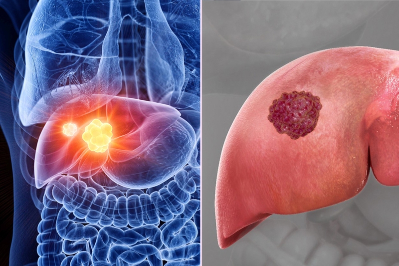
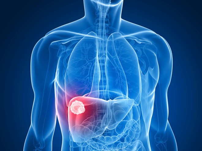
The term “liver cancer” most commonly refers to hepatocellular carcinoma (HCC), the type that originates in the liver’s own cells and accounts for approximately 90 percent of primary liver cancers. HCC is distinct from metastatic liver cancer — cancer that has spread to the liver from another organ, such as the colon, breast, or lung — which is actually far more common than primary liver cancer but is categorized by its site of origin, not the liver. This article focuses on HCC and other primary liver cancers: the diseases that begin in the liver and are treated as liver cancer.
Types of Primary Liver Cancer
Primary liver cancer arises in the cells of the liver itself. There are several types, but hepatocellular carcinoma dominates the clinical picture.
Hepatocellular carcinoma (HCC) accounts for approximately 85 to 90 percent of all primary liver cancers. It develops from hepatocytes, the main functional cells of the liver. HCC almost always occurs in livers that have been damaged by chronic disease — cirrhosis from hepatitis B, hepatitis C, alcohol, fatty liver disease, or hereditary metabolic disorders. Hepatitis B is unique in its ability to cause HCC even in the absence of cirrhosis.
Intrahepatic cholangiocarcinoma (iCCA) arises from the bile duct cells within the liver and accounts for approximately 10 to 15 percent of primary liver cancers. It is biologically and clinically distinct from HCC, with different risk factors, imaging characteristics, and treatment approaches. Unlike HCC, iCCA is not typically cirrhosis-related; its risk factors include primary sclerosing cholangitis and liver fluke infection in endemic regions.
Fibrolamellar HCC is a variant that occurs in younger patients without underlying liver disease. It has a distinctive histologic appearance and generally better prognosis than conventional HCC, though advanced disease remains difficult to treat.
Risk Factors for Liver Cancer
The risk factors for hepatocellular carcinoma divide broadly into infectious, metabolic, toxic, and genetic categories. Most cases are traceable to a relatively small number of causes, many of which are modifiable.
Chronic hepatitis B virus (HBV) infection is the single largest cause of HCC globally, accounting for approximately 50 to 55 percent of all cases worldwide. HBV integrates viral DNA into the hepatocyte genome, directly causing genetic alterations that promote malignant transformation — a mechanism that can drive HCC development even in the absence of cirrhosis. This is a critical distinction: HBV is the only major HCC risk factor that does not require established cirrhosis. Globally, HBV-related HCC is most prevalent in sub-Saharan Africa and East Asia, where perinatal and childhood HBV transmission was common before vaccination programs.
Chronic hepatitis C virus (HCV) infection is the leading cause of HCC in the United States and Europe. Unlike HBV, HCV causes HCC almost exclusively through cirrhosis — decades of inflammation and regeneration that create the fertile ground for malignant transformation. Approximately 1 to 5 percent of patients with HCV-related cirrhosis develop HCC each year.
Liver cirrhosis from any cause is the most important risk factor for HCC in the developed world. Approximately 80 to 90 percent of HCC occurs in cirrhotic livers, regardless of the cause of cirrhosis. The constant cycle of hepatocyte death and regeneration, combined with chronic inflammation and oxidative stress, creates conditions in which genetic mutations accumulate and malignant clones emerge. Common causes include alcohol-related liver disease, metabolic dysfunction-associated steatohepatitis (MASH), hereditary hemochromatosis, primary biliary cholangitis, and Wilson’s disease.
Non-alcoholic fatty liver disease (NAFLD) and MASH represent the fastest-growing cause of HCC in Western countries. As obesity and type 2 diabetes have become epidemic, fatty liver disease and its progressive inflammatory form have surged. MASH-related HCC is particularly concerning because it can develop in patients without established cirrhosis — a subset who may not be enrolled in surveillance programs and may present with more advanced tumors.
Alcohol-related liver disease causes cirrhosis that predisposes to HCC. Long-term heavy alcohol consumption accelerates liver damage from any cause: patients with both HCV infection and heavy alcohol use have dramatically higher HCC risk than either factor alone.
Aflatoxin B1 is a mycotoxin produced by Aspergillus mold that grows on improperly stored grain, corn, and peanuts in warm, humid climates. It is a potent liver carcinogen and a major cause of HCC in parts of sub-Saharan Africa and Southeast Asia, where it acts synergistically with HBV to dramatically increase HCC risk.
Male sex and older age are independent risk factors. HCC occurs 3 to 4 times more frequently in men than women. Hereditary conditions that cause chronic liver disease — hereditary hemochromatosis, alpha-1 antitrypsin deficiency, Wilson’s disease — also predispose to HCC through cirrhosis. Diabetes and obesity independently increase HCC risk beyond their role in causing fatty liver disease. Understanding how these modifiable risk factors compare to inherited predispositions helps with personalized prevention — much as understanding ovarian cancer risk factors helps individuals stratify their individual risk profile.
~90% of primary liver cancers are hepatocellular carcinoma (HCC). 80–90% of HCC occurs in cirrhotic livers. Only ~20% 5-year overall survival, but up to 70–80% for patients who receive liver transplantation within Milan criteria. Hepatitis B vaccination is the most effective known intervention to prevent a major solid tumor cancer.
Symptoms of Liver Cancer
One of the greatest clinical challenges with liver cancer is that it rarely causes symptoms in early stages. HCC frequently develops in a background of cirrhosis, and patients with cirrhosis often have baseline symptoms — fatigue, mild abdominal discomfort, occasional swelling — that can mask the emergence of a tumor. This is why surveillance programs exist: to detect liver cancer before it causes symptoms, when curative options are still available.
Symptoms that develop with more advanced disease include:
- Right upper quadrant pain or pressure: The liver is located under the right rib cage. As a tumor grows, it can cause dull aching or pressure in the upper right abdomen, or referred pain to the right shoulder blade.
- Abdominal swelling (ascites): A growing tumor can worsen portal hypertension in a cirrhotic liver, causing new or worsening fluid accumulation in the abdomen.
- Unexplained weight loss and loss of appetite: Common in many cancers; in liver cancer these occur both from the metabolic demands of the tumor and from impaired liver function.
- Jaundice: Yellow discoloration of the skin and whites of the eyes, indicating that bilirubin is not being processed effectively. In liver cancer, jaundice is typically a late finding, either from direct bile duct invasion by the tumor or from severe liver decompensation.
- Fatigue and weakness: General malaise that can be difficult to distinguish from the fatigue that often accompanies underlying cirrhosis.
- New decompensation in a previously stable cirrhotic patient: One of the most clinically important patterns — a patient with well-controlled cirrhosis who suddenly develops new ascites, hepatic encephalopathy (confusion from liver failure), or a variceal bleed may be experiencing these as the first sign of HCC disrupting previously compensated liver function.
Paraneoplastic syndromes are rare but recognizable: some HCC tumors produce hormones or hormone-like substances that cause hypoglycemia (the tumor consumes glucose), polycythemia (elevated red blood cell count from erythropoietin production), or hypercalcemia. These systemic manifestations are uncommon but occasionally the first clue to an underlying liver tumor.
How Liver Cancer Is Diagnosed
For patients enrolled in surveillance programs because of cirrhosis or HBV infection, liver cancer is often detected at an early stage before symptoms develop. The surveillance protocol recommended by major hepatology societies consists of abdominal ultrasound every 6 months, with or without measurement of AFP (alpha-fetoprotein), a tumor marker produced by many HCC tumors.
CT or MRI with contrast provides the definitive imaging characterization of liver masses in cirrhotic patients. Hepatocellular carcinoma has a characteristic imaging appearance: during the arterial phase, the tumor enhances more brightly than surrounding liver tissue (arterial hyperenhancement); in the portal venous or delayed phase, the tumor loses contrast faster than normal liver parenchyma (portal venous washout). This pattern — arterial enhancement followed by washout — is so specific for HCC in a cirrhotic liver that international guidelines allow diagnosis of HCC for lesions greater than 1 cm with this pattern, without requiring biopsy.
LI-RADS (Liver Imaging Reporting and Data System) categorizes liver observations from LR-1 (definitely benign) to LR-5 (definitely HCC) based on imaging features. A LR-5 lesion in a cirrhotic patient is treated as HCC without further tissue confirmation in most clinical scenarios.
Alpha-fetoprotein (AFP) is a serum protein elevated in approximately 60 to 70 percent of HCC cases. An AFP level above 400 ng/mL in a patient with cirrhosis and an appropriate liver mass is highly supportive of HCC diagnosis. However, AFP also rises in benign liver conditions and in pregnancy, and is not sensitive enough to be used alone for screening. It is most valuable for monitoring treatment response and detecting recurrence after treatment.
Liver biopsy is performed when imaging is non-diagnostic (lesion does not meet LI-RADS 5 criteria) or when the patient does not have established cirrhosis. CT-guided or ultrasound-guided percutaneous biopsy of a liver mass is safe in experienced hands. This diagnostic approach differs from pancreatic cancer diagnosis, where tissue via EUS is nearly always required before treatment — while HCC can be managed based on imaging alone in the right clinical context.
Staging Liver Cancer — The BCLC System
Liver cancer staging is uniquely complex because it requires accounting not just for the extent of the cancer but for the underlying liver function. A patient with the same-size tumor but severely impaired liver function cannot safely undergo the same treatments as a patient with well-preserved liver function. This dual assessment is incorporated into the Barcelona Clinic Liver Cancer (BCLC) staging system, the most widely used framework for HCC globally.
The Child-Pugh score quantifies liver function using five parameters: degree of ascites, bilirubin level, albumin level, prothrombin time, and degree of hepatic encephalopathy. Total scores of 5–6 = Child-Pugh A (well-compensated), 7–9 = Child-Pugh B (significant impairment), 10–15 = Child-Pugh C (severely impaired). Only patients with Child-Pugh A or B liver function are generally candidates for active HCC treatment.
- BCLC 0 (Very Early): Single tumor smaller than 2 cm, Child-Pugh A. Curative treatment is highly effective; 5-year survival approaches 70–80%.
- BCLC A (Early): Single tumor or up to 3 tumors each 3 cm or smaller, Child-Pugh A or B. All curative treatments are considered: resection, transplantation, or ablation.
- BCLC B (Intermediate): Multinodular disease without vascular invasion or extrahepatic spread, Child-Pugh A or B. Standard treatment is TACE (transarterial chemoembolization).
- BCLC C (Advanced): Portal vein tumor invasion or extrahepatic spread. Treatment is systemic therapy.
- BCLC D (Terminal): Severely impaired liver function (Child-Pugh C). Treatment is supportive care focused on quality of life.
Treatment Options for Early-Stage Liver Cancer
For patients with early-stage HCC (BCLC 0 or A) and adequate liver function, three potentially curative treatments are available.
Surgical resection removes the portion of the liver containing the tumor with a margin of normal tissue. It is most appropriate for patients with a single tumor, preserved liver function (Child-Pugh A), and no clinically significant portal hypertension. The liver has exceptional regenerative capacity — up to 70 to 80 percent can be removed and the remnant will typically regenerate over several months. Five-year survival rates after resection of early HCC are 50 to 70 percent. The main risk is liver failure in patients with underlying cirrhosis, because the cirrhotic remnant may not regenerate adequately.
Liver transplantation is considered the optimal treatment when applicable, because it simultaneously removes the HCC and replaces the diseased liver — addressing both the cancer and its underlying cause. Transplant eligibility for HCC is governed by the Milan criteria: single tumor 5 cm or smaller, or up to 3 tumors each 3 cm or smaller, with no vascular invasion and no extrahepatic disease. Within Milan criteria, five-year post-transplant survival rates are 70 to 80 percent with HCC recurrence rates below 15 percent. Patients with HCC receive MELD exception points to increase their priority on the transplant waiting list, acknowledging that their cancer may progress while waiting.
Thermal ablation (RFA or MWA) uses heat energy to destroy tumor tissue. Radiofrequency ablation (RFA) delivers electrical current to a needle electrode within the tumor; microwave ablation (MWA) uses microwave energy to heat tumor tissue more rapidly and create larger ablation zones. Ablation is preferred for tumors 3 cm or smaller that are not surgically accessible or when the patient’s liver function makes surgery too risky. For very small HCC (under 2 cm), ablation achieves similar long-term outcomes to surgical resection with far less procedural risk.
Locoregional Treatments — TACE, TARE, and Radiation
For patients with intermediate-stage HCC (BCLC B) who have multiple tumors and preserved liver function but are not candidates for curative treatments, locoregional therapies delivered through the hepatic arteries provide tumor control and survival benefit.
TACE (transarterial chemoembolization) is the standard treatment for intermediate-stage HCC. It exploits the fact that liver tumors derive most of their blood supply from the hepatic artery rather than the portal vein — unlike normal liver parenchyma, which relies primarily on portal blood flow. An interventional radiologist advances a catheter through the hepatic arteries and delivers chemotherapy directly to tumor vessels, followed by embolization particles that block blood flow. This delivers high local chemotherapy concentration while cutting the tumor’s blood supply. TACE can provide tumor response sufficient to bring a patient within transplant criteria (downstaging) and is used as bridge therapy while awaiting transplant.
TARE (transarterial radioembolization) with Y-90 microspheres delivers radioactive yttrium-90 spheres through the same arterial route, permanently lodging in tumor vessels and delivering localized internal radiation over weeks. TARE is an increasingly used alternative to TACE, particularly for patients with limited portal vein flow.
SBRT (stereotactic body radiation therapy) delivers precise, high-dose external beam radiation to liver tumors in 3 to 5 sessions. It is used for patients who are not candidates for ablation or TACE due to tumor location, size, or vascular involvement.
Systemic Therapy for Advanced Liver Cancer
For patients with advanced-stage HCC — those with portal vein tumor invasion, lymph node involvement, or distant metastases — systemic treatment is the standard approach. This field has been transformed in recent years by immunotherapy combinations.
Atezolizumab plus bevacizumab is the current first-line standard of care for patients with advanced HCC and Child-Pugh A liver function. This combination pairs atezolizumab (an anti-PD-L1 immune checkpoint inhibitor) with bevacizumab (an anti-VEGF antibody that reduces tumor blood vessel formation). The IMbrave150 clinical trial demonstrated significantly improved overall survival compared to sorafenib alone — median overall survival of approximately 19 months vs. approximately 13 months — establishing this combination as the preferred first-line therapy. Bevacizumab carries a risk of gastrointestinal bleeding, so patients with significant esophageal or gastric varices require endoscopic evaluation before starting treatment.
Sorafenib is a multikinase inhibitor that was the first systemic therapy approved for HCC (in 2007) and was the standard of care for a decade. It inhibits tumor angiogenesis and tumor cell proliferation. It remains relevant for patients in whom bevacizumab is contraindicated due to bleeding risk.
Lenvatinib is an alternative multikinase inhibitor shown to be non-inferior to sorafenib and is also used as a first-line option. Second-line options for patients who progress include regorafenib, cabozantinib, and ramucirumab (specifically for patients with AFP above 400 ng/mL). Pembrolizumab and nivolumab have received approvals for previously treated HCC.
For updated information on liver cancer clinical trials and treatment options, the National Cancer Institute’s liver cancer page provides current trial listings and evidence summaries.
Prevention and Surveillance — Lowering Your Risk
Liver cancer is one of the most preventable major cancers because so many of its risk factors are modifiable.
Hepatitis B vaccination is the most powerful lever for preventing HCC globally. The hepatitis B vaccine is safe, effective, and universally recommended in childhood immunization schedules. Widespread vaccination in Taiwan beginning in 1984 has produced a dramatic reduction in HCC incidence in children and young adults — a model for global HCC prevention. Adults in high-risk groups who were not vaccinated in childhood should be vaccinated if not already immune.
Hepatitis C treatment has transformed HCC prevention in countries with high HCV prevalence. Direct-acting antivirals (DAAs) cure hepatitis C in greater than 95 percent of patients after 8 to 12 weeks of oral treatment, with minimal side effects. Curing HCV eliminates ongoing liver inflammation and substantially reduces HCC risk — though risk remains elevated in patients who already have cirrhosis at the time of viral clearance, who should continue surveillance.
Alcohol reduction or cessation prevents alcohol-related cirrhosis and its associated HCC risk. This is particularly important in patients with concurrent viral hepatitis, where alcohol acts synergistically to accelerate liver damage.
Management of metabolic risk factors — losing weight in the setting of NAFLD/MASH, controlling blood glucose in diabetes, and addressing metabolic syndrome — reduces hepatic fat and inflammation. Weight loss of even 5 to 10 percent body weight can meaningfully improve liver histology in MASH. The American Liver Foundation provides lifestyle resources for patients with fatty liver disease.
Surveillance programs for high-risk patients — those with cirrhosis from any cause and chronic HBV carriers meeting risk criteria — provide the best opportunity to catch liver cancer at an early, curable stage. The standard is abdominal ultrasound every 6 months. High-risk patients should discuss enrollment in a surveillance program with their gastroenterologist or hepatologist rather than waiting for symptoms to develop.
Prognosis and Survival
Liver cancer outcomes vary dramatically by stage and treatment. Overall five-year survival for all stages combined is approximately 20 percent, reflecting the fact that the majority of patients are diagnosed at advanced stages. However, within subgroups, outcomes differ strikingly.
For patients with early-stage HCC who undergo liver transplantation within Milan criteria, five-year survival approaches 70 to 80 percent. Resection of early-stage HCC in patients with well-preserved liver function achieves five-year survival rates of 50 to 70 percent. For intermediate-stage HCC treated with TACE, median survival is typically 16 to 26 months. For advanced-stage HCC treated with atezolizumab plus bevacizumab, median overall survival is approximately 19 months — a significant improvement from the 12 to 13 months achieved with sorafenib alone, representing the progress immunotherapy has brought to this disease.
Two diseases are always present simultaneously in most HCC patients: the cancer and the underlying liver disease. Prognosis depends on both. A patient may have a small, technically resectable tumor but liver function too impaired to safely undergo surgery. This integration of liver function into staging and treatment decisions is what makes the multidisciplinary liver tumor board essential for every patient with HCC.
Getting the Right Care for Liver Cancer
Liver cancer management requires a multidisciplinary team: hepatology, surgical oncology, interventional radiology, transplant medicine, and medical oncology must coordinate around each patient’s unique profile. No single specialist manages all the treatment options for HCC, and the optimal plan requires integrating decisions about transplant listing, locoregional therapy, systemic treatment, and palliative care.
Patients navigating a liver cancer diagnosis alongside other GI cancer concerns — such as context from a prior pancreatic cancer workup or concern about overlapping GI symptoms — benefit from coordinated care by a team that can review all relevant diagnoses together. For high-risk individuals who have not yet developed cancer, the most valuable step is enrollment in a formal surveillance program with a hepatologist experienced in HCC risk management.
The trajectory of liver cancer treatment has changed substantially in recent years. Immunotherapy combinations have extended survival for advanced disease. Better understanding of MASH-related HCC has expanded surveillance eligibility. And above all, the tools to prevent most hepatitis B and C-related HCC already exist. For patients and families facing this diagnosis, connecting with a specialized liver cancer program and understanding the full staging picture provides the foundation for every treatment decision that follows.
Sources: National Cancer Institute — Liver Cancer | NCCN Guidelines for Patients — Hepatocellular Carcinoma | American Association for the Study of Liver Diseases (AASLD)