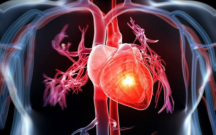
Understanding how the heart works is more than an anatomy lesson. It is the foundation for understanding why every cardiovascular health recommendation exists — why blood pressure matters, why coronary artery blockages cause heart attacks, why electrical malfunctions produce dangerous rhythm disorders, and why the lifestyle choices made in ordinary life either protect or damage the heart over decades. The heart is among the most remarkable structures in the body: a four-chambered muscular pump that beats approximately 100,000 times per day, every day, for a lifetime, without rest and without conscious direction.
The Four Chambers and What They Do
The heart is divided into four chambers arranged in two pairs, each serving a distinct function in the two-circuit cardiovascular system.
The right atrium is the receiving chamber for deoxygenated blood returning from the body. Blood arrives via two large veins: the superior vena cava, which drains the upper body (head, arms, thorax), and the inferior vena cava, which drains the lower body and abdominal organs. The right atrium serves primarily as a holding and transit chamber — it collects blood and passes it through the tricuspid valve into the right ventricle.
The right ventricle receives blood from the right atrium and pumps it through the pulmonary valve into the pulmonary arteries, which carry it to the lungs. The right ventricle operates against relatively low resistance — the pulmonary circulation is a low-pressure system — so its walls are thinner than those of the left ventricle.
The left atrium receives freshly oxygenated blood returning from the lungs through the four pulmonary veins. Like the right atrium, it serves as a receiving chamber, passing blood through the mitral valve into the left ventricle.
The left ventricle is the most muscular and powerful chamber of the heart. It must generate enough pressure to drive blood through the aorta and distribute it throughout the entire body — a vastly higher resistance circuit than the pulmonary system. The left ventricular wall is approximately three times thicker than the right ventricular wall. When doctors speak of “heart failure” in most clinical contexts, they are primarily referring to impairment of left ventricular function.
The Four Heart Valves — One-Way Doors
The heart’s four valves ensure that blood flows in only one direction through each chamber. They open and close passively in response to pressure gradients — they require no active muscle to operate.
Tricuspid valve (right atrioventricular valve): positioned between the right atrium and right ventricle; has three leaflets. Opens during diastole to allow filling of the right ventricle; closes during ventricular systole to prevent backflow into the right atrium.
Pulmonary valve (right semilunar valve): positioned at the outlet of the right ventricle into the pulmonary artery. Opens during ventricular systole to allow ejection into the pulmonary circulation; closes during diastole.
Mitral valve (left atrioventricular valve, bicuspid valve): positioned between the left atrium and left ventricle; has two leaflets supported by chordae tendineae (fibrous cords) attached to papillary muscles within the left ventricle. This structural complexity makes the mitral valve prone to regurgitation (leaking) when the left ventricle enlarges or when chordae rupture.
Aortic valve (left semilunar valve): positioned at the outlet of the left ventricle into the aorta. Opens during ventricular systole; closes during diastole, preventing blood from flowing back from the aorta into the ventricle. The aortic valve is subject to calcification with age, which can progress to aortic stenosis — a narrowing that impedes outflow and places enormous strain on the left ventricle.
The two heart sounds heard through a stethoscope correspond to valve closures. The first heart sound (S1, “lub”) represents the closure of the mitral and tricuspid valves at the beginning of ventricular systole. The second heart sound (S2, “dub”) represents the closure of the aortic and pulmonary valves at the end of ventricular systole. Abnormal heart sounds — murmurs — are produced by turbulent blood flow through stenotic or regurgitant valves.
The Journey of Blood — Step by Step
Following a single red blood cell through the complete circuit illustrates how the heart and circulation work together:
- Deoxygenated blood arrives at the right atrium via the superior and inferior vena cava, having delivered oxygen to the body’s tissues
- The right atrium contracts, pushing blood through the tricuspid valve into the right ventricle
- The right ventricle contracts, pushing blood through the pulmonary valve into the pulmonary artery
- Blood flows through the pulmonary arteries to the lungs, where capillaries surround the alveoli (air sacs). Carbon dioxide diffuses from blood to alveoli (to be exhaled); oxygen diffuses from alveoli to blood
- Oxygenated blood flows through the pulmonary veins to the left atrium
- The left atrium contracts, pushing blood through the mitral valve into the left ventricle
- The left ventricle contracts powerfully, pushing blood through the aortic valve into the aorta
- The aorta distributes blood through successively smaller arteries → arterioles → capillaries in every organ and tissue
- In capillaries, oxygen and nutrients are delivered to cells; carbon dioxide and metabolic waste products are absorbed
- Blood drains from capillaries → venules → veins → superior/inferior vena cava → right atrium, completing the circuit
At rest, this entire circuit completes approximately once per minute. During intense exercise, the circuit can complete more than four times per minute.
The Cardiac Cycle — What Happens in Each Heartbeat
Each heartbeat consists of a precisely coordinated sequence of filling and contraction.
Diastole (relaxation phase): The ventricles relax after the previous contraction. Pressure inside the ventricles falls below atrial pressure, causing the mitral and tricuspid valves to open. Blood flows passively from the atria into the ventricles — accounting for approximately 80 percent of ventricular filling.
Atrial systole (atrial contraction): Near the end of diastole, the atria contract, actively pushing the remaining 20 percent of blood into the ventricles. This atrial contribution becomes more important as people age, because ventricular filling efficiency declines with aging. This is why atrial fibrillation — which eliminates coordinated atrial contraction — can cause heart failure symptoms more readily in older patients than in younger ones.
Ventricular systole (ventricular contraction): Ventricular pressure rises sharply. When it exceeds atrial pressure, the mitral and tricuspid valves close (S1 sound). Pressure continues rising until it exceeds arterial pressure (80 mmHg diastolic in the aorta), causing the aortic and pulmonary valves to open. Blood is ejected. When the ventricles begin to relax, aortic and pulmonary valve pressure exceeds ventricular pressure, causing these valves to close (S2 sound).
Ejection fraction (EF) quantifies how effectively the left ventricle ejects blood with each beat. A normal EF is 55 percent or greater — meaning more than half the blood present in the left ventricle at the end of diastole is ejected with each beat. An EF below 40 percent indicates significantly reduced left ventricular function (heart failure with reduced ejection fraction, HFrEF) and is associated with increased mortality and the need for specific medications including ACE inhibitors, beta-blockers, and mineralocorticoid receptor antagonists.
The Electrical System — The Heart’s Own Pacemaker
The coordinated contraction sequence of the heart’s four chambers is driven by an intrinsic electrical conduction system that generates and distributes electrical impulses without input from the brain. This system has its own built-in redundancy: if the primary pacemaker fails, backup pacemakers at lower levels of the conduction system activate — though at slower rates.
The sinoatrial (SA) node, located in the upper right atrium, is the heart’s primary pacemaker. It spontaneously generates electrical impulses at a rate of 60–100 per minute in a resting adult — a rate modified by the autonomic nervous system. The SA node’s impulse spreads through the atrial muscle, causing both atria to contract simultaneously.
The impulse reaches the atrioventricular (AV) node, located at the junction between atria and ventricles. The AV node deliberately delays the impulse by approximately 0.1 seconds — a critical pause that allows the atria to complete their contraction and finish filling the ventricles before ventricular contraction begins.
From the AV node, the impulse enters the Bundle of His, a specialized conduction pathway that splits into the left bundle branch and right bundle branch. These branches rapidly distribute the impulse through the ventricular myocardium via the Purkinje fiber network, triggering near-simultaneous contraction of both ventricles for maximum pumping efficiency.
The electrocardiogram (ECG) records this electrical activity externally: the P wave represents atrial depolarization; the QRS complex represents ventricular depolarization; the T wave represents ventricular repolarization. When the electrical system malfunctions, the result is an arrhythmia. Atrial fibrillation (the most common arrhythmia) occurs when the atria fire chaotically at 400–600 times per minute, producing irregular ventricular response and creating high stroke risk from atrial blood pooling.
Cardiac Output — How Much Blood the Heart Moves
Cardiac output (CO) is the total volume of blood the heart pumps per minute:
CO = Heart Rate × Stroke Volume
At rest, a typical adult has a heart rate of 70 bpm and a stroke volume of approximately 70 mL, producing a cardiac output of approximately 5 liters per minute. During maximal exercise, a fit adult can achieve a cardiac output of 20 or more liters per minute.
Stroke volume is determined by three physiological variables: preload (the degree of ventricular filling before contraction; more filling within physiological limits means more forceful contraction — the Frank-Starling mechanism); afterload (the resistance the ventricle must overcome; high blood pressure increases afterload); and contractility (the intrinsic force of myocardial contraction, increased by sympathetic activation and decreased by heart failure).
Regular aerobic exercise increases stroke volume — which is why aerobically fit adults have lower resting heart rates (50–60 bpm) than sedentary adults (70–80 bpm) at equivalent cardiac outputs.
Coronary Circulation — The Heart Feeds Itself
The heart muscle (myocardium) cannot absorb oxygen from the blood inside its chambers — it depends entirely on the coronary arteries, which branch from the aorta immediately above the aortic valve.
The left coronary artery (LCA) branches into two major vessels: the left anterior descending artery (LAD), which supplies the front and apex of the heart (including the anterior left ventricle, the ventricular septum, and the bundle branches), and the left circumflex artery (LCx), which supplies the lateral and posterior left ventricle.
The right coronary artery (RCA) supplies the right ventricle, the inferior portion of the left ventricle, and (in most individuals) the SA and AV nodes — which explains why RCA blockage can cause heart block or bradycardia (abnormally slow heart rate).
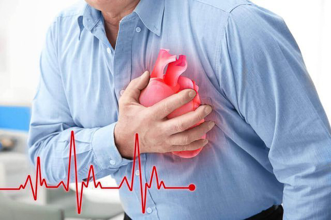
Coronary blood flow primarily occurs during diastole. This matters clinically: very fast heart rates shorten diastolic time and can impair coronary perfusion, provoking angina in patients with coronary artery disease. Atherosclerosis — the accumulation of cholesterol-laden plaques — progressively narrows the coronary lumen. When a plaque ruptures and triggers clot formation that abruptly blocks a coronary artery, a heart attack (myocardial infarction) occurs.
Blood Pressure and the Vascular System
The blood vessels through which the heart distributes blood are active, structurally diverse channels essential to cardiovascular function.
Arteries carry blood under high pressure from the heart to organs. Arterial walls contain elastic tissue that stretches during systole and recoils during diastole, smoothing the pulsatile flow from the heart into a more continuous peripheral stream.
Arterioles are the primary resistance vessels — small arteries with muscular walls that regulate blood distribution to different tissues by adjusting their diameter. When arterioles constrict, total peripheral resistance rises and blood pressure increases.
Capillaries are the site of all physiological exchange between blood and tissues. Their walls are only one cell thick (endothelium), allowing oxygen, carbon dioxide, nutrients, and metabolic waste to diffuse rapidly.
Veins return blood to the heart under low pressure. Venous one-way valves — particularly prominent in the leg veins — prevent backflow and assist venous return against gravity. When these valves fail, blood pools in the legs, producing varicose veins and increasing the risk of deep vein thrombosis.
Autonomic Regulation of Heart Rate and Blood Pressure
The cardiovascular system is continuously regulated by the autonomic nervous system, which adjusts heart rate, stroke volume, and vascular tone in response to changing physiological demands.
Sympathetic nervous system (fight-or-flight): releases norepinephrine, which increases heart rate, contractility, and vasoconstriction — raising cardiac output and blood pressure. Critical during exercise, acute illness, and physiological stress.
Parasympathetic nervous system (rest-and-digest): the vagus nerve releases acetylcholine, which slows heart rate and reduces contractility. Dominant at rest; high vagal tone in aerobically fit individuals accounts for low resting heart rates.
Baroreceptors in the carotid sinus and aortic arch detect changes in blood pressure and reflexively adjust autonomic tone. When blood pressure rises, baroreceptor activation reduces sympathetic drive and increases parasympathetic tone, lowering heart rate and causing vasodilation.
The renin-angiotensin-aldosterone system (RAAS) provides long-term blood pressure regulation through fluid balance. When blood pressure or kidney perfusion falls, the kidneys release renin, triggering a cascade that produces angiotensin II (a potent vasoconstrictor) and aldosterone (which promotes sodium and water retention). ACE inhibitors and ARBs — two of the most widely prescribed cardiovascular medications — block this system to lower blood pressure and protect the heart and kidneys.
For a comprehensive overview of what can go wrong with this system, see our guide to what is cardiovascular disease. For the role of heart health monitoring in adults, see what is heart health. And for how coronary artery narrowing causes the most common form of heart disease, see our article on coronary artery disease.
Sources: American Heart Association | National Heart, Lung, and Blood Institute | CDC — Heart Disease
What Happens When the Heart’s Anatomy Goes Wrong
Each component of the heart’s anatomy — chambers, valves, electrical system, coronary arteries, myocardium — is a potential site of disease. Understanding normal structure clarifies why specific disease processes produce the clinical consequences they do.
Atherosclerosis and coronary artery disease represent the accumulation of cholesterol-laden plaques within coronary artery walls over years and decades. The process begins with endothelial injury — microscopic damage to the inner lining of blood vessels — caused by high blood pressure, elevated LDL cholesterol, smoking, blood glucose dysregulation, and chronic inflammation. LDL particles infiltrate the arterial wall at sites of injury, become oxidized, and trigger an inflammatory response. Macrophages engulf the oxidized LDL and become foam cells. Over years, these accumulations form fibrous plaques that progressively narrow the coronary lumen.
When a plaque is stable, the lumen narrows gradually, limiting blood flow during exertion before limiting it at rest. A patient with a 70 percent narrowing of the LAD may have chest pain (angina) only when walking uphill or climbing stairs. When a plaque becomes unstable — its fibrous cap thins and ruptures — the lipid core contacts circulating blood, triggering massive platelet aggregation and clot formation. Blood flow through the artery can stop within seconds, producing a myocardial infarction (heart attack). The myocardial cells supplied by that artery begin to die within 20 to 40 minutes of complete occlusion if blood flow is not restored.
Valve disease develops through several mechanisms. Aortic stenosis, the most common valvular heart disease in developed countries, typically results from calcium deposition on the valve leaflets in adults over 70 — the same atherosclerotic risk factors that cause coronary artery disease accelerate aortic valve calcification. The progressively narrowing valve opening forces the left ventricle to work harder and harder to eject blood against the obstruction. Over years, the left ventricle hypertrophies (its walls thicken), diastolic filling becomes impaired, and eventually systolic function declines. Classic symptoms — angina, syncope (fainting), and heart failure — typically appear late in the disease course, after decades of hemodynamic stress.
Mitral regurgitation (leaking of the mitral valve) is the most common valvular disease overall. When the mitral valve fails to close completely, blood leaks backward from the left ventricle into the left atrium during systole, reducing forward cardiac output and progressively dilating both the left atrium and left ventricle. Causes include mitral valve prolapse (common, often benign), ischemic heart disease (papillary muscle dysfunction from prior heart attack), and heart failure itself (dilated left ventricle distorting valve geometry).
Hypertension and left ventricular hypertrophy: When the left ventricle must chronically eject blood against elevated blood pressure (high afterload), it responds by increasing its wall thickness — the same adaptive response as skeletal muscle to resistance exercise, but in the heart it is pathological rather than beneficial. Hypertrophied ventricular walls are stiffer, fill less efficiently, and their increased muscle mass demands more oxygen from the coronary circulation — while the coronary supply per gram of myocardium decreases because the blood vessel density fails to keep pace with muscle growth. This combination — impaired filling (diastolic dysfunction) and impaired coronary perfusion — produces the syndrome of heart failure with preserved ejection fraction (HFpEF), which accounts for at least half of all heart failure in adults over 65.
Atrial fibrillation and structural heart disease: AF is not merely an electrical problem — it reflects structural remodeling of the atria. Chronically elevated left atrial pressure (from hypertension, mitral valve disease, or diastolic dysfunction) stretches and scars the atrial myocardium over years, creating conditions that promote the multiple re-entrant electrical circuits that sustain AF. Once established, AF itself causes further atrial remodeling, and the disease becomes self-perpetuating. This is why rhythm control (restoring sinus rhythm) is most effective early in the disease course, and progressively less effective after years of established AF have produced significant atrial fibrosis.
How Exercise Changes the Heart’s Anatomy and Function
Chronic aerobic exercise produces measurable structural and functional adaptations in the heart — collectively called the “athlete’s heart” — that are physiological and beneficial, in contrast to the pathological remodeling of hypertension and heart disease.
In response to sustained endurance training, the heart adapts primarily through eccentric hypertrophy: the ventricular chambers enlarge (increased chamber volume) with proportional increases in wall thickness. The result is a larger heart capable of accommodating and ejecting more blood with each beat — a larger stroke volume. This is fundamentally different from the concentric hypertrophy of hypertension, in which the walls thicken without chamber enlargement in response to the pressure burden.
The larger stroke volume of the trained heart means that at any given cardiac output requirement, the trained heart beats fewer times per minute — explaining the resting bradycardia (heart rates of 40–55 bpm) commonly seen in endurance athletes. This bradycardia is driven by increased vagal (parasympathetic) tone and intrinsic SA node adaptations. The trained heart is also more efficient: it extracts more oxygen per unit of blood flow and operates with less wall tension per unit of output at any given workload.
Resistance (strength) training produces different cardiac adaptations, primarily modest increases in wall thickness without significant chamber enlargement — more similar to the concentric pattern but within normal physiological limits. The clinical challenge is that an athlete presenting with marked cardiac enlargement and bradycardia can be difficult to distinguish from someone with pathological cardiomyopathy — a distinction that requires careful clinical evaluation, as the management implications are entirely different.
Even moderate exercise — 150 minutes per week of brisk walking — produces measurable improvements in cardiac function: lower resting heart rate, modestly improved stroke volume, reduced arterial stiffness, lower blood pressure, and favorable changes in autonomic tone. These functional benefits are observable within weeks of beginning a regular exercise program and are partially reversible within weeks of stopping — supporting the importance of sustained, lifelong physical activity rather than episodic intense training.