
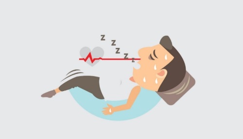
Trouble breathing when lying down — medically termed orthopnea — is one of the most clinically significant cardiac symptoms and a hallmark feature of heart failure. When the body assumes the horizontal position and breathing becomes suddenly more difficult, requiring the use of extra pillows or a reclining position to achieve comfort, the underlying cause is most often congestion in the pulmonary circulation from a failing heart. Understanding why trouble breathing when lying down occurs, which cardiac and non-cardiac conditions produce it, and what the symptoms indicate about the severity of underlying disease is important for patients, caregivers, and anyone experiencing this increasingly disruptive symptom pattern.
What Causes Trouble Breathing When Lying Down
The physiological reason that lying flat makes breathing more difficult in people with heart failure relates to the redistribution of blood volume when posture changes. In the upright position, gravity causes blood to pool in the legs and the lower body, reducing the amount of blood returning to the heart and lungs at any given moment. When a person with heart failure lies flat, this gravitational pooling is eliminated: the blood volume previously sequestered in the legs and splanchnic circulation is redistributed toward the thorax, increasing the total blood volume within the pulmonary circulation by approximately 300–500 milliliters.
In a healthy person, this redistribution is handled without difficulty — the heart increases output slightly and the lungs accommodate the modest change without impairing gas exchange. In a person with left heart failure, the left ventricle is already unable to pump all the blood returning to it efficiently, and the pulmonary circulation is already operating at elevated pressures. The additional venous return from postural change pushes the pulmonary system beyond its compensatory capacity, causing acute worsening of pulmonary congestion — excess fluid is forced from the pulmonary capillaries into the alveolar spaces, flooding the gas-exchanging units of the lung. The result is shortness of breath that develops within minutes of lying flat and is relieved within minutes of sitting upright.
Heart Failure: The Most Common Cardiac Cause
Heart failure is by far the most common cardiac cause of trouble breathing when lying down. Both heart failure with reduced ejection fraction (HFrEF — weakened heart muscle) and heart failure with preserved ejection fraction (HFpEF — stiffened heart muscle with elevated filling pressures) can produce orthopnea, because both lead to elevated left-sided filling pressures transmitted backward into the pulmonary circulation.
Clinicians grade orthopnea by the number of pillows a patient must use to sleep comfortably — “two-pillow orthopnea,” “three-pillow orthopnea” — as a simple, reproducible measure of disease severity tracked over time. A patient progressing from two to three pillows is experiencing worsening heart failure that typically requires medication adjustment or urgent review. Some patients with severe heart failure cannot lie flat at all and sleep sitting upright in a chair — the most extreme expression of orthopnea. Weight gain of two to three kilograms over 24–48 hours from fluid retention often precedes worsening orthopnea and is one of the earliest indicators of acute decompensation, providing a window for pre-emptive diuretic adjustment before hospitalization becomes necessary.
Orthopnea vs. Paroxysmal Nocturnal Dyspnea
Orthopnea and paroxysmal nocturnal dyspnea (PND) are related but distinct symptoms, both arising from pulmonary congestion in the supine position but differing in timing and severity. Orthopnea occurs very soon after lying flat — within minutes — and is relieved promptly by sitting upright. Paroxysmal nocturnal dyspnea occurs after the patient has already been asleep for one to two hours, awakening them with sudden, severe breathlessness — often described as a frightening sensation of suffocation — accompanied by cough and sometimes audible wheezing. The delayed onset of PND reflects the gradual nature of fluid redistribution from the peripheral tissues into the pulmonary circulation during sleep.
The wheezing component of PND — caused by peribronchial edema producing airway narrowing — has historically been called “cardiac asthma” because it is clinically indistinguishable from a nocturnal asthma attack without additional evaluation. The resolution of PND requires 15 to 30 minutes of sitting upright, slower than the resolution of orthopnea, reflecting greater pulmonary congestion. PND is generally considered a marker of more severely impaired cardiac function than orthopnea alone and should prompt prompt clinical reassessment and medication optimization.
Valve Disease and Other Cardiac Causes
Beyond heart failure from myocardial dysfunction, several structural cardiac abnormalities produce trouble breathing when lying down. Mitral stenosis — narrowing of the mitral valve — produces a fixed obstruction that raises left atrial pressure and secondarily elevates pulmonary venous pressure, causing pulmonary congestion and orthopnea even with a normally functioning left ventricle. Severe mitral regurgitation produces volume overload of the left atrium and pulmonary veins. Cardiac tamponade — pericardial fluid compressing the heart — is notably worsened in the supine position and partially relieved by sitting forward (the tripod position), leading patients with tamponade to instinctively lean forward to breathe more comfortably. Constrictive pericarditis — a rigid, scarred pericardium restricting cardiac filling — causes systemic and pulmonary venous congestion that produces orthopnea as part of a broader picture of elevated venous pressures.
Non-Cardiac Causes of Trouble Breathing When Lying Down
Not all orthopnea has a cardiac origin. Severe COPD and difficult-to-control asthma can produce positional dyspnea because the diaphragm — the primary respiratory muscle — must work against the weight of the abdominal organs when supine, a burden substantially reduced in the upright position. Patients with severe COPD often prefer the tripod position (leaning forward, arms supported) to optimize diaphragm mechanics. Severe obesity produces orthopnea through the same mechanism: abdominal fat compresses the diaphragm supine, reducing functional residual capacity (FRC) and causing hypoventilation. Large pleural effusions redistribute when supine and can compress previously functioning lung tissue. Massive ascites elevates the diaphragm substantially when supine, reducing lung volumes — this responds to ascites drainage (paracentesis). Gastroesophageal reflux with laryngeal involvement can produce cough-predominant dyspnea supine through acid-triggered laryngospasm, mimicking true orthopnea.
How Doctors Evaluate Trouble Breathing When Lying Down
The evaluation begins with a detailed history: when the symptom began, how many pillows are required, whether accompanied by PND, and what associated symptoms are present (leg swelling, fatigue, reduced exercise tolerance, cough). The physical examination looks for elevated jugular venous pressure (elevated right-sided filling pressures), bilateral fine crackles at lung bases (alveolar fluid), a displaced cardiac apex (ventricular dilation), S3 gallop (volume overload), and bilateral pitting ankle edema. BNP or NT-proBNP levels differentiate cardiac from pulmonary dyspnea — markedly elevated levels strongly support heart failure, while normal levels argue against it. Chest X-ray reveals cardiomegaly, pulmonary vascular redistribution (cephalization), Kerley B lines (thickened interlobular septa from interstitial fluid), and pleural effusions. Echocardiography provides definitive assessment of ejection fraction, valve function, and diastolic filling pressures (through E/e’ ratio).
Treatment and Day-to-Day Management
The cornerstone of treating cardiac orthopnea is decongestion — removing the excess fluid volume congesting the pulmonary circulation. Loop diuretics, particularly furosemide, dramatically increase urine output and reduce total body fluid volume. For acute decompensated heart failure with severe orthopnea, intravenous furosemide provides faster diuresis alongside vasodilators (IV nitroglycerin) that reduce cardiac filling pressures. Non-invasive positive pressure ventilation (CPAP or BiPAP) reduces the work of breathing and can reverse acute pulmonary edema without intubation in most cases.
For chronic management, the four pillars of guideline-directed medical therapy (GDMT) for heart failure — ACE inhibitors/ARBs/ARNIs (sacubitril-valsartan), beta-blockers, mineralocorticoid receptor antagonists (spironolactone), and SGLT2 inhibitors (dapagliflozin, empagliflozin) — address the underlying cardiac dysfunction rather than simply managing symptoms, reducing the risk of hospitalization and death while decreasing the severity of orthopnea and PND over months. Practical day-to-day measures include elevating the head of the bed 30–45 degrees, restricting dietary sodium to less than two grams per day to minimize fluid retention, and monitoring daily morning weight to detect early fluid accumulation before symptoms worsen.
For those who also experience waking up short of breath at night — PND at its most acute — these management strategies are similarly applicable. For broader context on breathlessness and cardiac causes, shortness of breath and heart health provides additional background. Monitoring key heart health numbers remains essential for managing cardiovascular risk over time. More information on heart failure is available from the American Heart Association, the National Heart, Lung, and Blood Institute, and the CDC.
When Trouble Breathing When Lying Down Requires Emergency Care
Most patients with chronic orthopnea from stable heart failure manage their symptoms predictably with elevated pillows and diuretic therapy — the breathlessness is uncomfortable but controlled. However, several presentations of trouble breathing when lying down represent acute decompensation requiring emergency evaluation rather than watchful management. Sudden-onset severe orthopnea — particularly in a patient who was previously able to sleep with fewer pillows — accompanied by rapidly worsening breathlessness at rest, the inability to complete a sentence without stopping for breath, or a sensation of air hunger despite being upright, suggests acute pulmonary edema requiring immediate hospital-level care.
Flash pulmonary edema — a dramatic, rapid onset of severe pulmonary congestion — is a specific acute emergency that can develop within minutes in patients with severe hypertension, acute mitral regurgitation (from papillary muscle rupture in MI), or severe ischemia causing acute diastolic dysfunction. Patients with flash pulmonary edema typically appear in extreme respiratory distress, are coughing frothy or pink-tinged sputum (from alveolar flooding), have oxygen saturations in the 80s despite breathing rapidly, and require immediate vasodilator therapy, high-dose IV diuretics, and often non-invasive positive pressure ventilation to avoid intubation. The chest X-ray in flash pulmonary edema characteristically shows a bilateral “butterfly” or “bat-wing” pattern of perihilar alveolar infiltrates. Any patient with sudden severe orthopnea plus these features should call 911 immediately rather than attempting to drive or arrange transport themselves.
Additional emergency presentations include trouble breathing when lying down accompanied by new palpitations or irregular pulse (suggesting arrhythmia complicating heart failure decompensation), accompanied by chest pain or new ECG changes (suggesting ischemia precipitating decompensation), accompanied by significantly reduced urine output despite taking diuretics (suggesting worsening renal function in the setting of cardiorenal syndrome), or accompanied by confusion or excessive somnolence (suggesting critically reduced cardiac output with impaired cerebral perfusion). The combination of orthopnea plus any of these features, or the development of orthopnea for the first time in a person without prior cardiac diagnosis, should prompt urgent medical evaluation.
Advanced and Device-Based Treatments for Refractory Orthopnea
For patients whose orthopnea persists despite optimal medical therapy — including maximum tolerated doses of all four pillars of GDMT — several advanced therapeutic options address the underlying cardiac dysfunction more directly. Cardiac resynchronization therapy (CRT) — a biventricular pacemaker that coordinates the contraction of the right and left ventricles simultaneously — is indicated in patients with heart failure, ejection fraction below 35%, and a wide QRS complex (typically left bundle branch block ≥150 ms) whose symptoms persist on maximally tolerated GDMT. CRT improves the mechanical efficiency of the failing left ventricle by restoring synchronous contraction, increasing effective stroke volume, and reducing functional mitral regurgitation — improvements that directly reduce pulmonary congestion and orthopnea. Approximately 70% of CRT recipients experience functional improvement, and many report significant reduction in their pillow count and nocturia within weeks of device activation.
The CardioMEMS implantable pulmonary artery pressure sensor is an FDA-approved device for patients with heart failure (NYHA Class III) who have been hospitalized for HF within the prior 12 months. Placed in the left pulmonary artery during a catheterization procedure, the sensor transmits daily pulmonary artery pressure measurements to the patient’s care team. Elevated PA pressures predict HF decompensation and worsening orthopnea by days to weeks before the patient develops overt symptoms, enabling pre-emptive diuretic adjustment that prevents hospitalization — a benefit demonstrated in the CHAMPION trial, which showed a 37% reduction in HF hospitalization in the CardioMEMS group. For patients with end-stage heart failure refractory to all medical and device therapies, left ventricular assist devices (LVADs) and heart transplantation represent definitive treatments that eliminate pulmonary congestion at its source by restoring adequate forward cardiac output.
Practical Sleeping Strategies for People with Orthopnea
While medical and device therapy address the underlying pathophysiology, practical adaptations in sleeping position and environment can substantially improve quality of life for patients living with chronic orthopnea. Elevating the head of the bed — using a wedge pillow, hospital-style bed adjustment, or bricks under the head end of the bed frame — provides a gradual incline that partially limits the fluid shift from the legs to the lungs compared to lying completely flat. A 30–45 degree incline is generally sufficient for mild to moderate orthopnea; patients with severe HF may require a 60–75 degree elevation or a recliner chair for adequate symptom control during sleep.
Timing of diuretic dosing can also influence nightly symptoms. Taking the morning dose of furosemide shortly after waking allows peak diuresis to occur during the day when the patient is upright and active, rather than during the night when prolonged supine positioning without diuretic activity can allow fluid to accumulate. A second smaller afternoon dose (if prescribed) helps prevent nocturnal fluid redistribution without producing nocturia that disrupts sleep at an inconvenient hour. Patients should discuss diuretic timing with their cardiologist or heart failure nurse rather than adjusting independently, as changes in diuretic timing and dose affect electrolyte balance, renal function, and blood pressure and require monitoring. Daily morning weight measurement — taken after first urination, before eating, wearing the same clothing — remains the most reliable home indicator of fluid balance changes and should prompt contact with the care team when weight increases by two or more kilograms in 24–48 hours.
Distinguishing Orthopnea from Other Causes of Nighttime Breathing Difficulty
Several other common conditions disrupt breathing during the night and can be confused with orthopnea from cardiac causes. Obstructive sleep apnea (OSA) — the most common sleep-related breathing disorder — causes repeated episodes of upper airway obstruction during sleep, producing oxygen desaturations, arousal, and gasping or choking that can awaken the patient and feel like difficulty breathing. Unlike orthopnea (which occurs immediately on lying flat and is positional) and PND (which occurs one to two hours into sleep from fluid redistribution), OSA-related breathing difficulty occurs throughout the sleep period, is associated with snoring, witnessed apneas, excessive daytime sleepiness, and morning headache, and is not relieved by sitting upright — it is relieved by CPAP therapy that splints the upper airway open.
OSA and heart failure frequently coexist — OSA is both a risk factor for and a consequence of heart failure, and the two conditions amplify each other’s severity. Central sleep apnea (CSA), a pattern where the brain periodically fails to send breathing signals during sleep, is specifically associated with advanced heart failure and produces the Cheyne–Stokes breathing pattern — cycles of progressively deeper breathing followed by temporary breathing cessation, which then restarts with gradually increasing depth. CSA in heart failure indicates significantly elevated left ventricular filling pressures and is associated with worse prognosis; treatment of the underlying heart failure (optimized GDMT, CRT where indicated) is the most effective approach to CSA in this setting. Nocturnal asthma — bronchospasm occurring during sleep — is aggravated by supine position, cool air, and circadian patterns of airway resistance, and can closely mimic cardiac asthma from PND; spirometry and bronchodilator responsiveness testing help distinguish these conditions when the clinical presentation is ambiguous.
Monitoring Progress and Knowing When to Escalate Care
For patients living with chronic heart failure and orthopnea, an effective monitoring strategy is the most reliable way to prevent acute decompensation from catching them off guard. Daily weight monitoring — the most sensitive home indicator of fluid balance — should be performed every morning after using the bathroom and before eating, in minimal clothing, on the same scale. A weight gain of more than one kilogram overnight or two kilograms or more over 48 hours indicates significant fluid retention that typically precedes worsening orthopnea and dyspnea by one to three days, providing a window to adjust diuretics and prevent hospitalization. Most heart failure programs provide patients with a written “action plan” that specifies which weight thresholds trigger a phone call to the heart failure nurse, which trigger a dose adjustment, and which trigger an emergency room visit.
Beyond weight monitoring, patients can track orthopnea progression by noting changes in pillow requirements over days to weeks. A patient who gradually needs an additional pillow over one to two weeks is experiencing a gradual accumulation of fluid that requires evaluation — this is less alarming than the acute overnight change but should not be ignored for more than a day or two before contacting the heart failure care team. Ankle and leg swelling, assessed by pressing a thumb into the skin above the ankle bone and noting how long an indentation takes to fill in, provides additional evidence of fluid retention. Symptoms of reduced cardiac output — increasing fatigue with activities that were previously well-tolerated, reduced appetite, new confusion or poor concentration — can accompany worsening orthopnea and indicate more severely impaired cardiac function that warrants urgent assessment.
Regular follow-up appointments with a cardiologist or heart failure specialist, periodic echocardiography to reassess ejection fraction and filling pressures, and adherence to the full regimen of GDMT medications provide the structural framework within which stable orthopnea is maintained and deterioration is detected early. For patients with access to implantable hemodynamic monitoring (CardioMEMS), remote pulmonary artery pressure data enables the clinical team to make proactive medication adjustments based on objective hemodynamic data rather than waiting for symptoms to develop. The combination of engaged patient self-monitoring, reliable medication adherence, and regular clinical oversight offers the best available strategy for minimizing the burden of trouble breathing when lying down on daily life and reducing the risk of life-threatening acute decompensation.
Orthopnea in Special Populations
Older adults with orthopnea face particular challenges because multiple conditions frequently overlap: heart failure, COPD, obesity, and chronic kidney disease often coexist, making the primary driver of positional breathlessness difficult to determine without systematic evaluation. In this population, diuretic-induced orthostatic hypotension — a drop in blood pressure on standing that causes dizziness and fall risk — can complicate aggressive diuretic therapy for orthopnea, requiring careful titration. Frailty further limits both the tolerance of upright sleeping positions (which can worsen back and joint pain) and the patient’s ability to self-monitor and report symptom changes reliably. Heart failure programs tailored to older adults often incorporate home health nurses, telehealth weight and symptom monitoring, and simplified medication regimens to address these barriers.
Pregnant women in the third trimester commonly experience positional breathlessness that can mimic orthopnea, arising from compression of the diaphragm by the growing uterus and compression of the inferior vena cava in the supine position — the latter causing reduced venous return and compensatory tachycardia (supine hypotensive syndrome). True cardiac orthopnea can develop during pregnancy in women with peripartum cardiomyopathy — a rare but serious condition in which the heart muscle weakens in the last month of pregnancy or in the first months after delivery, producing new-onset heart failure with orthopnea, PND, and dyspnea on exertion. Peripartum cardiomyopathy requires prompt echocardiographic evaluation and cardiac care; many cases recover full or near-full cardiac function with appropriate treatment, but the condition can be life-threatening without diagnosis and management.