
Mini-Stroke (TIA): Symptoms and Why It Matters
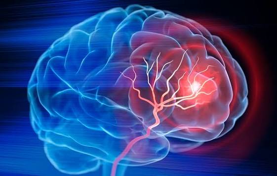
A mini-stroke — the common name for a transient ischemic attack (TIA) — is one of the most important neurological events a person can experience, not because of what it causes in the moment, but because of what it predicts: up to 10 to 15 percent of patients who have a TIA will have a major stroke within 90 days, with much of this risk concentrated in the first 48 hours after the event. The name “mini-stroke” is clinically misleading in a dangerous way — it suggests something minor and self-limited, when in reality a TIA is a neurological emergency that requires the same urgent evaluation as a completed stroke. The critical difference between TIA and ischemic stroke is not in the management urgency but in the outcome: TIA causes transient symptoms without permanent brain infarction (or only very small infarcts), while ischemic stroke produces permanent neurological deficit from irreversible infarction.
Understanding what TIA is, how to recognize its symptoms, why it demands immediate medical attention, and what evaluation and treatment it requires can prevent a major disabling stroke in a significant proportion of patients who experience a warning event — making TIA recognition among the highest-impact health knowledge an individual can possess.
What Is a TIA — The Definition That Changed Over Time
The original definition of TIA, established in the 1960s, was purely time-based: neurological symptoms of presumed vascular origin lasting less than 24 hours. This definition was established before MRI became available and before the pathophysiology of transient ischemia was well understood. It was clinically convenient but scientifically problematic, because many patients with symptoms lasting less than 24 hours had visible brain infarcts on early MRI — meaning they had completed strokes by pathological definition, even if their symptoms resolved.
The modern tissue-based definition, recommended by the American Heart Association/American Stroke Association since 2009, defines TIA as a transient episode of neurological dysfunction caused by focal brain, spinal cord, or retinal ischemia, without acute infarction. This definition shifts the diagnostic criterion from duration to imaging — a patient with symptoms lasting 4 hours but no infarct on MRI has a TIA; a patient with symptoms lasting 20 minutes but a small DWI-positive lesion has a minor ischemic stroke. Approximately 10 to 20 percent of clinically diagnosed TIAs (symptoms resolving within 24 hours) have DWI-positive lesions on MRI, representing completed micro-infarcts — these patients have higher recurrent stroke risk than DWI-negative TIA patients.
In practice, this distinction matters for risk stratification and communication: a DWI-positive patient should be counseled that they have had a small stroke, not just a warning event. But for emergency management, the distinction is secondary — all patients with transient neurological symptoms consistent with TIA require the same urgent evaluation because the symptom-onset-to-evaluation interval is the strongest predictor of identifying causative pathology (like high-grade carotid stenosis or newly detected atrial fibrillation) in time to prevent recurrence.
TIA Symptoms — Recognizing the Warning Signs
TIA produces the same neurological symptoms as ischemic stroke — the only difference is that they resolve completely (by definition) rather than persisting. This means TIA symptoms are identical to the FAST warning signs used for stroke recognition:
Face drooping: Sudden unilateral facial weakness or numbness, most noticeable when the patient smiles (one side droops) or tries to show their teeth. Facial droop in TIA/stroke reflects ischemia in the motor cortex of the opposite hemisphere or, less commonly, in the ipsilateral facial nucleus in the brainstem.
Arm weakness: Sudden weakness or heaviness in one arm (or one side of the body including both arm and leg), often accompanied by numbness. The patient cannot lift the arm normally or it drifts downward with eyes closed (pronator drift). This reflects ischemia in the corticospinal motor tracts supplying the opposite arm.
Speech difficulty: Either difficulty producing speech (expressive aphasia — the patient knows what they want to say but cannot get the words out, or words come out garbled), difficulty understanding speech (receptive aphasia — the patient cannot comprehend what is being said to them), or slurred speech (dysarthria — the words are correctly formed but articulation is impaired, often from motor weakness affecting the tongue, lips, or palate). These symptoms reflect ischemia in different language-processing regions of the dominant hemisphere (usually left).
Vision changes: Transient monocular blindness (amaurosis fugax) — sudden, temporary loss of vision in one eye, often described as a “shade coming down” over the eye — is a TIA of the retinal artery and a high-risk warning sign of internal carotid artery stenosis. Sudden loss of half the visual field in both eyes (homonymous hemianopia) reflects posterior cerebral artery territory ischemia. Double vision (diplopia) from brainstem ischemia affecting cranial nerve nuclei controlling eye movements.
Posterior circulation TIA symptoms: Ischemia in the vertebrobasilar territory produces different symptoms than anterior circulation TIA — vertigo, dizziness, ataxia (unsteady gait), diplopia, dysphagia (swallowing difficulty), dysarthria, or combinations of these. The classic teaching is that isolated vertigo alone is unlikely to be TIA, but vertigo combined with any other brainstem symptom (diplopia, facial numbness, ataxia) should be treated as TIA until proven otherwise, because posterior circulation strokes have higher mortality than anterior circulation strokes when they progress.
Symptoms NOT typical of TIA: Isolated symptoms that TIA rarely or never causes include: isolated loss of consciousness (syncope is not TIA — transient global ischemia from syncope affects both hemispheres symmetrically, without focal deficits); isolated amnesia without other focal deficits (transient global amnesia, which does cause TIA-mimicking episodes of confusion and memory loss, is not thought to be ischemic in origin in most cases); isolated vertigo without other brainstem symptoms; and slowly progressive symptoms over hours to days (TIA/stroke is almost always maximal at onset or builds over seconds to minutes, not gradually over hours). These distinguishing features help identify TIA mimics — conditions that produce transient neurological symptoms through non-vascular mechanisms.
Why TIA Is an Emergency — The 48-Hour Risk Window
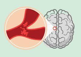
The most critical fact about TIA is that it is a neurological emergency, not a benign resolved event. The 90-day stroke risk after TIA is approximately 10 to 15 percent overall — but population studies consistently show that the risk is dramatically front-loaded: approximately half of all 90-day strokes following TIA occur within the first 48 hours of the TIA. This temporal clustering reflects the fact that the causative pathology (large artery thrombus, cardiac embolism, carotid plaque rupture) is acutely active and unstable at the time of TIA — the temporary restoration of blood flow that terminated the TIA symptoms does not resolve the underlying vascular disease that caused it.
The EXPRESS study (Early use of eXisting PREventive Strategies for Stroke) in Oxford, UK, demonstrated the magnitude of benefit from rapid TIA treatment with extraordinary clarity. In this quasi-experimental study, patients seen in a same-day TIA clinic with immediate initiation of antiplatelet therapy, statin therapy, antihypertensive therapy, and anticoagulation (for AF) experienced 80 percent fewer strokes in the 90 days after TIA compared to patients treated in the previous era with standard deferred outpatient evaluation. 80 percent — meaning that 4 out of every 5 strokes after TIA in the previous era were preventable with same-day treatment initiation. This is one of the most compelling demonstrations of the real-world impact of TIA evaluation urgency in all of preventive medicine.
The specific causes of TIA that carry the highest early recurrent stroke risk are: high-grade ipsilateral carotid stenosis (70 to 99 percent) with symptomatic amaurosis fugax or hemispheric TIA; cardioembolic TIA from atrial fibrillation; and crescendo TIA (multiple TIAs in rapid succession, each with the same territory involved), which predicts very high short-term risk of stroke from the same mechanism. These high-risk etiologies require inpatient evaluation and treatment rather than outpatient management.
TIA Evaluation — What Needs to Happen Urgently
The evaluation of TIA is directed at two goals: confirming the diagnosis (ruling out TIA mimics and determining whether MRI shows an acute infarct) and identifying the causative mechanism (which determines secondary prevention therapy). Both goals must be accomplished urgently — ideally within 24 hours of symptom onset and certainly within 48 hours:
Brain MRI with DWI: The most sensitive test for acute ischemia and the standard evaluation for TIA. Identifies small infarcts (reclassifying clinical TIA as minor stroke), characterizes infarct pattern (cortical suggests cardioembolic or large artery source; small deep infarct suggests small vessel occlusion), and detects alternative diagnoses (tumor, abscess, demyelinating disease, CAA microbleeds). If MRI is not available, CT of the head excludes hemorrhagic stroke and identifies large established infarcts but misses acute ischemic lesions.
Vascular imaging of the cervical and intracranial arteries: CTA or MRA of the head and neck identifies carotid stenosis (which requires urgent endarterectomy if high-grade and symptomatic), intracranial large vessel stenosis, vertebral artery stenosis, and dissection. The finding of high-grade ipsilateral carotid stenosis in a patient with hemispheric TIA or amaurosis fugax is one of the highest-risk TIA findings — carotid endarterectomy in this setting reduces 5-year ipsilateral stroke risk from approximately 26 percent to 9 percent, with benefit concentrated in the first weeks to months after the symptomatic event (benefit declines sharply if surgery is delayed more than 2 weeks).
Cardiac evaluation: ECG is performed in all TIA patients to identify atrial fibrillation. Prolonged cardiac monitoring (24 to 72 hours of telemetry as inpatient, or 30-day to long-term ambulatory monitoring as outpatient) detects paroxysmal AF that is missed on a single ECG — studies show that up to 20 percent of cryptogenic TIA patients have AF detected by prolonged monitoring. Echocardiography evaluates for structural cardiac sources of embolism (left atrial thrombus, valvular disease, intracardiac masses) and is performed in most TIA patients, particularly those with cardioembolic clinical features.
Laboratory evaluation: Blood glucose (TIA mimic exclusion), complete blood count (polycythemia and thrombocytopenia both affect stroke risk), lipid panel, coagulation studies, and additional hypercoagulable workup in younger patients or those with recurrent events without identified cause. HbA1c identifies undiagnosed diabetes.
The American Stroke Association’s TIA resource explains mini-stroke symptoms and urgency of evaluation for patients and families. The CDC mini-stroke information page covers TIA warning signs, risk factors, and what to do when symptoms occur. The NHLBI TIA information provides educational content on the relationship between TIA and subsequent stroke risk.
Related reading: What Is a Stroke? | Stroke Warning Signs: FAST | Ischemic vs Hemorrhagic Stroke | High Blood Pressure and Stroke | Atrial Fibrillation
Sources
- Rothwell PM, et al. Effect of urgent treatment of transient ischaemic attack and minor stroke on early recurrent stroke (EXPRESS study). Lancet. 2007;370(9596):1432-1442.
- Johnston SC, et al. Validation and Refinement of Scores to Predict Very Early Stroke Risk After Transient Ischaemic Attack (ABCD2). Lancet. 2007;369(9558):283-292.
- Easton JD, et al. Definition and Evaluation of Transient Ischemic Attack: A Scientific Statement for Healthcare Professionals. Stroke. 2009;40(6):2276-2293.
- Kleindorfer D, et al. Incidence and Short-term Prognosis of TIA Among Adults. JAMA. 2005;294(12):1515-1521.
- Barnett HJ, et al. Benefit of Carotid Endarterectomy in Patients With Symptomatic Moderate or Severe Stenosis. N Engl J Med. 1998;339(20):1415-1425.
TIA Treatment — What Secondary Prevention Means in Practice
Once the causative mechanism of TIA has been identified, secondary prevention therapy must be initiated immediately — not scheduled for a follow-up appointment in days or weeks. The treatment depends critically on the mechanism identified:
Antiplatelet therapy for non-cardioembolic TIA: For TIA caused by large artery atherosclerosis or small vessel disease (not atrial fibrillation or other cardioembolic sources), antiplatelet therapy is the cornerstone of secondary prevention. The POINT trial and the CHANCE trial both demonstrated that dual antiplatelet therapy with aspirin plus clopidogrel, started within 12 to 24 hours of TIA or minor stroke, reduces recurrent stroke risk by approximately 25 to 32 percent compared to aspirin alone during the first 21 to 90 days — with the benefit concentrated in the high-risk early period. After 21 to 90 days, dual antiplatelet therapy is stopped (because the bleeding risk then outweighs the benefit) and single antiplatelet therapy (clopidogrel or aspirin) is continued long-term. The THALES trial confirmed benefit for aspirin plus ticagrelor in minor stroke and high-risk TIA, providing an alternative dual antiplatelet regimen.
Anticoagulation for cardioembolic TIA: When TIA is caused by atrial fibrillation or another cardioembolic source, anticoagulation is substantially more effective than antiplatelet therapy for preventing recurrence — direct oral anticoagulants (DOACs) are preferred over warfarin for AFib-related TIA/stroke due to their superior net clinical benefit (similar or better stroke prevention with lower major bleeding risk, particularly lower ICH risk). The timing of anticoagulation initiation after TIA is simpler than after major ischemic stroke — most guidelines support initiation within 24 hours for TIA (where there is no significant brain infarct to undergo hemorrhagic transformation), with INR- or DOAC-level management confirming therapeutic anticoagulation rapidly.
Carotid revascularization for high-grade stenosis: For patients with ipsilateral carotid stenosis of 70 to 99 percent causing hemispheric TIA or amaurosis fugax, carotid endarterectomy (CEA) is one of the most time-sensitive interventions in vascular medicine. The landmark NASCET (North American Symptomatic Carotid Endarterectomy Trial) demonstrated that CEA reduces 5-year ipsilateral stroke risk from 26 percent to 9 percent (absolute risk reduction 17 percent) in patients with 70 to 99 percent symptomatic stenosis — a number needed to treat of approximately 6 to prevent one major stroke in 5 years. Crucially, this benefit is time-dependent: the majority of preventable strokes occur within the first 2 weeks, and the benefit of CEA declines sharply for surgeries performed more than 2 weeks after the index TIA. Carotid stenting (CAS) is an alternative to CEA for high-risk surgical candidates, with similar long-term outcomes in trials comparing the two revascularization approaches.
Statin therapy: High-intensity statin therapy (atorvastatin 40 to 80 mg or rosuvastatin 20 to 40 mg) reduces recurrent stroke risk by approximately 15 to 20 percent in patients with TIA or ischemic stroke and is recommended for all patients regardless of baseline LDL level. The SPARCL trial demonstrated atorvastatin 80 mg reduced recurrent stroke by 16 percent over 5 years in patients with prior stroke or TIA. Statins provide both LDL-lowering benefit (through reduced atherosclerotic plaque progression and stabilization of existing plaque) and pleiotropic anti-inflammatory and endothelial-stabilizing effects that may contribute to stroke risk reduction beyond LDL reduction alone.
Blood pressure management: Hypertension management is critical for secondary stroke prevention after TIA — the PROGRESS trial established that blood pressure lowering with ACE inhibitor plus thiazide-like diuretic reduces recurrent stroke by 28 percent in patients with prior cerebrovascular disease. Target blood pressure after TIA is generally below 130/80 mmHg. In the acute phase (within the first 24 to 48 hours of TIA), aggressive blood pressure lowering is avoided in most patients because some degree of hypertension may maintain cerebral perfusion in the setting of recent ischemia — blood pressure management is initiated at 24 to 48 hours and titrated over weeks to reach long-term targets.
TIA Mimics — Conditions That Can Look Like a Mini-Stroke
Because TIA is diagnosed clinically based on symptom history (without objective neurological findings at presentation, since symptoms have resolved), distinguishing true TIA from TIA mimics — conditions that cause transient neurological symptoms through non-ischemic mechanisms — requires careful clinical evaluation. Approximately 30 to 40 percent of patients referred to TIA clinics with “presumed TIA” ultimately have alternative diagnoses:
Hypoglycemia in diabetic patients is the most common and most important TIA mimic to exclude. Severe hypoglycemia can cause focal neurological deficits — hemiparesis, aphasia, visual changes — that are clinically indistinguishable from TIA or stroke. Blood glucose is measured immediately in all suspected TIA/stroke patients, and prompt glucose replacement reverses the neurological symptoms when hypoglycemia is the cause. Patients with diabetes who experience sudden focal neurological symptoms should have their blood glucose checked without delay — if below normal range, immediate glucose administration by mouth (if conscious) or IV dextrose (if not) before calling 911 or en route to the emergency department.
Complex migraine with aura (hemiplegic migraine, basilar migraine, visual aura migraine) can produce focal neurological deficits — typically visual symptoms (scintillating scotoma, spreading visual disturbance), sensory symptoms (spreading tingling in face and arm), or motor weakness — that may superficially resemble TIA. The key distinguishing feature is the temporal evolution of migraine aura: aura symptoms gradually develop and spread over 20 to 30 minutes (the “march” of aura), whereas TIA symptoms are maximal at onset. Prior history of similar migraine episodes, age under 40, and the typical throbbing headache following aura are supportive of migraine — but these features are not sufficiently specific to reliably exclude TIA in a patient presenting to the emergency department. Patients over 40 with new “migraine with aura” without prior history should receive full TIA evaluation.
Focal seizure with Todd’s paralysis: A focal (partial) seizure can be followed by Todd’s paralysis — transient focal weakness lasting minutes to hours after the seizure stops. If the seizure was not witnessed (as commonly occurs with nocturnal seizures), the patient may present with the postictal weakness as the only complaint, mimicking a TIA. EEG findings, a reliable witness history of the seizure, and the temporal pattern of weakness onset after a period of altered consciousness or involuntary movements help distinguish Todd’s paralysis from TIA.
Transient global amnesia (TGA): TGA is a striking clinical syndrome of sudden onset, profound anterograde amnesia (inability to form new memories) lasting 1 to 24 hours, with repetitive questioning and intact neurological function otherwise. Despite appearing alarming, TGA is benign and not of ischemic origin in most cases — the recurrence risk is low and it does not carry the early stroke risk of TIA. The distinction from TIA is important: TGA does not require the urgent vascular evaluation and secondary prevention treatment that TIA demands, though neuroimaging is typically performed to exclude other pathology.
Functional neurological disorder (FND): Functional (psychogenic) neurological symptoms can mimic TIA — sudden onset focal weakness, sensory changes, or speech difficulty that resolves spontaneously. Features that suggest functional etiology include: symptom inconsistency on examination, positive Hoover sign (hip extension weakness that resolves when hip flexion is resisted on the contralateral side), la belle indifférence (inappropriate lack of concern about the symptoms), and psychiatric comorbidity — but these features are insufficiently specific to exclude TIA without neuroimaging. Emergency evaluation remains appropriate for suspected TIA, with functional etiology established after TIA is excluded by imaging and vascular workup.
The 80 Percent That Is Preventable — Acting on the Warning
The EXPRESS study’s finding that 80 percent of strokes following TIA were preventable with same-day evaluation and treatment initiation carries a profound implication: TIA is medicine’s most actionable stroke warning. Unlike most risk factors for stroke (hypertension, which requires years to cause stroke; atrial fibrillation, which may be asymptomatic; atherosclerosis, which is silent until it is not), TIA announces the causative pathology at the moment of highest risk, when intervention can most dramatically reduce outcome. The patient who calls 911 when TIA symptoms begin, receives urgent evaluation, has carotid stenosis or AF identified, and receives carotid surgery or anticoagulation within 24 to 48 hours may never experience the major stroke that would otherwise follow in the next 48 hours to 2 weeks.
The barriers to achieving this benefit are almost entirely on the patient side — not the medical system side. The medical system, in most developed countries with stroke-capable hospitals, is prepared to evaluate TIA urgently when patients present. The barriers are: patients waiting to see if symptoms resolve before calling 911 (understandable, but defeats the purpose of the warning — symptoms resolving IS the TIA, and is the moment to call); patients or families deciding the symptoms “weren’t that bad” or “probably weren’t a stroke”; and primary care systems that schedule TIA follow-up in days rather than hours. The solution is community TIA education — teaching that transient, resolved stroke-like symptoms are more dangerous in the hours and days following resolution than during the event itself, and require immediate emergency evaluation.
Among patients who survive TIA without subsequent stroke, the event itself can be life-changing in a positive sense if it prompts the evaluation that identifies and treats the underlying vascular disease. A patient who has a TIA from 85 percent carotid stenosis, receives carotid endarterectomy within a week, starts dual antiplatelet therapy and high-intensity statin, and achieves blood pressure below 130/80 mmHg has substantially reduced their long-term stroke risk — potentially from 26 percent over 5 years to under 5 percent. TIA, approached correctly, is a second chance the vascular system rarely offers — one that demands it be taken.
Came across this while researching mini-stroke (tia): symptoms and why it for a family member. The connection between diet, lifestyle, and long-term outcomes is explained clearly here. Exactly the kind of evidence-based information that is hard to find in one place.
I have been reading about mini-stroke (tia): symptoms and why it for weeks and this is the most thorough guide I have found. What I liked most was that the article didn’t just say what to avoid — it also gave alternatives. Thank you for making complex medical information accessible without dumbing it down.
Came across this while researching mini-stroke (tia): symptoms and why it for a family member. The article answered questions I didn’t even know I had until I started reading. Keep up this kind of thorough health journalism — it genuinely helps patients like me.
Bookmarked this article on mini-stroke (tia): symptoms and why it immediately — going to reference it regularly. I especially valued the explanation of why these recommendations exist, not just what they are. Keep up this kind of thorough health journalism — it genuinely helps patients like me.
I never fully understood mini-stroke (tia): symptoms and why it until I read this. I especially valued the explanation of why these recommendations exist, not just what they are. This gave me real confidence going into my next specialist appointment.
Really well-written article on mini-stroke (tia): symptoms and why it. The article answered questions I didn’t even know I had until I started reading. Appreciate the effort that went into researching and writing this — it shows.