
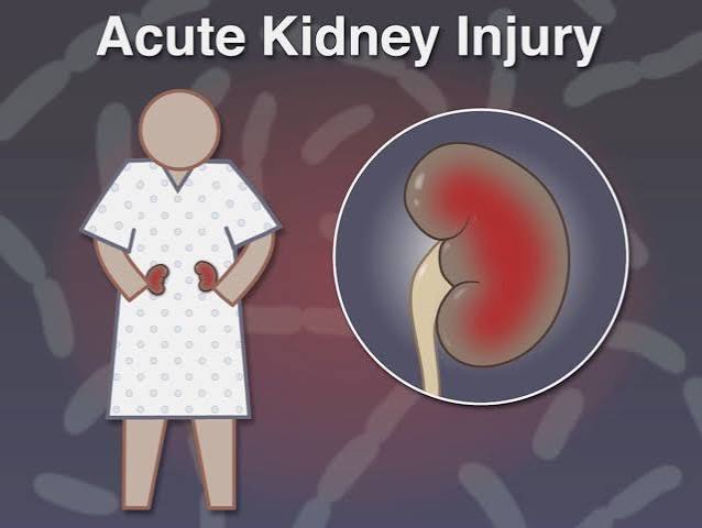
Acute kidney injury is a sudden, rapid decline in kidney function that develops over hours to days, in contrast to the slow, years-long progression of chronic kidney disease. Where chronic kidney disease reflects permanent, irreversible nephron loss, acute kidney injury is often — though not always — reversible if the underlying cause is identified and corrected quickly. AKI affects approximately 10 to 15 percent of all hospitalized adults and up to 50 percent of those admitted to intensive care units, making it one of the most common and consequential complications of serious illness. Despite its frequency, AKI is underrecognized outside hospital settings, and many adults who have experienced an episode of acute kidney injury are unaware of the implications it carries for their long-term kidney health. This guide explains what acute kidney injury is, how it is diagnosed, what causes it, how it is treated, and what its consequences are over time.
The Medical Definition of Acute Kidney Injury
Acute kidney injury is defined by the KDIGO 2012 guidelines using three diagnostic criteria: a rise in serum creatinine of 0.3 mg/dL or more within 48 hours; a rise in serum creatinine to 1.5 times or more the baseline level within 7 days; or urine output below 0.5 mL per kilogram of body weight per hour for 6 or more consecutive hours. Meeting any one of these criteria is sufficient for the AKI diagnosis. The shift from the older term “acute renal failure” to “acute kidney injury” reflects a clinical recognition that kidney damage occurs across a spectrum of severity — not only at the point of complete failure — and that even milder episodes carry prognostic significance.
AKI staging: Stage 1 AKI: creatinine 1.5 to 1.9 times baseline, or a rise of 0.3 mg/dL or more, or urine output below 0.5 mL/kg/h for 6 to 12 hours. Stage 2: creatinine 2.0 to 2.9 times baseline, or urine output below 0.5 mL/kg/h for 12 or more hours. Stage 3: creatinine 3 times or more the baseline, or rising to 4.0 mg/dL or above, or initiation of kidney replacement therapy, or urine output below 0.3 mL/kg/h for 24 or more hours. Higher stages carry progressively greater risk of non-recovery and long-term kidney impairment.
The Three Categories of Acute Kidney Injury
AKI is classified by mechanism into three broad categories: pre-renal (reduced blood flow to the kidneys), intrinsic renal (direct damage to kidney tissue), and post-renal (blocked urine outflow). This classification directs treatment, because the intervention for a pre-renal AKI — restoring blood flow — is different from the treatment for a post-renal AKI — relieving obstruction — and neither applies to intrinsic renal AKI, which is primarily managed by removing the offending cause and providing supportive care.
Pre-renal AKI accounts for approximately 55 to 60 percent of hospital-acquired AKI cases. In pre-renal AKI, the kidney tissue itself is initially undamaged — the problem is insufficient blood flow. Common causes include severe dehydration from vomiting, diarrhea, or poor intake; blood loss from hemorrhage; burns; heart failure with reduced cardiac output; liver failure with hepatorenal syndrome; and sepsis. NSAIDs cause pre-renal AKI by blocking prostaglandin-mediated dilation of the afferent arteriole, reducing glomerular perfusion pressure — a risk amplified in the setting of dehydration, heart failure, or pre-existing CKD. ACE inhibitors and ARBs can precipitate AKI in pre-renal states by blocking efferent arteriole constriction, causing glomerular perfusion pressure to fall below the filtration threshold.
Intrinsic renal AKI refers to direct damage to kidney structures — tubules (ATN), interstitium (AIN), glomeruli (glomerulonephritis), or renal vasculature. Post-renal AKI results from obstruction of urine outflow — bilateral ureteral obstruction from pelvic malignancy or stones, or bladder outlet obstruction from BPH or prostate cancer — and is rapidly reversible if relieved promptly.
Acute Tubular Necrosis — The Most Common Intrinsic AKI
Acute tubular necrosis (ATN) is the most common cause of intrinsic renal AKI. Ischemic ATN occurs when a prolonged pre-renal state causes tubular cells to become ischemic and undergo necrosis. The proximal tubules and thick ascending limb of the loop of Henle are particularly vulnerable because of their high metabolic demands. Once tubular cells die, they slough off into the tubular lumen, forming “muddy brown” granular casts visible on urine microscopy — diagnostic of ATN.
Nephrotoxic ATN is caused by substances toxic to tubular cells. Common nephrotoxins include aminoglycoside antibiotics (gentamicin, tobramycin), which accumulate in proximal tubular cells and cause mitochondrial dysfunction; cisplatin and other platinum-based chemotherapy agents; amphotericin B; myoglobin released from damaged muscle in rhabdomyolysis (from trauma, prolonged immobility, or statin toxicity); hemoglobin from hemolysis; and iodinated contrast agents, which cause direct tubular toxicity amplified by medullary vasoconstriction. Unlike cardiac muscle cells, tubular cells can regenerate — recovery from ATN is driven by tubular cell regeneration over days to weeks, depending on the severity and duration of the insult.
How Acute Kidney Injury Is Diagnosed
Diagnosis of AKI depends on comparing current serum creatinine to a known baseline — a single value without context cannot confirm AKI. Urinalysis and urine sediment are the most informative early tests. Muddy brown granular casts are characteristic of ATN. White blood cell casts with urine eosinophils suggest acute interstitial nephritis from drug hypersensitivity. Red blood cell casts are hallmarks of glomerulonephritis.
Urine sodium and fractional excretion of sodium (FENa) help distinguish pre-renal from intrinsic AKI in patients not yet receiving diuretics. In pre-renal AKI, intact tubules retain sodium — urine sodium below 20 mEq/L and FENa below 1%. In ATN, damaged tubules cannot retain sodium — urine sodium above 40 mEq/L and FENa above 2%. FENa is unreliable after diuretics, in contrast nephropathy, and in myoglobinuria. Kidney ultrasound excludes obstruction by checking for hydronephrosis. BUN-to-creatinine ratio above 20:1 suggests pre-renal AKI or upper GI bleeding.
Symptoms of Acute Kidney Injury
Most AKI episodes, particularly Stage 1 and many Stage 2 cases, produce no symptoms and are detected only by blood and urine testing. When symptoms occur, they reflect impaired filtration and fluid regulation. Reduced urine output (oliguria below 400 mL/day, or anuria) is the most visible sign but is absent in non-oliguric AKI — common with nephrotoxic causes. Fluid retention causes peripheral edema and, in severe cases, pulmonary edema with dyspnea. Nausea, fatigue, and loss of appetite reflect early uremia. Confusion indicates severe uremia. Chest pain from pericarditis occurs in AKI severe enough to cause systemic uremia.
Treatment of Acute Kidney Injury
Pre-renal AKI is treated with volume replacement — intravenous balanced crystalloids (lactated Ringer’s, Plasmalyte) preferred over normal saline in sepsis, based on the SMART and SALT-ED trials, which showed reduced adverse kidney events. Heart failure-related AKI requires careful diuresis rather than fluid administration. Hepatorenal syndrome requires vasoconstrictors (terlipressin or norepinephrine) plus albumin infusion.
Intrinsic AKI (ATN) has no treatment to accelerate recovery — the intervention is: immediately discontinue the nephrotoxin or reverse the ischemic insult; maintain euvolemia; correct electrolyte abnormalities; and avoid further kidney insults. Furosemide does not protect kidneys or accelerate ATN recovery and is used only for managing fluid overload. Acute interstitial nephritis from drug hypersensitivity: stop the offending drug; corticosteroids for severe cases. Post-renal AKI: bladder outlet obstruction relieved by urethral catheterization; ureteral obstruction by stenting or nephrostomy.
Electrolyte management: severe hyperkalemia (above 6.0–6.5 mEq/L or with EKG changes) requires IV calcium gluconate, IV insulin with dextrose, and dialysis if unresponsive. Kidney replacement therapy is initiated for life-threatening hyperkalemia, severe metabolic acidosis (pH below 7.15), pulmonary edema unresponsive to diuretics, pericarditis, or uremic encephalopathy. Dialysis supports the patient while tubular cell regeneration occurs — it does not repair kidney function.
Recovery from Acute Kidney Injury
Stage 1 AKI from a promptly reversed pre-renal cause often shows creatinine returning to baseline within 24 to 72 hours. Stage 2 ATN may take 1 to 3 weeks to recover as tubular cells regenerate. Stage 3 AKI requiring dialysis may take 4 to 6 weeks or longer, and a proportion of patients remain dialysis-dependent. Approximately 15 to 30 percent of AKI episodes do not result in full creatinine recovery — these patients develop new CKD or experience acceleration of pre-existing CKD. KDIGO recommends checking creatinine and UACR 3 months after an AKI episode to establish whether recovery is complete and to diagnose CKD if it has developed.
AKI and Long-Term Kidney Health
AKI and CKD are not separate diseases but interconnected states along a continuum. Each AKI episode damages some proportion of tubular cells beyond the capacity for full regeneration, contributing cumulatively to CKD risk. Survivors of hospital-acquired AKI have significantly elevated risk of subsequent CKD, ESRD, and cardiovascular events compared to matched hospitalized patients without AKI — and the risk scales with AKI severity and the number of episodes. For adults who have experienced AKI, annual eGFR and UACR checks are important for catching any CKD that develops. For the broader relationship between acute and chronic kidney disease, see our article on what is chronic kidney disease.
How to Reduce Your Risk of Acute Kidney Injury
Many AKI episodes are preventable. Stay well-hydrated during illness, heat exposure, or strenuous exercise. Avoid NSAIDs when dehydrated, when you have CKD, when you have heart failure, or in older age where renal reserve is reduced. Before any procedure using iodinated contrast dye, inform your physician if you have CKD or diabetes, and ask whether pre-procedure hydration is indicated. For adults with eGFR below 30, contrast procedures should be accompanied by IV saline pre-hydration when possible and the minimum contrast dose should be used. Review all medications during acute illness — many common drugs become nephrotoxic or accumulate to dangerous levels when kidney function is acutely reduced. For context on what markers indicate healthy kidney function, see our article on signs of healthy kidney function. For the full range of kidney conditions, see our guide on common kidney problems in adults. For the key lab values used to detect and monitor kidney injury, see our article on kidney health numbers every adult should know.
Acute Interstitial Nephritis — A Frequently Missed Cause
Acute interstitial nephritis (AIN) is an underdiagnosed cause of intrinsic AKI, responsible for 10 to 15 percent of AKI cases in some series. AIN is an inflammatory reaction in the renal interstitium — the supporting tissue surrounding the tubules — typically triggered by a drug hypersensitivity reaction rather than a direct toxic effect. The most common culprits are NSAIDs, proton pump inhibitors (omeprazole, pantoprazole), beta-lactam antibiotics (amoxicillin, cephalosporins), sulfonamides, and diuretics. Less commonly, AIN results from infections (hantavirus, leptospirosis, CMV) or systemic inflammatory conditions (sarcoidosis, Sjögren’s syndrome).
The classic triad of AIN — fever, rash, and eosinophilia — appears in only 10 to 30 percent of drug-induced cases, making it an unreliable diagnostic screen. The diagnosis is suggested by a rising creatinine 1 to 3 weeks after starting a new medication, mild pyuria, urine eosinophils on Wright’s or Hansel’s stain, and the absence of muddy brown casts (which would suggest ATN instead). Confirmation requires kidney biopsy in ambiguous cases. Treatment is prompt discontinuation of the offending drug. If creatinine does not begin improving within 3 to 5 days of drug removal, a short course of oral prednisone (1 mg/kg/day) is often given, though the evidence base for steroids in AIN is observational rather than from randomized trials. Full recovery occurs in the majority of patients who stop the causative drug early — delays in recognition increase the risk of interstitial fibrosis and permanent CKD.
Contrast-Induced AKI — Risk, Prevention, and Controversy
Contrast-induced acute kidney injury (CI-AKI), also called contrast-induced nephropathy, refers to AKI occurring within 48 to 72 hours of iodinated contrast exposure, in the absence of another explanation. The mechanisms include direct tubular toxicity from contrast agents and medullary ischemia from contrast-induced vasoconstriction. CI-AKI is generally transient — creatinine typically peaks at 2 to 5 days post-contrast and returns toward baseline by 10 to 14 days — but in high-risk patients it can result in permanent kidney injury and, in severe cases, dialysis.
Risk factors for CI-AKI include pre-existing CKD (eGFR below 30 is the strongest risk factor), diabetes, dehydration, heart failure, high contrast volume, repeat contrast procedures within 24 to 48 hours, and the use of nephrotoxic medications around the time of contrast. Low-osmolality or iso-osmolality contrast agents are preferred over high-osmolality agents in high-risk patients. The cornerstone of prevention is adequate hydration — IV isotonic saline (1 mL/kg/h for 6 to 12 hours before and after the procedure) reduces CI-AKI risk in high-risk patients. N-acetylcysteine was historically given for CI-AKI prophylaxis, but subsequent high-quality trials showed no benefit beyond adequate hydration alone. Metformin should be withheld 48 hours before contrast-enhanced procedures in patients with eGFR below 60, as contrast-induced AKI can impair metformin excretion and increase the risk of lactic acidosis. Note: oral contrast (barium) used for GI procedures does not carry CI-AKI risk — the concern applies exclusively to intravenous iodinated contrast.
Rhabdomyolysis-Induced AKI — A Special Cause Requiring Aggressive Hydration
Rhabdomyolysis is the breakdown of skeletal muscle, releasing myoglobin into the circulation. Myoglobin is directly nephrotoxic — it causes ATN through direct tubular toxicity, tubular obstruction by myoglobin casts, and renal vasoconstriction. Common causes of rhabdomyolysis include crush injuries, prolonged immobility (being found down for hours — a common scenario in elderly fall victims and those with altered consciousness), statin toxicity (particularly at higher doses and in combination with fibrates or CYP3A4 inhibitors), severe heat stroke, seizures, and extreme exercise (exercise-induced rhabdomyolysis).
The hallmark laboratory finding is a markedly elevated serum creatine kinase (CK) — often exceeding 10,000 U/L, with severe cases reaching 100,000 U/L or more. Urine dipstick may be positive for blood (false positive from myoglobin) with no red blood cells on microscopy — this discrepancy should raise suspicion for myoglobinuria. Treatment is aggressive IV fluid resuscitation with isotonic saline, targeting urine output of 200 to 300 mL/hour until myoglobin has cleared (CK trending down, urine clearing). Urinary alkalinization with sodium bicarbonate is sometimes used to prevent myoglobin precipitation in acidic tubules, though evidence is mixed. The aggressiveness of hydration in rhabdomyolysis — often 10 to 15 liters in the first 24 hours — is substantially greater than for other AKI types, reflecting the need to dilute and flush nephrotoxic myoglobin from the tubular lumen before necrosis becomes irreversible.
Post-AKI Care — What Happens After Discharge
The period after hospital discharge following an AKI episode is a high-risk time that many patients and even some providers underappreciate. The kidneys may still be recovering, nephrotoxic exposures are especially dangerous during this window, and medications that were renally dose-adjusted during the hospitalization may need re-evaluation as function normalizes — or may need further adjustment if function does not recover fully. KDIGO recommends that all patients who experienced AKI during hospitalization have serum creatinine and UACR checked at 3 months post-discharge. If creatinine has returned to baseline and UACR is normal, the AKI is considered fully resolved. If creatinine remains elevated above baseline or UACR is now abnormal, the patient has developed new CKD and should be managed accordingly — including nephrology referral for significant elevations.
In the post-AKI period: restart ACE inhibitors and ARBs cautiously — withhold if creatinine has not stabilized; resume metformin only after confirming renal recovery; avoid NSAIDs for at least 6 to 12 weeks; and ensure follow-up is scheduled specifically to address kidney function status rather than relying on incidental future testing. A short hospitalization-related AKI that is not followed with a 3-month creatinine check is a missed opportunity — the transition from AKI to CKD is the period when early intervention has the greatest potential to prevent or slow long-term kidney damage.
Community-Acquired AKI — AKI Outside the Hospital
While hospital-acquired AKI receives more attention in clinical literature, community-acquired AKI — AKI presenting at the time of hospital admission rather than developing during the hospitalization — is increasingly recognized as common and consequential. Studies suggest that 20 to 40 percent of all AKI cases are community-acquired, with dehydration from gastroenteritis, NSAID use during acute illness, uncontrolled diabetes, and urinary obstruction being the most frequent causes. Community-acquired AKI in otherwise healthy adults often follows a predictable trigger — a weekend of gastroenteritis with poor fluid intake and continued NSAID use for fever, for instance, or a hot week of physical labor without adequate fluid replacement. In adults with pre-existing CKD, the threshold for community-acquired AKI is lower and the triggers are often less dramatic — a two-day flucloxacillin course for a skin infection, a day of poor intake during a cold, or a brief hypotension episode during a dental procedure can tip a kidney already operating at reduced reserve into AKI.
Community-acquired AKI that resolves promptly with oral or IV rehydration may never require hospitalization — creatinine normalizes within days and the episode passes without formal diagnosis. This under-diagnosis has an important consequence: the cumulative effect of multiple sub-clinical AKI episodes accelerating CKD is not captured in conventional disease statistics but may explain why CKD progression in some individuals is faster than their risk factors predict. If you have CKD or diabetes and experience significant dehydrating illness, vomiting, or prolonged fever, checking a creatinine within 48 to 72 hours of recovery is a reasonable and low-cost precaution — and one that can catch an AKI episode before it passes without documentation.
Hepatorenal Syndrome — AKI in Liver Disease
Hepatorenal syndrome (HRS) is a specific form of pre-renal AKI occurring in patients with advanced liver disease (cirrhosis or acute liver failure). In HRS, splanchnic vasodilation from portal hypertension causes effective arterial underfilling — the kidneys perceive a decrease in perfusion pressure and respond with intense renal vasoconstriction and RAAS activation. The kidney itself is structurally normal — kidneys from patients who died of HRS have been successfully transplanted into people with normal livers, and those kidneys function normally, confirming that the kidney damage in HRS is functional rather than structural.
HRS type 1 (now classified as HRS-AKI under the KDIGO framework) is the rapidly progressive form — creatinine rises sharply over days and carries a very poor short-term prognosis without liver transplant. HRS type 2 is more gradual and associated with refractory ascites. Terlipressin plus albumin infusion is the most effective pharmacological bridge therapy for HRS-AKI. The definitive treatment is liver transplantation, after which kidney function typically recovers without the need for long-term dialysis — provided severe prolonged ischemia has not caused irreversible tubular damage. This underscores the principle that in HRS, identifying and reversing the trigger (infection, GI bleeding, hypovolemia) can sometimes prevent the full syndrome from developing.
Sources: National Institute of Diabetes and Digestive and Kidney Diseases (NIDDK), niddk.nih.gov; National Kidney Foundation, kidney.org; American Kidney Fund, kidneyfund.org. KDIGO AKI Guidelines 2012; SMART Trial 2018; SALT-ED Trial 2018.
Pingback: Chronic Kidney Disease vs Acute Kidney Injury
Finally a resource that explains what is acute kidney injury? in plain language. I have tried following advice from several sources but this is most consistent with what my specialist told me. Looking forward to reading more articles from this website.
I never fully understood what is acute kidney injury? until I read this. This is the kind of evidence-based writing that actually changes how people approach their health. Exactly the kind of evidence-based information that is hard to find in one place.
I have been reading about what is acute kidney injury? for weeks and this is the most thorough guide I found. I have tried following advice from several sources but this is most consistent with what my specialist told me. Forwarding this to others in my support group who are dealing with similar issues.