
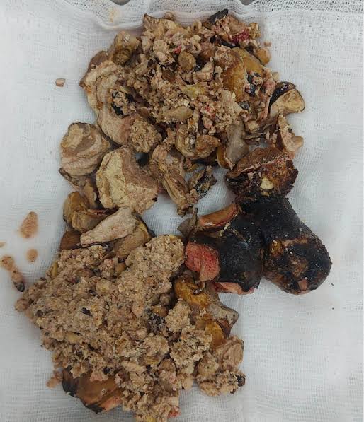
Kidney Stones: Symptoms, Causes, and Treatment
Kidney stones are one of the most common — and most painful — conditions affecting the urinary tract. Each year, approximately 600,000 Americans visit emergency departments because of kidney stones, and the lifetime prevalence of developing at least one kidney stone has risen to roughly 11% in men and 9% in women in the United States, up from around 3 to 4% in the 1970s. Despite how common they are, kidney stones remain widely misunderstood: many people do not recognize the symptoms until a stone is already causing significant obstruction, and many do not understand why stones form or what changes in diet and lifestyle could prevent them. This guide covers what kidney stones are, how they form, what they feel like, and what treatments are available — from watchful waiting to surgical intervention.
A kidney stone is a hard, crystalline mineral deposit that forms within the kidney when certain substances in the urine — including calcium, oxalate, uric acid, and phosphate — become too concentrated for the liquid volume of urine to keep them dissolved. When their concentration exceeds the saturation threshold, these substances begin to crystallize and aggregate into particles that can grow over weeks to months into stones ranging in size from a grain of sand to a golf ball. Most stones form in the kidney but can migrate into the ureter (the tube connecting the kidney to the bladder), the bladder, or the urethra, and it is typically during this passage through the ureter that the most severe pain occurs.
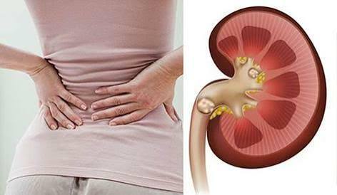
Types of Kidney Stones: Why Composition Matters
Kidney stones are not all the same — they differ in chemical composition, and this difference matters because it determines what caused the stone, what treatment is most appropriate, and what dietary or medical interventions will be most effective at preventing recurrence. The four main types are calcium oxalate stones, calcium phosphate stones, uric acid stones, and struvite (infection) stones, with a fifth, rare type — cystine stones — occurring in people with a hereditary disorder of amino acid transport.
Calcium oxalate stones are by far the most common, accounting for 70 to 80% of all kidney stones. They form when calcium and oxalate combine in the urine. Despite the name “calcium” stones, reducing dietary calcium is almost never the right prevention strategy — low dietary calcium actually increases oxalate absorption from the gut (because calcium normally binds oxalate in the intestine and carries it out in the stool), raising urinary oxalate and increasing stone risk. The correct approach is matching calcium intake with meals and reducing dietary oxalate from high-oxalate foods such as spinach, almonds, and beets.
Uric acid stones account for approximately 5 to 10% of kidney stones and form when urine is persistently acidic (low pH). Unlike calcium stones, they are often radiolucent — meaning they do not appear on plain X-ray — and are most common in patients with gout, chronic diarrhea, diabetes, or metabolic syndrome. Uric acid stones are uniquely amenable to dissolution therapy: because they form in acidic urine, alkalinizing the urine with potassium citrate or sodium bicarbonate can dissolve existing stones and prevent new ones — making them the most medically treatable stone type.
Struvite stones (also called infection stones or triple phosphate stones) form as a direct consequence of infection with urease-producing bacteria — most commonly Proteus mirabilis — which split urea into ammonia, alkalinizing the urine and enabling struvite crystal formation. They tend to grow rapidly and can fill the entire collecting system of the kidney as staghorn calculi. Complete stone removal is the cornerstone of treatment because residual stone fragments harbor bacteria and perpetuate infection. Calcium phosphate stones are associated with conditions causing abnormally alkaline urine, most notably renal tubular acidosis and primary hyperparathyroidism. Understanding which type of stone a patient has is the foundation of prevention — which is why all first-time stone formers should have their stone analyzed if it is recovered.
Causes and Risk Factors
Kidney stones form when the chemical composition of urine creates an environment favorable for crystal formation. The three fundamental drivers are: supersaturation (the crystallizing substance is present at concentrations exceeding its solubility), low urine volume (insufficient dilution to keep solutes dissolved), and imbalance between promoters and inhibitors of crystallization (citrate, pyrophosphate, and certain urinary proteins normally inhibit crystal formation; their deficiency promotes it). Any combination of factors that tilts this balance toward crystal formation increases stone risk.
Dehydration is the single most modifiable risk factor across all stone types. When fluid intake is insufficient — from inadequate drinking, excessive sweating (from heat, exercise, or fever), or increased gastrointestinal losses from diarrhea or vomiting — urine volume drops and solute concentrations rise proportionally. People living in hot climates or working in physically demanding outdoor occupations have substantially higher stone risk than those who are sedentary and well-hydrated. The landmark clinical evidence: increasing daily urine output above 2 liters per day reduces recurrent stone formation by approximately 50% compared to low urine output.
Dietary factors beyond hydration play important roles. High dietary sodium raises urinary calcium excretion by reducing calcium reabsorption in the kidney tubules — a direct mechanism linking high-salt diets to calcium stone formation. High dietary oxalate (from spinach, nuts, chocolate, and certain grains and legumes) increases urinary oxalate in susceptible individuals. Very high protein intake — particularly animal protein — raises urinary uric acid and reduces urinary citrate, increasing risk for both uric acid and calcium oxalate stones. The DASH diet (Dietary Approaches to Stop Hypertension), characterized by high fruit and vegetable intake, adequate dairy, and low sodium and animal protein, has been associated with significantly reduced kidney stone risk in large prospective studies.
Medical conditions that substantially increase kidney stone risk include: primary hyperparathyroidism (excess PTH drives high urinary calcium), renal tubular acidosis (impaired urinary acidification leads to calcium phosphate and calcium oxalate stone formation and low urinary citrate), inflammatory bowel disease and bowel surgery (fat malabsorption leads to enteric hyperoxaluria — excess oxalate absorption from the gut), gout and hyperuricemia (elevated uric acid production and excretion predisposes to uric acid stones), and obesity and metabolic syndrome (associated with insulin resistance, low urinary pH, and reduced urinary citrate). A first kidney stone, particularly at a young age, should prompt evaluation for these underlying conditions.
Genetic predisposition is significant: the risk of kidney stones is 2.5 times higher in individuals with a family history of stones compared to those without. Specific hereditary conditions — cystinuria, primary hyperoxaluria, and Dent’s disease — cause early-onset, severe, recurrent stone disease and require specialized metabolic evaluation and management. For the majority of patients without these rare disorders, genetic risk is polygenic (multiple genes each contributing modest risk) and is expressed through intermediate phenotypes such as hypercalciuria (high urinary calcium), hypocitraturia (low urinary citrate), and hyperoxaluria (high urinary oxalate) that are measurable through a 24-hour urine collection.
Symptoms: What a Kidney Stone Feels Like
A kidney stone can exist in the kidney for months or even years without causing any symptoms — sitting silently in the renal pelvis or a calyx, growing slowly, until it migrates into the ureter. The transition from silent stone to symptomatic stone typically occurs abruptly, and the resulting pain — renal colic — is among the most severe that humans experience. Studies consistently find that kidney stone pain is rated as severely as or more severely than childbirth and post-surgical pain by patients who have experienced both.
Renal colic is the classic pain of kidney stone passage. It is caused by ureteral peristalsis attempting to push the stone through the ureter, combined with ureteral spasm and dilation above the obstruction. Unlike the constant aching of kidney infection pain, renal colic is typically colicky — it waxes and wanes in waves of severe, cramping pain lasting 20 to 60 minutes, interspersed with periods of partial relief. The pain is classically severe, located in the flank, and radiates toward the groin, genitalia, or inner thigh as the stone moves distally down the ureter. Patients often cannot find a comfortable position — they may pace, shift, or writhe, in contrast to peritonitis patients who lie still to minimize movement.
Hematuria (blood in the urine) — either visible to the naked eye or detectable only on urinalysis — occurs in approximately 85% of patients with kidney stones and results from the stone’s abrasion of the urothelium as it moves through the ureter. Visible hematuria produces pink, red, or brown urine and can be alarming to patients, but it is not in itself dangerous. Importantly, hematuria alone does not confirm a kidney stone — it is a feature shared with kidney infection, bladder cancer, and other urological conditions, and its cause should always be established rather than assumed.
Nausea and vomiting accompany acute renal colic in the majority of patients and are driven by the intense pain and vagal stimulation from the inflamed ureter. Urinary symptoms — frequency, urgency, and dysuria — appear when the stone has migrated to the distal ureter near the bladder, where it mimics the irritative symptoms of a lower UTI. A patient who suddenly develops what feels like a UTI after hours or days of severe flank pain likely has a stone approaching or entering the bladder. Fever accompanying stone pain is a critical warning sign: it suggests concurrent infection proximal to the obstruction — a urological emergency that requires urgent drainage.
Diagnosis
The evaluation of a suspected kidney stone combines clinical assessment, urine testing, and imaging. A urinalysis showing hematuria and, in some cases, crystals, supports the diagnosis. A non-contrast CT scan of the abdomen and pelvis (CT-KUB) is the gold standard imaging study — it detects virtually all stone types regardless of composition (with the exception of small uric acid stones, which may be invisible on plain X-ray but are visible on CT), reveals stone size, number, and location, and identifies any proximal hydronephrosis (ureteral dilation from obstruction). A CT scan also evaluates for alternative diagnoses that can mimic renal colic — appendicitis, ovarian cyst pathology, diverticulitis, and aortic aneurysm among them.
Renal ultrasound is an alternative first-line imaging study in certain populations — particularly pregnant women (in whom CT radiation is avoided where possible), children, and patients in whom reducing radiation exposure is a priority. Ultrasound has lower sensitivity for ureteral stones than CT but is highly effective at detecting hydronephrosis, which indirectly confirms significant obstruction even when the stone itself is not visualized. Plain abdominal X-ray (KUB) is insensitive for many stone types and is not recommended as a first-line tool, though it has a role in monitoring known radiopaque stones during treatment.
For patients with a first kidney stone, or those with recurrent stones, a 24-hour urine collection is the cornerstone of metabolic evaluation. This test measures daily excretion of calcium, oxalate, uric acid, citrate, phosphate, sodium, creatinine, and total urine volume — providing a comprehensive chemical profile of the urinary environment and identifying specific modifiable risk factors (hypercalciuria, hyperoxaluria, hypocitraturia, hyperuricosuria, low urine volume). Stone analysis — determining the stone’s chemical composition — is performed on any recovered stone and guides targeted prevention. Blood tests including serum calcium, uric acid, creatinine, parathyroid hormone (PTH), and bicarbonate assess for systemic metabolic disorders contributing to stone formation.
Treatment Options
Conservative management (watchful waiting) is appropriate for small stones — typically less than 5 mm in diameter — in patients without obstruction, infection, or intractable pain. Approximately 68% of stones smaller than 5 mm pass spontaneously within four weeks, compared to about 47% of stones 5 to 10 mm. Patients managed conservatively should drink adequate fluids (aiming for 2 to 3 liters of urine output daily), strain their urine to capture the stone for analysis, and take analgesics for pain control. NSAIDs (such as ibuprofen or ketorolac) are the preferred analgesics because they reduce prostaglandin-mediated ureteral spasm and have been shown in clinical trials to control renal colic pain as effectively as opioids in most patients.
Medical expulsive therapy (MET) with alpha-blockers — most commonly tamsulosin — has been widely used to facilitate stone passage by relaxing ureteral smooth muscle. Multiple meta-analyses show that tamsulosin increases the stone passage rate for distal ureteral stones, particularly those 5 to 10 mm, though the effect size is modest and the evidence is stronger for larger stones in this size range. Alpha-blockers are generally well tolerated (the main side effects are dizziness and retrograde ejaculation in men) and are a reasonable adjunct for patients managing stones conservatively, particularly when the stone is in the lower ureter.
Shock wave lithotripsy (SWL) is a non-invasive procedure that uses high-energy ultrasound waves to fragment stones into smaller pieces that can then pass through the ureter. It is most effective for stones smaller than 2 cm in the kidney or upper ureter, and for softer stone compositions (calcium oxalate dihydrate and uric acid stones fragment more easily than calcium oxalate monohydrate or brushite stones). SWL is performed under sedation or analgesia, requires no incisions, and is typically performed in an outpatient setting. Multiple sessions may be required for complete fragmentation, and stone fragments may cause “steinstrasse” (a column of fragments blocking the ureter) in a minority of cases.
Ureteroscopy (URS) involves passing a thin flexible or semi-rigid scope through the urethra and bladder into the ureter or kidney to directly visualize and treat the stone. Laser lithotripsy (most commonly using a holmium:YAG or thulium fiber laser) is used to fragment the stone, and fragments are extracted using a small basket or allowed to pass. URS is highly effective regardless of stone location or composition, and has largely replaced SWL as the preferred treatment for ureteral stones in many centers due to its higher stone-free rates and single-session efficacy. It requires general or spinal anesthesia and is performed as an outpatient or short-stay procedure.
Percutaneous nephrolithotomy (PCNL) is the treatment of choice for large kidney stones (typically greater than 2 cm), staghorn calculi (stones filling the kidney’s collecting system), and stones in locations inaccessible to ureteroscopy. It involves creating a percutaneous tract through the flank directly into the kidney under fluoroscopic and ultrasound guidance, through which a nephroscope is introduced to visualize and fragment the stone. PCNL has the highest stone-free rates of any stone removal technique for large stones but requires general anesthesia and a one to two day hospital stay, with a recovery period of one to two weeks. Mini and ultra-mini PCNL variants use smaller-caliber equipment to reduce trauma and recovery time.
Prevention: The Long Game
Kidney stones have a high recurrence rate — approximately 50% of patients who form a first stone will form another within five to seven years without preventive intervention. This makes prevention the most important phase of kidney stone management for most patients. Prevention begins with the universal measures that apply to all stone types, and layers on type-specific strategies guided by stone analysis and 24-hour urine results.
The universal prevention pillar is high fluid intake. The target is a urine output of 2.0 to 2.5 liters per day, which typically requires a total daily fluid intake of 2.5 to 3.5 liters depending on climate, activity level, and sweat losses. Water is the preferred fluid — high intakes of sugar-sweetened beverages (particularly those sweetened with fructose) are associated with increased stone risk, and grapefruit juice specifically increases stone risk in epidemiological studies. Lemon juice and other citrus juices increase urinary citrate, which inhibits calcium crystal formation, and lemonade therapy (diluted fresh lemon juice) is a palatable way to increase citrate intake between medical visits.
For patients with recurrent calcium oxalate or calcium phosphate stones confirmed on metabolic evaluation, targeted pharmacological therapy — thiazide diuretics for hypercalciuria, potassium citrate for hypocitraturia and uric acid stones, and allopurinol for hyperuricosuria — has been shown in randomized controlled trials to meaningfully reduce recurrence rates. These medications are not appropriate for all stone formers and should be prescribed based on 24-hour urine findings rather than empirically.
For more detail on recognizing the earliest signs before a stone causes acute pain, see the early signs of kidney stones guide on Horizon Health Guide. The kidney stone pain article on this site describes the pain experience in clinical detail. For urinary tract infection complications that can coexist with stone disease, the kidney infection article at Horizon Health Guide covers symptoms and warning signs. External references include the NIDDK kidney stones patient guide, the National Kidney Foundation kidney stone resource, and the StatPearls clinical review of nephrolithiasis.
When to Seek Emergency Care
Most kidney stones, while extremely painful, are not life-threatening and can be managed with outpatient or urgent care evaluation. However, certain presentations require immediate emergency department evaluation. Fever combined with stone pain is a urological emergency — it indicates infection behind an obstructed ureter, which can progress to urosepsis and septic shock within hours if not drained. Intractable pain and vomiting that cannot be controlled with oral medications requires IV analgesics and antiemetics and urgent urological consultation. Bilateral ureteral stones or a stone in a solitary kidney causing complete obstruction threatens acute kidney injury and renal failure. Anuria (absence of urine output) in a patient with known stone disease is a medical emergency. Patients with a single kidney, a renal transplant, or significantly reduced baseline kidney function require a lower threshold for urgent intervention than those with two normal kidneys.
For uncomplicated renal colic without fever or vomiting and with adequate pain control at home, a scheduled visit to a urologist within one to two weeks is appropriate. Most urologists will arrange imaging to confirm the stone’s location, assess for obstruction, and estimate the likelihood of spontaneous passage before deciding whether intervention is warranted. Patients should be instructed to return immediately to the emergency department if fever, rigors, inability to tolerate fluids, worsening pain unresponsive to analgesics, or no urine output develops during conservative management.
Sources: NIDDK — Kidney Stones · National Kidney Foundation · StatPearls — Nephrolithiasis
Living With Kidney Stones: What Recurrence Means for Long-Term Health
For many patients, a first kidney stone is also the last — particularly if it prompts lifestyle changes such as significantly increasing fluid intake and moderating sodium and animal protein. For others, stone formation is a chronic and recurrent condition that requires ongoing medical management. The key distinction is between patients who form stones occasionally despite basic measures (relatively low-risk recurrence) and those who form frequent stones despite apparently adequate hydration and dietary modification — the latter group warrants comprehensive metabolic evaluation and often requires targeted pharmacological prevention.
Long-term untreated recurrent stone disease carries meaningful risks beyond the acute episodes of pain and obstruction. Repeated passage of stones and the instrumentation required to treat them can cause ureteral scarring and stricture — a narrowing of the ureter that itself can cause chronic obstruction and hydronephrosis. Recurrent obstructive episodes and chronic pyelonephritis from struvite stones cause progressive renal parenchymal damage, leading to chronic kidney disease over years to decades. Patients with recurrent stones have a measurably higher rate of chronic kidney disease compared to the general population, which is one of the strongest arguments for investing in systematic prevention rather than treating each episode in isolation. Working with a urologist and, for complex cases, a nephrologist with expertise in stone disease provides the most comprehensive approach to both acute management and long-term kidney health preservation.
I’ve had three kidney stones in the past five years, all calcium oxalate. Each time the pain was so severe I ended up in the ER. I always assumed I needed to cut out calcium from my diet but my urologist told me the opposite — cutting calcium actually made things worse because it let more oxalate get absorbed. This article explains that mechanism perfectly. I’m now on a normal calcium diet matched with meals and drinking about 3 liters of water a day, and I haven’t had a stone in 18 months.
The section on when to go to the ER is so important. My husband had a kidney stone last year and he tried to tough it out at home for two days with increasing pain and then developed a fever. We didn’t realize fever plus stone pain meant an emergency — he ended up with a kidney infection behind the blocked ureter and needed an emergency stent procedure. If we had read something like this, we would have gone in immediately when the fever started. Please share this widely.
We’re so glad your husband recovered well, Michelle — and your experience is exactly why we emphasize the fever-plus-stone-pain combination so strongly. That presentation — obstructed ureter with infection above the blockage — is a urological emergency because the infection cannot drain naturally past the stone, and bacteria accumulate rapidly in the trapped urine. Unlike an uncomplicated kidney infection that can often be managed outpatient, an infected obstructed kidney requires emergency stenting or nephrostomy drainage before antibiotics can be fully effective. Your message is a valuable reminder of why knowing when to escalate care can be genuinely life-saving. Thank you for sharing your story.