

Kidney Infection: Symptoms and Warning Signs
A kidney infection — medically known as pyelonephritis — is a serious bacterial infection that begins in the lower urinary tract and ascends to one or both kidneys. Unlike a bladder infection (cystitis), which is confined to the lower urinary tract and is usually uncomfortable but manageable, a kidney infection involves organ tissue and carries a risk of permanent kidney damage, bloodstream infection (urosepsis), and in severe cases, life-threatening septic shock. Recognizing the symptoms and warning signs of a kidney infection — and understanding how they differ from those of a simple bladder infection — can mean the difference between a timely recovery and a medical emergency.
Kidney infections are among the most common serious bacterial infections in adults, with an incidence of approximately 25 cases per 10,000 women and 8 cases per 10,000 men annually in the United States. Women are disproportionately affected due to their shorter urethra, which allows bacteria to reach the bladder and ascend to the kidneys more easily. Most kidney infections are caused by Escherichia coli — the same bacterium responsible for the majority of bladder infections — but other gram-negative organisms including Klebsiella pneumoniae, Proteus mirabilis, and Pseudomonas aeruginosa can also cause pyelonephritis, particularly in patients who have had recent hospitalization, urological procedures, or antibiotic exposure.
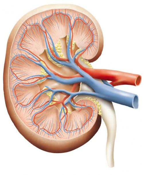
How Kidney Infections Develop: The Ascending Pathway
The vast majority of kidney infections develop through an ascending pathway: bacteria that colonize the periurethral area enter the urethra, migrate to the bladder (causing cystitis), and — if not cleared by the immune system or treatment — continue ascending through the ureters to reach the renal pelvis and kidney parenchyma. The ureters normally prevent this ascending movement through their peristaltic action and the competence of the vesicoureteral junction, which acts as a one-way valve. When this mechanism is compromised — by obstruction, reflux, or an overwhelming bacterial load — infection reaches the kidney.
Vesicoureteral reflux (VUR), a condition in which urine flows backward from the bladder into the ureters and sometimes the kidneys, is a major predisposing factor for kidney infections, particularly in children and young adults. Even mild VUR can carry bacteria-laden urine directly into the renal collecting system. Urinary tract obstruction from kidney stones, ureteral strictures, or external compression (such as from an enlarged prostate or pelvic mass) creates stasis that allows bacteria to multiply rapidly. Urinary catheterization and other instrumentation introduce bacteria directly into the urinary tract, bypassing its natural defenses. Any of these conditions transforms a potentially self-limited bladder infection into a serious renal infection requiring urgent treatment.
Core Symptoms: What a Kidney Infection Feels Like
The hallmark symptoms of a kidney infection are the combination of lower urinary tract symptoms (burning urination, urinary frequency and urgency) with systemic signs of infection. The systemic component is what distinguishes pyelonephritis from cystitis — a simple bladder infection does not cause fever, chills, or flank pain.
Flank pain is the most characteristic symptom of kidney infection. It presents as an aching or throbbing pain in the lower back or side, typically on one side (though bilateral infection can cause bilateral pain). The pain often radiates toward the groin or lower abdomen and can be severe enough to be mistaken for a kidney stone. Unlike the sharp, colicky pain of ureteral stone passage, the pain of pyelonephritis tends to be constant rather than wave-like. Costovertebral angle (CVA) tenderness — pain elicited by gentle percussion of the back at the junction of the lowest rib and the spine — is a classic physical examination finding in kidney infection and is often the most reliable sign clinicians use to distinguish pyelonephritis from cystitis during the physical exam.
Fever and chills are present in the majority of patients with kidney infection, though their severity varies. Fever in pyelonephritis is often high — temperatures of 38.5°C (101.3°F) or above are common, and temperatures exceeding 39.5°C (103.1°F) are not unusual in moderate to severe cases. Rigors (severe, uncontrolled shaking chills) signal that bacteria have entered the bloodstream — a sign of bacteremia that requires urgent evaluation and IV antibiotics rather than outpatient oral treatment. Even without rigors, persistent high fever in the context of urinary symptoms should prompt consideration of urosepsis and evaluation in an emergency setting.
Nausea and vomiting occur in roughly half of patients with kidney infection, driven both by the systemic inflammatory response and by direct stimulation of the vagal nerve by the adjacent inflamed kidney tissue. Significant vomiting that prevents oral fluid intake and oral antibiotic absorption is itself an indication for intravenous antibiotic therapy and hospitalization, regardless of the patient’s overall clinical appearance.
Lower urinary tract symptoms — burning with urination (dysuria), increased frequency and urgency, and cloudy or foul-smelling urine — accompany the upper tract symptoms in most but not all patients. In some cases, particularly in elderly patients, the classic lower urinary tract symptoms may be absent or minimal, making the diagnosis more challenging and increasing reliance on the systemic features (fever, altered mental status, flank pain) for clinical recognition.
Warning Signs That Require Emergency Evaluation
Not all kidney infections are the same in severity, and certain warning signs should trigger immediate emergency department evaluation rather than a scheduled doctor’s visit or urgent care visit. Recognizing these red flags can be critical to preventing life-threatening complications.
High fever with rigors (uncontrolled shaking chills) indicates likely bacteremia — bacteria in the bloodstream — and substantially raises the risk of septic shock. Patients presenting this way typically require blood cultures, IV antibiotics, and close monitoring for hemodynamic instability.
Confusion or altered mental status in the context of urinary symptoms and fever is a red flag for urosepsis progressing toward septic encephalopathy. This is especially important to recognize in elderly patients, for whom confusion may be the most prominent presenting symptom of a severe systemic infection.
Signs of septic shock — including a rapid heart rate (above 100 beats per minute), low blood pressure (systolic below 90 mmHg), rapid breathing, and cold or mottled extremities — constitute a medical emergency. Urosepsis is one of the most common causes of septic shock in adults and carries a mortality rate of 10 to 30% once shock develops. Immediate IV fluids, broad-spectrum antibiotics, and intensive care monitoring are required.
Inability to keep fluids or medications down due to nausea and vomiting prevents adequate oral rehydration and oral antibiotic absorption, making outpatient treatment ineffective and requiring IV access for both fluids and antibiotics.
Pregnancy dramatically alters the risk profile of a kidney infection. Pyelonephritis in pregnancy carries a risk of preterm labor, sepsis, and acute respiratory distress syndrome far exceeding that in non-pregnant women with the same severity of infection. Pregnant women with any features of pyelonephritis should be evaluated urgently, and most guidelines recommend hospitalization for IV antibiotics during pregnancy even for moderate-severity presentations.
Underlying structural abnormalities including known kidney stones, ureteral obstruction, polycystic kidney disease, or a solitary kidney increase the risk of rapid progression and poor outcomes. Similarly, immune compromise from diabetes, HIV, chemotherapy, or long-term corticosteroid use impairs the body’s ability to contain the infection and warrants a lower threshold for hospitalization.
Diagnosis: What Clinicians Look For
The diagnosis of kidney infection is primarily clinical — based on the combination of typical symptoms and physical examination findings — supported by laboratory confirmation. A urinalysis showing pyuria (white blood cells in the urine), bacteriuria (bacteria), and often nitrites is nearly always present. A urine culture is essential in pyelonephritis — unlike uncomplicated cystitis, where empiric treatment without culture is often appropriate, kidney infection requires culture-guided therapy because of the importance of selecting an effective antibiotic and the potential for resistant organisms.
Blood tests including a complete blood count (CBC) and basic metabolic panel assess the severity of systemic inflammation and renal function. Elevated white blood cell count (leukocytosis), particularly with a left shift (increased immature neutrophils), confirms systemic infection. An elevated serum creatinine may indicate acute kidney injury secondary to infection, sepsis-related hypoperfusion, or obstructive uropathy. Blood cultures are indicated whenever bacteremia is suspected — particularly in patients with high fever, rigors, or hemodynamic instability — and should be drawn before antibiotics are started.
Imaging is not required for uncomplicated pyelonephritis in otherwise healthy adults, but is indicated when obstruction, abscess, or anatomical abnormality is suspected. A renal ultrasound can identify hydronephrosis (ureteral dilation from obstruction), gross abscesses, and renal stones. CT scan with contrast (CT urogram) provides more detailed anatomical information and can detect perinephric abscess, emphysematous pyelonephritis (a rare gas-forming infection requiring urgent intervention), and ureteral stones with much greater sensitivity than ultrasound. CT is the preferred imaging modality when the clinical picture is atypical or fails to improve with initial treatment.
Treatment: Antibiotics, Hospitalization, and Follow-Up
The cornerstone of kidney infection treatment is antibiotics, selected to cover gram-negative uropathogens and guided — ultimately — by urine culture and sensitivity results. For mild to moderate outpatient pyelonephritis in otherwise healthy, non-pregnant adults, oral fluoroquinolones (ciprofloxacin or levofloxacin) remain highly effective where local resistance rates are acceptable. Trimethoprim-sulfamethoxazole is an alternative for susceptible organisms but should not be used empirically in areas with high resistance rates. Oral beta-lactam antibiotics (such as amoxicillin-clavulanate or cephalosporins) are effective but generally have lower urinary tissue penetration than fluoroquinolones and are considered second-line options.
The typical duration of antibiotic therapy for pyelonephritis is 7 to 14 days for outpatient treatment, depending on the antibiotic selected and the patient’s response. Shorter courses (5 to 7 days) with fluoroquinolones have been shown in clinical trials to be as effective as 14-day courses for uncomplicated pyelonephritis. Extended courses may be required for complicated pyelonephritis — infection in the setting of obstruction, structural abnormalities, or resistant organisms. Failure to complete the full antibiotic course, or inadequate treatment with an antibiotic to which the organism is resistant, risks treatment failure, persistent infection, and the development of renal abscess or chronic pyelonephritis.
Indications for hospitalization and intravenous antibiotics include: high fever with rigors, hemodynamic instability, inability to take oral medications due to nausea and vomiting, pregnancy, known or suspected obstruction, immune compromise, failure to respond to outpatient treatment within 48 to 72 hours, and social circumstances that preclude reliable outpatient follow-up. IV antibiotics used in the inpatient setting commonly include extended-spectrum cephalosporins (such as ceftriaxone), aminoglycosides (such as gentamicin), carbapenems (such as meropenem or ertapenem) for multidrug-resistant organisms, and piperacillin-tazobactam. Once the patient is clinically improving and able to tolerate oral medications, step-down to oral antibiotics guided by culture sensitivities is standard practice.
Complications of Untreated or Undertreated Kidney Infection
Prompt treatment of kidney infection is essential because complications — though avoidable in most cases — can be severe. Renal abscess (a collection of pus within the kidney) develops when infection is inadequately treated or involves an obstructed collecting system. Small abscesses may respond to prolonged antibiotic therapy, but larger abscesses typically require percutaneous drainage or surgical intervention. Emphysematous pyelonephritis — a rare, rapidly progressive necrotizing infection characterized by gas production within the kidney — carries a mortality rate of up to 50% without surgical management (usually nephrectomy) and occurs predominantly in patients with poorly controlled diabetes.
Chronic pyelonephritis results from repeated or poorly treated kidney infections that cause progressive renal scarring. In children and young adults with vesicoureteral reflux and recurrent pyelonephritis, chronic renal scarring can lead to hypertension and chronic kidney disease over years to decades. Urosepsis — systemic infection originating from the urinary tract — is responsible for approximately 25% of all sepsis cases and is associated with a mortality rate of 10 to 30% once septic shock develops. Early recognition and aggressive IV antibiotic therapy are the primary determinants of outcome in urosepsis.
For more on how kidney infections relate to UTI recurrence, the recurrent UTIs article on Horizon Health Guide provides detailed prevention strategies. The UTI vs kidney infection comparison guide on this site helps clarify the clinical distinctions between upper and lower tract disease. For comprehensive information on urosepsis and sepsis management, the Surviving Sepsis Campaign guidelines provide current evidence-based protocols. The StatPearls review of pyelonephritis offers detailed clinical reference material.
Kidney Infection in Special Populations
Kidney infection presents and behaves differently across different patient populations, and these differences have important implications for recognition and management.
In pregnant women, the anatomical and hormonal changes of pregnancy — including ureteral dilation and smooth muscle relaxation from progesterone — predispose to ascending infection. Pyelonephritis complicates approximately 1 to 2% of pregnancies and is one of the most common non-obstetric reasons for hospital admission during pregnancy. The consequences extend beyond maternal illness: pyelonephritis is associated with preterm labor, fetal growth restriction, and neonatal complications. Antibiotics safe in pregnancy include cephalosporins, azithromycin, and amoxicillin-clavulanate; fluoroquinolones and tetracyclines are generally avoided. Screening for and treating asymptomatic bacteriuria in pregnancy is standard of care precisely because of the high risk of progression to pyelonephritis.
In elderly patients, the classic triad of fever, flank pain, and dysuria is often absent or muted. Confusion, agitation, functional decline, and falls may be the dominant presenting features, making diagnosis challenging. Atypical presentations in older adults with baseline cognitive impairment are especially difficult to evaluate — and because asymptomatic bacteriuria is common in this population, positive urine cultures must be interpreted in clinical context rather than triggering automatic treatment. That said, true pyelonephritis in elderly patients carries higher morbidity and mortality than in younger adults, and a high index of suspicion is warranted when any systemic signs of infection are present.
In men, a kidney infection without an identifiable cause (such as catheterization, recent instrumentation, or structural abnormality) should prompt evaluation for prostate involvement. Prostatitis frequently accompanies or precedes pyelonephritis in men and may serve as a bacterial reservoir that drives recurrence. Imaging to evaluate prostate size and urinary tract anatomy is commonly recommended for men with a first episode of pyelonephritis, particularly if there are features suggesting incomplete bladder emptying or structural abnormality.
In patients with diabetes, kidney infections tend to be more severe, more likely to involve resistant organisms, and more likely to lead to complications such as emphysematous pyelonephritis and renal abscess. Poor glycemic control impairs neutrophil function and immune surveillance, allowing bacterial infections to progress more rapidly. Diabetic patients with pyelonephritis warrant a lower threshold for imaging, hospitalization, and extended treatment courses, and their glucose management should be optimized concurrently with antibiotic therapy.
Prevention of Kidney Infections
Prevention of kidney infections is largely synonymous with preventing the lower urinary tract infections that precede them. Key evidence-based prevention strategies include adequate hydration (at least 2 liters of water daily), regular voiding, post-coital urination, and avoiding urinary catheterization unless absolutely necessary. In women with recurrent UTIs, prophylactic antibiotic regimens — either daily low-dose antibiotics or post-coital single-dose antibiotics — have been shown in clinical trials to reduce both UTI and pyelonephritis recurrence by 80 to 95%.
Cranberry products (juice or concentrated capsules) have been studied extensively for UTI prevention with mixed results. The proposed mechanism — proanthocyanidins inhibiting E. coli adhesion to uroepithelial cells — is biologically plausible, and some trials show modest benefit in premenopausal women and elderly institutionalized women. However, effect sizes are generally small and the evidence is not strong enough to recommend cranberry products as a primary prevention strategy, though their use is safe and reasonable as an adjunct.
For postmenopausal women, topical intravaginal estrogen restores the vaginal Lactobacillus-dominant flora and significantly reduces recurrent lower UTI — and by extension, pyelonephritis risk. Addressing structural risk factors — treating kidney stones, managing benign prostatic hyperplasia in men, correcting vesicoureteral reflux — removes the anatomical conditions that predispose to ascending kidney infection. Any patient who has experienced one episode of pyelonephritis should have a discussion with their clinician about risk factor modification to reduce the likelihood of recurrence.
Sources: StatPearls — Pyelonephritis · Surviving Sepsis Campaign · CDC — Urinary Tract Infections
Distinguishing Kidney Infection Pain From Other Causes of Back and Flank Pain
Flank pain is not unique to kidney infection, and correctly identifying its cause is important for timely and appropriate treatment. Musculoskeletal back pain — from muscle strain, herniated discs, or facet joint disease — is far more common than pyelonephritis in the general population and often presents with similar distribution. However, musculoskeletal pain typically worsens with movement, changes position or posture, and is not associated with fever, urinary symptoms, or systemic signs of infection. Kidney infection pain, by contrast, is usually constant, does not change significantly with position, and is accompanied by fever and urinary symptoms in most cases.
Kidney stone pain (renal colic) is often the first condition considered when a patient presents with severe flank pain. Ureteral stones classically produce severe, colicky (wave-like) pain that radiates from the flank toward the groin, often with nausea and vomiting. Hematuria (blood in the urine) is common with kidney stones. While kidney stones can coexist with or precipitate kidney infection — particularly if the stone causes obstruction — the pain character differs: renal colic tends to be episodic and excruciating, whereas pyelonephritis pain is typically constant and aching. In clinical practice, CT urogram is the definitive imaging study to distinguish between these two conditions when the diagnosis is uncertain.
Other conditions that can mimic kidney infection include ovarian pathology in women (ovarian cyst rupture or torsion), acute appendicitis (right-sided), diverticulitis (left-sided), and musculoskeletal injuries. The presence of urinary symptoms (dysuria, frequency, urgency), fever, CVA tenderness, and a positive urinalysis together provide a clinical picture that is sufficiently specific to diagnose pyelonephritis with reasonable confidence, reserving imaging for atypical or treatment-refractory cases. The key clinical principle is that fever plus flank pain plus urinary symptoms together is kidney infection until proven otherwise — and prompt antibiotic treatment should not be delayed while awaiting confirmatory results.
What to Expect During Recovery From a Kidney Infection
Most patients with uncomplicated pyelonephritis treated with appropriate antibiotics begin to feel meaningfully better within 48 to 72 hours. Fever typically resolves within two to three days of starting effective antibiotic therapy, and flank pain and urinary symptoms usually improve substantially within the first few days. Persistent fever or worsening symptoms beyond 48 to 72 hours of antibiotic treatment should prompt re-evaluation — either the chosen antibiotic is not covering the causative organism, there is an underlying obstruction preventing antibiotic delivery to the infected tissue, or a complication such as abscess has developed.
Even after symptoms resolve, completing the full prescribed antibiotic course is essential. Many patients feel well enough to stop antibiotics early once fever and pain have subsided, but stopping prematurely allows surviving bacteria to multiply, often leading to recurrence with a more resistant organism. A test-of-cure urine culture — a follow-up culture performed one to two weeks after completing antibiotic therapy — is recommended for pregnant women, patients with complicated pyelonephritis, and anyone whose initial culture showed a resistant organism. For otherwise healthy adults with a straightforward recovery, routine follow-up culture is not required after completing a full course, though monitoring for symptom recurrence over the following weeks is prudent.
Adequate hydration during recovery helps maintain urine flow, supporting bacterial clearance. Most patients can return to normal activity once fever has resolved and they feel well enough, typically within three to five days for outpatient cases. Those who required hospitalization may need a longer recovery period depending on the severity of infection and any complications. Follow-up with a primary care provider or urologist is recommended for any patient with structural risk factors, immune compromise, a first male UTI, or an unusually severe or complicated course — to identify modifiable factors and reduce recurrence risk.
Last month I had a kidney infection that came out of nowhere — I thought I just had a bad back at first because the pain was so low. I didn’t have much of the usual burning with urination. When the fever hit 103°F and I started getting chills that were almost violent, my husband drove me to the ER. They admitted me and put me on IV antibiotics for two days. I had no idea a UTI could turn into something so serious so fast. This article explains exactly what happened to me.
As an ER nurse I see pyelonephritis weekly, and the biggest issue is patients who wait too long because they think they just have a bad back or the flu. The combination of flank pain plus fever plus any urinary symptoms should be a trigger for same-day evaluation at minimum. The section on warning signs — especially rigors signaling bacteremia — is exactly the kind of information that helps patients make faster decisions. Well written and clinically accurate.
Thank you for sharing that perspective, Marcus — your point about patients misattributing the flank pain to back problems is so important. Many people don’t connect back pain with a kidney problem, especially when urinary symptoms are mild or absent (which happens more often than people expect, particularly in older patients). The combination of fever and flank pain together is the key pattern to recognize. We hope this article helps readers connect those dots faster and seek care before the infection progresses to bacteremia. Thank you for the work you do in the ER.