
Types of Kidney Stones: Composition, Causes, and Prevention
Kidney stones are not a single uniform condition — they are a family of related disorders that share the common feature of crystalline mineral deposits forming in the urinary tract but differ fundamentally in their chemical composition, underlying causes, and optimal prevention strategies. A patient who forms calcium oxalate stones requires entirely different dietary guidance and medical management than one who forms uric acid stones or struvite stones. Understanding which type of kidney stone a patient has — ideally through direct stone analysis after recovery or through metabolic evaluation of urine chemistry — is the foundation of evidence-based prevention and the reason that treating all kidney stones the same way (simply “drink more water and avoid calcium”) is an oversimplification that fails a significant proportion of stone formers.
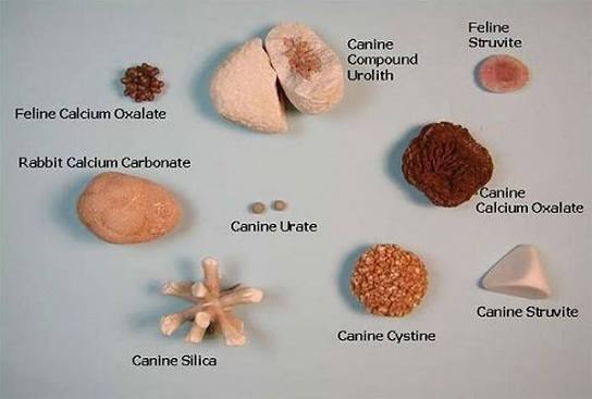
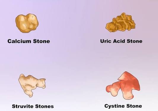
There are five main types of kidney stones: calcium oxalate, calcium phosphate, uric acid, struvite (infection-related), and cystine. Each has a distinct chemical composition, a different set of metabolic and anatomical risk factors, different radiographic characteristics (some are visible on plain X-ray, others are not), and different responses to dietary modification and pharmacological prevention. The distribution of stone types in the general population is heavily weighted toward calcium stones — calcium oxalate and calcium phosphate together account for approximately 80% of all kidney stones — but uric acid stones (5 to 10%), struvite stones (5 to 10%), and cystine stones (1 to 2%) are clinically significant and require type-specific management approaches that differ substantially from calcium stone protocols.
Calcium Oxalate Stones: The Most Common Type
Calcium oxalate stones are the most prevalent kidney stone type, responsible for approximately 70 to 75% of all stones in the United States. They form when calcium and oxalate — a naturally occurring compound found in many plant foods — combine in the urine at concentrations that exceed their solubility. There are two crystalline forms: calcium oxalate monohydrate (whewellite), which forms a darker, denser stone that is more resistant to shock wave lithotripsy; and calcium oxalate dihydrate (weddellite), a lighter, more friable stone that fragments more easily with lithotripsy. Stone analysis determines which form is present and can influence treatment selection.
The risk factors for calcium oxalate stone formation are well characterized. Hypercalciuria — elevated urinary calcium excretion — is present in up to 40 to 60% of calcium stone formers and can result from increased intestinal calcium absorption (absorptive hypercalciuria), impaired renal tubular calcium reabsorption (renal hypercalciuria), or elevated bone resorption from primary hyperparathyroidism or vitamin D excess. Hyperoxaluria — elevated urinary oxalate — can be dietary (from excessive oxalate intake), enteric (from fat malabsorption that increases colonic oxalate absorption, seen in inflammatory bowel disease and after bariatric surgery), or rarely primary (from inherited enzyme defects in oxalate metabolism). Hypocitraturia — low urinary citrate — is an important and often underappreciated risk factor: citrate is a natural inhibitor of calcium crystallization in the urine, and its deficiency (caused by systemic acidosis, potassium depletion, or excessive animal protein intake) allows calcium oxalate and calcium phosphate crystals to form more readily.
A counterintuitive but critically important point about calcium oxalate stones is that reducing dietary calcium increases stone risk rather than reducing it. This is because dietary calcium normally binds oxalate in the intestinal lumen and carries it out in the stool; when dietary calcium is reduced, more oxalate is available for intestinal absorption, raising urinary oxalate and increasing the risk of calcium oxalate crystal formation. Clinical trials have confirmed that a normal-calcium, low-oxalate, low-sodium diet produces significantly fewer recurrent calcium oxalate stones than a low-calcium diet. The correct dietary approach is maintaining adequate calcium intake (1,000 to 1,200 mg daily from food sources, ideally consumed with meals) while moderating oxalate intake and avoiding dietary patterns that elevate urinary sodium and animal protein.
Calcium Phosphate Stones
Calcium phosphate stones account for approximately 5 to 10% of all kidney stones and are most often associated with conditions that produce persistently alkaline urine — a urinary pH above 6.5 to 7.0. The two most common calcium phosphate mineral forms are hydroxyapatite and brushite (calcium hydrogen phosphate dihydrate). Brushite stones are notably harder and more resistant to shock wave lithotripsy than calcium oxalate stones, often requiring ureteroscopy or percutaneous nephrolithotomy for treatment.
The most important systemic condition causing calcium phosphate stones is renal tubular acidosis (RTA) — specifically Type 1 (distal) RTA, in which the kidney is unable to adequately acidify the urine. This results in persistently alkaline urine (pH above 6.0 to 6.5), low urinary citrate (because the acidic environment normally needed to excrete citrate as its tricarboxylate form is lost), and hypercalciuria — a combination that strongly promotes calcium phosphate crystallization. Primary hyperparathyroidism, by elevating serum calcium and stimulating calcitonin-independent bone resorption, also produces hypercalciuria that predisposes to calcium phosphate as well as calcium oxalate stones. Patients who form predominantly calcium phosphate stones should be evaluated for these underlying metabolic disorders with serum calcium, PTH, bicarbonate, chloride, and a 24-hour urine collection assessing pH, calcium, citrate, and phosphate.
Uric Acid Stones: A Metabolic Stone Type
Uric acid stones account for approximately 5 to 10% of kidney stones in the United States and represent a distinct pathophysiology from calcium stones. Unlike calcium stones, they are radiolucent — they do not absorb X-rays and therefore do not appear on plain abdominal X-rays or standard KUB films. CT scanning without contrast is the imaging modality that detects them reliably. The clinical implication of radiolucency is that a patient with flank pain and a negative KUB X-ray may still have a uric acid stone, and CT is required to confirm the diagnosis.
Uric acid stones form when urine pH is persistently low (acidic), typically below 5.5, in combination with elevated urinary uric acid concentration. The conditions most strongly associated with uric acid stone formation are: gout and hyperuricemia (from purine-rich diets, rapid cell turnover from chemotherapy, or primary disorders of uric acid metabolism); type 2 diabetes and metabolic syndrome (insulin resistance impairs renal tubular ammonium excretion, reducing urinary pH); chronic diarrheal states (fluid and bicarbonate loss produces concentrated, acidic urine); and high animal protein intake (which generates an acid load, lowers urinary pH, and increases urinary uric acid).
The most important clinical feature of uric acid stones is that they are uniquely amenable to dissolution therapy — complete medical dissolution of existing stones using urinary alkalinization. By raising urinary pH above 6.5 to 7.0 with potassium citrate or sodium bicarbonate, the solubility of uric acid in urine increases dramatically (uric acid solubility is 15-fold greater at pH 7.0 than at pH 5.0), allowing existing stones to dissolve over weeks to months and preventing new stone formation. This makes uric acid stones the only common stone type that can be treated without surgical or urological intervention — a significant advantage that makes correct type identification critically important.
Struvite Stones: Infection-Driven Calculi
Struvite stones — also called infection stones, triple phosphate stones, or magnesium ammonium phosphate stones — are caused by infection with urease-producing bacteria rather than by a metabolic disorder. The urease enzyme, produced by bacteria such as Proteus mirabilis, Klebsiella pneumoniae, Pseudomonas aeruginosa, and Staphylococcus saprophyticus, splits urea into ammonia and carbon dioxide. Ammonia alkalinizes the urine to a pH above 7.2, at which point struvite (magnesium ammonium phosphate) and carbonate apatite crystallize rapidly. Because this crystallization is driven by bacterial activity rather than slow metabolic accumulation, struvite stones can grow with remarkable speed — sometimes filling the entire collecting system of the kidney within months as staghorn calculi (stones shaped like a deer antler following the contours of the renal pelvis and calices).
Struvite stones are more common in women than men — a reflection of the higher prevalence of UTIs in women, which provide the bacterial inoculum that initiates struvite crystallization. They are also particularly common in patients with structural urinary tract abnormalities (neurogenic bladder, urinary diversion, vesicoureteral reflux), spinal cord injury, or indwelling urinary catheters — all conditions that predispose to chronic urinary tract infection. The management of struvite stones differs fundamentally from other stone types: complete stone removal is the treatment goal (not merely stone-free status in the renal pelvis, because bacterial biofilm within stone fragments perpetuates infection and stone regrowth), and long-term antibiotic therapy and urease inhibition (with acetohydroxamic acid) may be required to prevent recurrence in high-risk patients.
Cystine Stones: A Hereditary Stone Disease
Cystine stones are the least common of the five main types, accounting for approximately 1 to 2% of kidney stones in adults but a higher proportion in children — where they represent up to 6 to 8% of pediatric stone cases. They are caused by an autosomal recessive disorder of amino acid transport called cystinuria, in which mutations in the SLC3A1 or SLC7A9 genes impair the renal tubular reabsorption of cystine and three related amino acids (ornithine, lysine, and arginine). The resulting excess cystine in the urine exceeds its solubility (which is far lower than that of the common urinary ions), causing cystine crystals to form at any urinary pH but particularly in acidic urine.
Cystine stones are characteristically recurrent and difficult to treat: they are among the hardest kidney stones, resistant to shock wave lithotripsy, and require ureteroscopic laser lithotripsy or percutaneous nephrolithotomy for large stone removal. Medical management includes extremely high fluid intake (targeting urine output above 3 liters per day to dilute urinary cystine below its solubility threshold), urinary alkalinization with potassium citrate (cystine solubility increases markedly above pH 7.5), and in severe cases, thiol-binding drugs (d-penicillamine or tiopronin) that chemically bind cystine and increase its solubility. Cystinuria requires lifelong management and is best handled by urologists with specific expertise in complex stone disease and metabolic stone disorders.
How Stone Type Is Determined
Stone composition analysis is performed by infrared spectroscopy or X-ray diffraction on a stone that has been retrieved — either passed spontaneously (collected in a urine strainer), removed surgically (by ureteroscopy or percutaneous nephrolithotomy), or retrieved from an indwelling ureteral stent after stone fragmentation. The analysis identifies the mineral composition with high precision and, in the case of mixed stones, quantifies the proportion of each mineral. Stone analysis is recommended for all first-time stone formers and for any recurrent stone former whose prevention strategy is being adjusted.
When a stone is not available for analysis, stone type can often be inferred from: urinary pH (consistently acidic urine favors uric acid; consistently alkaline urine favors calcium phosphate or struvite); urine culture results (struvite stones are associated with positive cultures for urease-producing organisms); imaging characteristics (radiolucent stones on plain X-ray that are visible on CT are likely uric acid; very dense stones on CT — with high Hounsfield units — are likely calcium oxalate monohydrate or brushite); and serum chemistry (elevated calcium or PTH suggests calcium stones; elevated uric acid suggests uric acid stones). A 24-hour urine collection provides the most complete metabolic picture and often allows the likely stone type to be inferred from the combination of urinary risk factors even without direct stone analysis.
For a detailed discussion of calcium oxalate stones specifically — the most common type — see the calcium oxalate stones article on Horizon Health Guide. The kidney stones overview on this site covers treatment options across all stone types. For clinical reference, the American Urological Association kidney stone guidelines, the NIDDK kidney stone resource, and the StatPearls nephrolithiasis review provide comprehensive evidence-based information.
Sources: AUA Kidney Stone Guidelines · NIDDK — Kidney Stones · StatPearls — Nephrolithiasis
Dietary Factors That Influence Stone Type and Formation Risk
Diet plays a different role in the formation of each kidney stone type, and the dietary advice appropriate for one stone type can be counterproductive — or even harmful — for another. This is why stone type identification is not merely an academic exercise but has direct, practical implications for the dietary recommendations a patient receives. A one-size-fits-all approach to kidney stone prevention consistently underperforms type-specific dietary counseling in clinical trials.
For calcium oxalate stones, the key dietary targets are: limiting sodium intake (which increases urinary calcium by reducing tubular calcium reabsorption), maintaining adequate dietary calcium (not reducing it — see the oxalate-binding mechanism described above), moderating consumption of high-oxalate foods (dark leafy greens like spinach and Swiss chard, nuts and nut butters, chocolate, rhubarb, bran, and strong tea), and limiting animal protein intake (which acidifies urine, increases urinary uric acid, reduces urinary citrate, and increases urinary calcium). Vitamin C supplementation above 1,000 mg per day increases urinary oxalate and should be used cautiously in calcium oxalate stone formers. High fructose intake — from sweetened beverages and ultra-processed foods — independently increases urinary calcium and uric acid, and population studies consistently show associations between fructose consumption and kidney stone risk.
For uric acid stones, the dietary priority is reducing purine intake and limiting animal protein to reduce the acid load on the kidneys. High-purine foods include organ meats (liver, kidney, sweetbreads), shellfish (anchovies, sardines, mussels, scallops), red meat, and high-fructose foods (which independently raise uric acid levels by accelerating purine catabolism). Alcohol — particularly beer, which contains both purines and yeast fermentation products that raise uric acid — should be minimized. Conversely, vegetable sources of protein (legumes, tofu) are metabolically different from animal protein and do not raise urinary uric acid to the same degree; plant-based dietary patterns consistently show lower uric acid stone risk than omnivorous diets matched for total protein intake.
For struvite stones, diet is less directly relevant than for metabolic stone types — the primary driver is infection, and no dietary change eliminates urease-producing bacterial infection. However, adequate hydration to maintain high urine volumes and to flush the collecting system between antibiotic treatment courses is important. For cystine stones, dietary protein restriction (to reduce the intake of precursor amino acids including methionine, which is metabolized to cystine) is a component of prevention, but the dominant strategy is high fluid intake and urinary alkalinization; extreme protein restriction sufficient to meaningfully reduce cystine excretion is rarely achievable without compromise of nutritional adequacy.
Medications Used to Prevent Each Stone Type
Beyond dietary modification, pharmacological prevention is indicated for patients with recurrent stones, high stone burden, bilateral stones, solitary kidney, nephrocalcinosis, or identified metabolic disorders that cannot be adequately corrected by diet alone. The choice of medication depends almost entirely on stone type and the specific metabolic abnormality identified on 24-hour urine collection.
For calcium oxalate and calcium phosphate stones with hypercalciuria, thiazide diuretics (hydrochlorothiazide, chlorthalidone, indapamide) are the first-line pharmacological agent. They reduce urinary calcium by enhancing renal tubular calcium reabsorption, thereby reducing the driving force for calcium crystallization. For stones associated with hypocitraturia — low urinary citrate — potassium citrate raises urinary citrate levels, raises urinary pH, and provides an alkaline load that helps correct the mild systemic acidosis that often underlies citrate deficiency. For calcium oxalate stones associated with hyperuricosuria (elevated urinary uric acid, which seeds heterogeneous calcium oxalate nucleation), allopurinol (a xanthine oxidase inhibitor that reduces uric acid production) is effective even when serum uric acid is not elevated.
For uric acid stones, the treatment of choice is urinary alkalinization with potassium citrate — targeting a urinary pH of 6.5 to 7.0. In patients who cannot tolerate potassium citrate, sodium bicarbonate is an alternative, though it carries additional sodium load and may worsen hypercalciuria in calcium stone formers. Allopurinol is added for patients with significant hyperuricemia or hyperuricosuria in addition to alkalinization. For struvite stones, the medical adjuncts to surgical clearance include appropriate antibiotic therapy guided by stone culture and sensitivity, and in selected high-risk patients, acetohydroxamic acid (AHA), a urease inhibitor that blocks the enzymatic action responsible for struvite crystallization — though AHA has significant side effects (hemolytic anemia, thrombophlebitis, neurological symptoms) that limit its use to patients in whom stone clearance cannot otherwise be achieved. For cystine stones, the binding agents tiopronin (alpha-mercaptopropionylglycine) and d-penicillamine form mixed disulfides with cysteine that are far more soluble than cystine itself, effectively reducing the concentration of free cystine in the urine below the crystallization threshold.
Stone Type and Surgical Treatment Selection
Stone type influences not only medical prevention but also the selection of urological procedures when intervention is required. The hardness, radiopacity, and composition of a stone affect how readily it fragments under the acoustic pressure waves used in shock wave lithotripsy (SWL) and how effectively it can be dusted or fragmented with laser energy in ureteroscopy.
Calcium oxalate dihydrate and uric acid stones are generally the most amenable to SWL — they fragment relatively readily under acoustic shockwaves and, for uric acid stones, may even be amenable to dissolution before surgical intervention is required. Calcium oxalate monohydrate, brushite (calcium phosphate), and cystine stones are substantially harder — they have higher structural integrity, require more energy to fragment, and often produce incomplete fragmentation with SWL, leading to residual stone burden that may obstruct or require re-treatment. For these harder stone types, ureteroscopy with holmium laser lithotripsy — which delivers targeted laser energy directly to the stone — or percutaneous nephrolithotomy (PCNL) for large renal stones provides more reliable stone clearance. Struvite staghorn calculi, given their size and location filling the collecting system, almost universally require PCNL as the primary treatment modality, often performed in staged procedures when the stone burden is very large.
The density of the stone on non-contrast CT — measured in Hounsfield units (HU) — is one of the best preoperative predictors of SWL success available without stone composition analysis. Stones with CT density above 1,000 HU are substantially harder and less likely to fragment adequately with SWL than stones below 500 HU. Urologists use CT density alongside stone size (stones above 10 to 15 mm are generally poor candidates for SWL regardless of composition), stone location (upper ureter and renal pelvis are more favorable for SWL than lower pole calyx), and patient body habitus (skin-to-stone distance affects SWL efficacy) to select the most appropriate procedure for each individual patient. Understanding that stone type — which determines hardness and composition — is one driver of this surgical decision-making helps patients engage more meaningfully in conversations with their urologist about treatment options.
When to See a Specialist About Kidney Stone Type
A urologist should evaluate all patients who have passed or had a kidney stone removed, regardless of stone type. The initial evaluation includes imaging to assess for residual stone burden, stone analysis when stone material is available, and at minimum a focused history for the metabolic risk factors specific to the stone type identified. For first-time stone formers with a single small calcium stone and no identifiable risk factors, a simplified evaluation (serum calcium, uric acid, creatinine, and a spot urine for calcium-to-creatinine ratio) is often appropriate before deciding whether a full 24-hour urine collection is warranted. For recurrent stone formers, bilateral stone disease, nephrocalcinosis, any non-calcium stone type, or first presentation in children, a complete metabolic evaluation is indicated — this includes two 24-hour urine collections (to average day-to-day variability) measuring volume, pH, calcium, oxalate, uric acid, citrate, sodium, creatinine, and phosphate.
Patients with cystine stones should be referred to a center with specific expertise in hereditary stone disease, given the lifelong nature of the condition, the complexity of medical management, and the benefit of genetic counseling for first-degree relatives who may share the cystinuria mutation. Patients with struvite staghorn calculi should be evaluated urgently by a urologist with PCNL experience, as large infection stones carry risk of sepsis, progressive renal damage, and kidney loss if not appropriately treated. Patients with calcium stones and suspected primary hyperparathyroidism (elevated serum calcium or PTH) should be referred to endocrinology, as the underlying cause — most commonly a parathyroid adenoma — is surgically correctable, and parathyroidectomy is typically curative for the associated stone disease.
For additional context on kidney stone symptoms, causes, and general treatment approaches, see the complete kidney stones overview on Horizon Health Guide. For understanding the early warning signs that distinguish a kidney stone from other causes of flank and abdominal pain, the early signs of kidney stones article provides a detailed symptom-by-symptom review. The kidney stone pain guide describes the characteristic features of renal colic in detail and explains how it differs from other conditions in the clinical differential diagnosis.
I had a kidney stone last year and the ER doctor told me it was ‘calcium’ but never explained there are actually different types that need different management. I ended up just being told to drink more water and avoid calcium — which this article explains is actually wrong for calcium oxalate stones! I wish I had found this information before my follow-up appointment so I could have asked smarter questions about whether I actually needed stone analysis and whether my diet advice was correct for my specific stone type. The section on how dietary advice differs by stone type was particularly eye-opening.
As a nephrologist who manages complex stone disease, I appreciate this thorough breakdown of stone types. The point about uric acid stones being uniquely amenable to dissolution therapy is critically important and often underutilized — many patients with radiolucent stones on X-ray get sent straight to urology for procedures when a trial of urinary alkalinization with potassium citrate might dissolve the stone completely without any intervention. The section on cystinuria is also accurate — it is genuinely a lifelong disorder that benefits from subspecialty management, and the thiol-binding drugs have significant side effect profiles that require careful monitoring.
Thank you for adding that clinical perspective, Marcus — the point about uric acid stone dissolution being underutilized is one we wanted to emphasize precisely because it represents a category of kidney stone disease where identification of the correct type changes the entire management paradigm. A patient with a radiolucent stone and persistently acidic urine who is started on potassium citrate and achieves alkalinization to pH 6.5–7.0 may dissolve their stone entirely over weeks without any urological procedure — but this outcome requires recognizing that the stone is uric acid, not calcium. Rachel, your experience highlights why stone type identification matters so much beyond the acute episode — the dietary advice for calcium oxalate stones differs substantially from generic kidney stone advice, and stone analysis after recovery should be standard rather than optional for patients who want to prevent recurrence.