
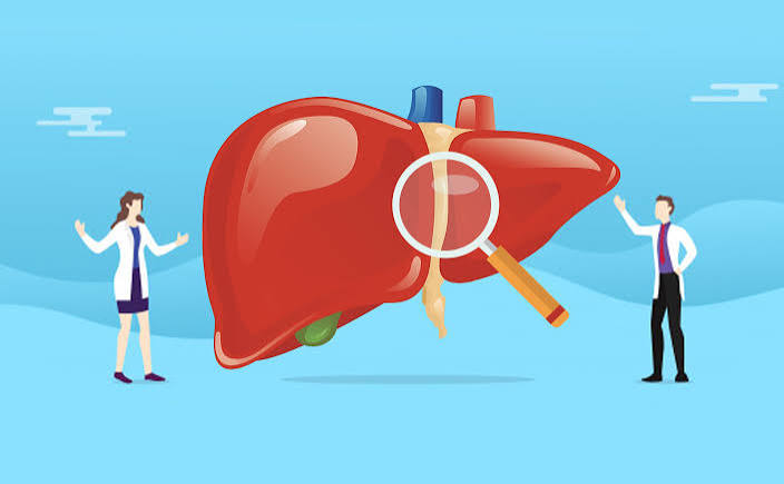
The liver is the body’s largest solid organ, weighing approximately 1.4 kilograms in adults and located in the upper right quadrant of the abdomen beneath the ribcage. It is one of the most complex organs in the body — performing over 500 distinct functions simultaneously, without pause, every moment of your life. Unlike the heart or lungs, the liver’s work is largely invisible: it does not beat, it does not move air in and out, it makes no sound. Yet it is performing continuous chemical transformations — processing everything absorbed from the gut, synthesizing the proteins that keep blood flowing and clotting, regulating blood sugar between meals, producing bile for fat digestion, and detoxifying a constant stream of substances that would otherwise accumulate to toxic levels. Understanding how the liver works at a mechanistic level makes clear why liver disease has such wide-ranging consequences — and why even early, silent liver damage is worth preventing.
The Liver’s Anatomy: Structure That Enables Function
The liver is divided into two main lobes — right (larger) and left — and further subdivided into eight functional segments, each with its own blood supply and bile drainage. This segmental organization means that surgical resection of one segment can occur without disrupting the function of adjacent segments — a property that makes liver surgery and partial resection technically possible.
The liver has a unique dual blood supply. Approximately 75% of its blood comes from the portal vein, which drains the entire gastrointestinal tract and spleen — delivering nutrient-rich, but also toxin- and bacteria-laden, blood from the gut directly to the liver for processing before it reaches general circulation. The remaining 25% comes from the hepatic artery, which delivers oxygenated blood. This arrangement gives the liver first-pass access to everything absorbed from the intestines, which is the basis of its gatekeeping and metabolic functions.
The functional units of the liver are called hepatic lobules — hexagonal structures visible under a microscope. Each lobule is organized around a central vein, with portal triads (containing a portal venule, hepatic arteriole, and bile ductule) at each corner. Hepatocytes — the main liver cells, making up approximately 80% of the liver’s mass — are arranged in plates radiating from the central vein outward to the portal triads. Blood flows inward from the portal triads to the central vein; bile flows in the opposite direction, from hepatocytes into bile canaliculi (tiny channels) that drain into bile ducts. This counter-current arrangement ensures both blood processing and bile collection can occur simultaneously in the same tissue.
Bile Production: The Digestive Role
The liver produces 600–800 mL of bile per day — a yellow-green digestive fluid containing bile acids (also called bile salts), phospholipids (primarily lecithin), cholesterol, bilirubin, water, and electrolytes. Bile’s primary digestive function is to emulsify dietary fat. Fat molecules are hydrophobic — they repel water and clump together in aqueous environments like the small intestine’s contents. Bile acids have both hydrophilic (water-attracting) and hydrophobic (fat-attracting) regions, allowing them to surround fat globules and break them into microscopic droplets (micelles) that can be accessed by pancreatic lipase. Without bile, fat digestion is severely impaired — producing the fatty, floating, foul-smelling stools (steatorrhea) characteristic of liver disease, bile duct obstruction, and fat malabsorption syndromes.
After performing their digestive function in the small intestine, approximately 95% of bile acids are reabsorbed in the terminal ileum and returned to the liver via the portal vein — a process called the enterohepatic circulation. The liver recaptures these bile acids and secretes them into bile again, recycling the same pool of bile acids 6–10 times per day. This efficient recycling is why the liver only needs to synthesize new bile acids to replace the small fraction lost in stool. Conditions that disrupt the terminal ileum (Crohn’s disease, surgical resection) break this cycle, causing bile acid malabsorption and watery diarrhea.
Detoxification and Drug Metabolism
The liver is the body’s primary detoxification organ. Every substance absorbed from the gut — including nutrients, medications, alcohol, bacterial endotoxins, environmental chemicals, and metabolic byproducts — passes through the liver via the portal vein before reaching systemic circulation. The liver transforms these compounds through a two-phase enzymatic process.
Phase I reactions are primarily carried out by the cytochrome P450 (CYP) enzyme family — particularly CYP3A4, CYP2D6, and CYP2C9, which together metabolize the majority of commonly used drugs. Phase I reactions typically convert lipid-soluble compounds (which would otherwise accumulate in fatty tissues) into more water-soluble, reactive intermediates. For some drugs, this phase produces active metabolites — the Phase I product is the pharmacologically active form. For others, Phase I produces toxic intermediates that Phase II must quickly process.
Phase II reactions conjugate (attach chemical groups to) Phase I products, making them highly water-soluble and suitable for excretion in bile or urine. Common conjugation reactions include glucuronidation, sulfation, and glutathione conjugation. Acetaminophen metabolism illustrates why both phases matter: at therapeutic doses, Phase II glucuronidation and sulfation handle the majority of the drug safely. At overdose doses, Phase II capacity is overwhelmed, and a toxic Phase I intermediate (NAPQI) accumulates faster than it can be conjugated, causing acute liver cell death. This is why acetaminophen overdose — even inadvertent — is the leading cause of acute liver failure in the United States.
The liver also clears ammonia — a byproduct of protein metabolism and bacterial activity — from the portal blood by converting it to urea via the urea cycle. Urea is harmless, water-soluble, and excreted in urine. When cirrhosis impairs this process, ammonia accumulates in the bloodstream and crosses the blood-brain barrier, causing hepatic encephalopathy — cognitive impairment, confusion, and in severe cases, coma. The NIDDK cirrhosis resource covers this complication in detail for patients managing advanced liver disease.
Protein Synthesis: Building the Body’s Infrastructure
The liver is the primary factory for plasma proteins — the proteins that circulate in the blood performing structural and functional roles throughout the body. The most clinically significant of these are:
Albumin (10–15 grams per day synthesized by the liver) is the most abundant plasma protein. Its roles include maintaining colloid osmotic pressure — the force that keeps fluid inside blood vessels rather than leaking into surrounding tissues. When albumin falls below approximately 25–30 g/L (normal: 35–50 g/L), osmotic pressure is insufficient to retain fluid in the vascular compartment, and fluid accumulates in the abdomen (ascites) and legs (peripheral edema). Albumin also serves as the main transport protein for fatty acids, hormones (thyroid hormones, cortisol), calcium, and many drugs. Drug dosing is affected by albumin levels because a drug bound to albumin is pharmacologically inactive — when albumin is low, the free (active) fraction of drug is higher.
Clotting factors. The liver synthesizes most of the coagulation cascade proteins — factors I (fibrinogen), II (prothrombin), V, VII, IX, X, and XI, along with protein C and protein S (anticoagulant proteins). Factors II, VII, IX, and X require vitamin K for synthesis. Prothrombin time (PT) and its derived measure INR (International Normalized Ratio) reflect the activity of these clotting factors — PT/INR is therefore one of the most sensitive blood tests for acute liver synthetic function. A prolonged PT in a person with liver disease indicates significant impairment of the liver’s synthetic capacity — it is incorporated into both the Child-Pugh score and the MELD score, which are the two main systems for assessing liver disease severity and prognosis.
Acute phase proteins. During infection or inflammation, the liver rapidly upregulates production of C-reactive protein (CRP), fibrinogen, and complement proteins — part of the innate immune response. This is why CRP rises in acute illness and why the liver is considered part of the immune system’s rapid-response infrastructure.
Glucose and Lipid Metabolism
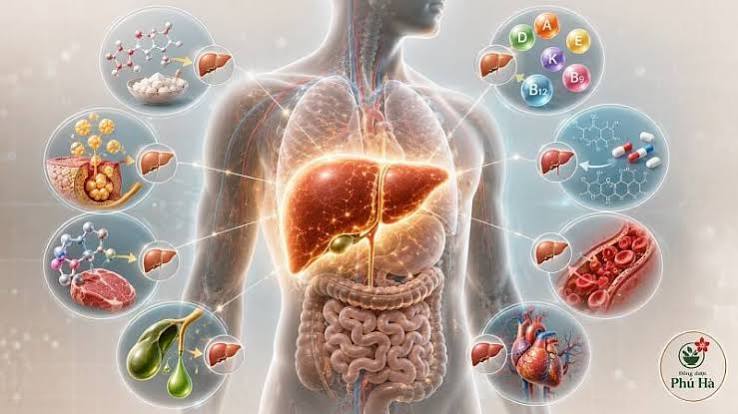
The liver is the body’s primary metabolic regulator — the organ that coordinates blood glucose and lipid levels between meals, after meals, and during exercise or fasting.
Glucose metabolism. After a carbohydrate-containing meal, absorbed glucose arrives at the liver via the portal vein. The liver removes approximately 50–60% of this portal glucose, converting it to glycogen (glycogenesis) for storage. The liver can store approximately 100 grams of glycogen. Between meals, as blood glucose falls, the liver releases glucose back into circulation by breaking down glycogen (glycogenolysis) and by synthesizing new glucose from non-glucose precursors — lactate, glycerol, and amino acids — via gluconeogenesis. This liver-driven glucose homeostasis prevents hypoglycemia during fasting. In fatty liver disease and insulin resistance, impaired hepatic glucose regulation contributes to fasting hyperglycemia — the liver continues gluconeogenesis even when blood glucose is already elevated, because insulin signaling is defective.
Lipid metabolism. The liver synthesizes approximately 80% of the body’s cholesterol and assembles the lipoprotein particles (VLDL, HDL) that transport lipids through the bloodstream. It converts excess cholesterol into bile acids for excretion — the only way the body eliminates cholesterol. When this conversion is impaired (in liver disease) or production exceeds elimination (in metabolic syndrome), cholesterol accumulates. The liver also converts excess dietary carbohydrate and protein into fatty acids via de novo lipogenesis — a process that is significantly upregulated in fatty liver disease, where excess fructose and refined carbohydrates drive hepatic fat production beyond the liver’s export capacity, causing fat to accumulate in hepatocytes.
Bilirubin Processing and Bile Excretion
Bilirubin is a yellow pigment produced when the body breaks down hemoglobin from old red blood cells — approximately 120 days old — which are dismantled by macrophages in the spleen and liver. The initial product, unconjugated (indirect) bilirubin, is insoluble in water and is transported to the liver bound to albumin. In the liver, hepatocytes conjugate it with glucuronic acid (conjugated/direct bilirubin), making it water-soluble. Conjugated bilirubin is secreted into bile and travels through bile ducts into the small intestine, where gut bacteria convert it to urobilinogen. Most urobilinogen is excreted in stool (giving stool its brown color); a small amount is reabsorbed and excreted in urine (giving urine its yellow color).
This pathway explains the specific patterns of jaundice. Pre-hepatic jaundice (excessive red blood cell breakdown — hemolysis) elevates unconjugated bilirubin. Hepatic jaundice (liver cell damage — hepatitis, cirrhosis) elevates both fractions as the liver’s capacity to conjugate and excrete is impaired. Post-hepatic (obstructive) jaundice — bile duct blockage from gallstones, tumor, or stricture — causes conjugated bilirubin to back up into the bloodstream, producing dark urine (bilirubin in urine), pale stool (no bilirubin reaching the gut), and itching (bile salts depositing in skin). The pattern of bilirubin fractionation guides clinical diagnosis.
Vitamin and Mineral Storage
The liver is the body’s primary storage organ for several essential nutrients. It stores approximately 1–2 years’ worth of vitamin B12 — which is why B12 deficiency from dietary inadequacy takes years to develop, even though the body cannot synthesize it. It stores fat-soluble vitamins A, D, E, and K, mobilizing them as needed. Iron is stored in the liver as ferritin — which is why ferritin blood levels reflect liver iron stores and why liver disease can disrupt iron balance in both directions (iron overload in hemochromatosis; iron deficiency from impaired storage in advanced cirrhosis).
The liver also stores glycogen (discussed above), copper (relevant in Wilson’s disease, where copper accumulation causes liver damage), and several B vitamins. This storage capacity means that depletion states — vitamin A deficiency, D deficiency, K deficiency leading to bleeding — typically only manifest when liver function is significantly compromised or when dietary intake has been extremely poor for an extended period.
The Liver’s Regenerative Capacity
The liver is uniquely capable of regeneration. After partial hepatectomy (surgical removal of up to 70% of the liver), the remaining tissue can regenerate to full mass within 6–8 weeks through compensatory hyperplasia of hepatocytes — existing liver cells multiply rather than new stem cells being recruited. This property enables living-donor liver transplantation, where a segment of a healthy donor’s liver is transplanted and both the donor’s remaining liver and the recipient’s new liver regenerate to adequate size.
This regenerative capacity is not unlimited. Chronic, repeated liver injury — from ongoing alcohol use, uncontrolled fatty liver disease, or untreated hepatitis — overwhelms regeneration. Instead of restoring normal tissue, the repair process lays down scar tissue (fibrosis). Over years, fibrosis replaces increasingly large areas of functional liver tissue, distorts the liver’s architecture, and impairs blood flow through the portal system (portal hypertension). Once fibrosis progresses to cirrhosis — replacement of most normal liver tissue with scar — regeneration can no longer restore function, and liver transplantation becomes the only curative option for end-stage disease. The American Liver Foundation’s overview of liver regeneration provides further detail on what this capacity means in practical patient terms.
How Liver Function Is Monitored
Blood tests can assess different aspects of liver function simultaneously, which is why a standard liver function panel provides so much diagnostic information. The key markers and what they reflect:
- ALT and AST — enzymes released when liver cells are damaged; indicate hepatocellular injury (damage to liver cells themselves)
- ALP and GGT — elevated in bile duct disease and cholestatic conditions; indicate biliary obstruction or inflammation
- Bilirubin — reflects how well the liver is processing and excreting bile pigment; elevated in cell damage and obstruction
- Albumin — reflects synthetic function; low albumin indicates significant impairment of protein synthesis
- PT/INR — reflects clotting factor synthesis; the most sensitive acute indicator of liver synthetic failure
Beyond blood tests, imaging (ultrasound, CT, MRI) assesses liver size, texture, and vascular flow. FibroScan (transient elastography) measures liver stiffness non-invasively, providing an estimate of fibrosis stage. Liver biopsy remains the gold standard for staging fibrosis and assessing the presence of inflammation in conditions like fatty liver disease where enzyme levels may not reflect histological severity. How to understand these tests and what abnormal results mean is covered in the companion article on what is liver health.
Frequently Asked Questions
What does the liver do in simple terms?
The liver filters blood from the gut, processes nutrients, removes toxins and drugs, produces bile for fat digestion, makes proteins the body needs to function, regulates blood sugar, and stores vitamins and minerals. It is simultaneously the body’s chemical processing plant, detoxification center, and metabolic regulator.
How does the liver clean the blood?
All blood from the digestive tract flows through the liver via the portal vein before entering general circulation. Liver cells (hepatocytes) extract toxins, metabolize drugs, convert ammonia to urea, and process bacterial endotoxins absorbed from the gut. The cytochrome P450 enzyme system carries out the primary chemical transformations, converting lipid-soluble compounds into water-soluble forms that can be excreted in bile or urine.
Can the liver heal itself?
Yes — to a significant degree. The liver can regenerate from injury if the cause is removed before extensive scarring occurs. Early fatty liver disease is reversible with weight loss. Hepatitis C is now curable, allowing liver inflammation to resolve. Even mild to moderate fibrosis has shown partial reversibility in patients who sustained the underlying condition’s treatment. Advanced cirrhosis, however, is not meaningfully reversible — the architecture disruption from widespread scar tissue is permanent.
Why does the liver have no pain receptors?
The liver parenchyma (functional tissue) lacks pain-sensitive nerve fibers. Pain from liver conditions typically originates from stretching the liver’s outer capsule (Glisson’s capsule), which does have sensory innervation — this is why rapidly enlarging conditions (acute hepatitis, congestive hepatopathy from heart failure) can cause right upper quadrant discomfort, while slowly progressive chronic liver disease often produces no pain until very late. The absence of pain in early disease is why blood test screening is the primary detection tool.
What is the liver’s role in the immune system?
The liver contains Kupffer cells — the body’s largest population of tissue macrophages — which filter pathogens, bacterial fragments, and cellular debris arriving via the portal blood. The liver also produces complement proteins and acute phase reactants (like CRP and fibrinogen) that are central to the innate immune response. Chronic stimulation of Kupffer cells by gut-derived bacterial products — increased in states of gut dysbiosis and increased gut permeability — contributes to the chronic liver inflammation seen in fatty liver disease and alcoholic liver disease.
Sources: National Institute of Diabetes and Digestive and Kidney Diseases (NIDDK), Cirrhosis; American Liver Foundation, How Your Liver Works; Trefts E et al., Current Biology 2017 (liver biology review); Rui L, Comprehensive Physiology 2011 (hepatic glucose and lipid metabolism); Jaeschke H, Toxicological Sciences 2002 (acetaminophen hepatotoxicity); Michalopoulos GK, Hepatology 2007 (liver regeneration).
The Liver’s Immune Role: Kupffer Cells and Innate Defense
One of the liver’s least appreciated roles is as a frontline immune organ. The liver contains approximately 80–90% of the body’s tissue-resident macrophages — specialized immune cells called Kupffer cells — which line the hepatic sinusoids (the liver’s tiny blood vessels) and continuously sample the blood arriving from the portal vein. Because portal blood from the gut carries bacterial fragments, endotoxins, and other immunostimulatory molecules, the liver is constantly engaged in low-level immune surveillance and response.
Kupffer cells phagocytose (engulf and destroy) bacteria, debris, and foreign particles. They also release cytokines that coordinate downstream immune responses. In a healthy liver, this activity is carefully regulated — just enough immune activation to clear threats, not so much that the liver damages itself. In states of gut dysbiosis, increased intestinal permeability (“leaky gut”), or alcohol use, excessive bacterial endotoxin floods through the portal vein, chronically overstimulating Kupffer cells. This sustained activation generates chronic liver inflammation — a mechanism central to the progression of alcoholic liver disease and nonalcoholic fatty liver disease (MASLD). Understanding why gut health directly affects liver health through this portal-immune pathway underscores the practical value of treating the liver and digestive system as one integrated system, as discussed in the overview articles on what is digestive health and why liver and digestive health matter after age 40.
What Damages the Liver and How to Protect It
The liver’s remarkable functional reserve means that significant liver damage can occur before blood tests or symptoms reflect it — up to 75% of liver function can be lost before overt liver failure becomes apparent. This reserve is protective in acute illness but dangerous in chronic disease, where silent progression allows conditions like fatty liver disease to advance from steatosis to fibrosis without the patient realizing it.
The most consistently protective behaviors are those that reduce the drivers of chronic liver inflammation: maintaining a healthy weight (the single strongest modifiable factor for fatty liver disease), limiting alcohol consumption, completing hepatitis B vaccination, attending hepatitis C screening if not yet done, reviewing medications and supplements with a pharmacist for hepatotoxic potential, and maintaining physical activity that independently reduces hepatic fat content. The American Association for the Study of Liver Diseases (AASLD) patient resources provide disease-specific guidance for patients who have received a liver disease diagnosis or have significant risk factors and want structured information on monitoring and management.
I was one of the people diagnosed with hepatitis C who had no idea I had it — my infection was found through a routine blood test during a pre-surgery workup. My ALT had been slightly elevated for years but always attributed to other causes. After treatment with a 12-week course of sofosbuvir-based antivirals, I achieved SVR (sustained virological response) — which means the virus is undetectable in my blood at 12 weeks post-treatment and is considered cured. The explanation in this article of how the liver clears ammonia and processes bilirubin finally made sense of why I had felt persistently fatigued for years — my liver was working harder than it should have been to compensate for HCV-related inflammation. Post-treatment, my liver enzymes normalized within weeks and my energy improved over the following months.
Nguyen, the trajectory you describe — years of mildly elevated ALT attributed to other causes before HCV diagnosis — represents a significant diagnostic gap. The CDC’s recommendation for universal one-time hepatitis C screening for all adults 18–79 exists precisely because the pre-treatment era produced a generation of adults with chronic HCV who have no awareness of their infection. The cure rates with current DAA regimens are remarkable — SVR at 12 weeks is considered virological cure, and post-treatment liver fibrosis regression has been documented in patients with pre-treatment F2–F3 fibrosis.
The section on Phase I and Phase II drug metabolism is one I explain to patients regularly when they ask why their medication doses change after a liver diagnosis. What many patients don’t realize is that CYP3A4 — which metabolizes approximately 50% of all drugs — is particularly sensitive to both inhibition and induction. Grapefruit juice inhibits intestinal and hepatic CYP3A4, which is why many cardiological and immunological drugs specifically contraindicate grapefruit. St. John’s Wort, by contrast, is a potent CYP3A4 inducer — it speeds up drug metabolism, reducing blood levels of medications like warfarin, oral contraceptives, and HIV antiretrovirals significantly enough to cause clinical failures. These herb-drug interactions are real, significant, and require the same pharmacological awareness as conventional drug interactions.