
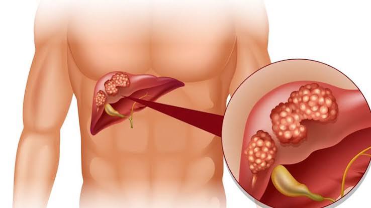
Right upper abdominal pain is one of the most common reasons adults seek medical evaluation — and one of the most diagnostically important, because the right upper quadrant contains two organ systems whose conditions range from entirely benign to life-threatening: the liver and the biliary system. The liver is the body’s largest internal organ, performing over 500 physiological functions including metabolizing drugs and toxins, synthesizing proteins essential for blood clotting, producing bile for fat digestion, and regulating glucose and cholesterol metabolism. The gallbladder and bile ducts store and deliver bile to the small intestine to assist fat absorption. Both systems produce pain through capsular stretch, inflammation, obstruction, or infection — and the patterns they generate in the right upper quadrant are among the most clinically distinctive in abdominal medicine. This guide covers the full range of biliary and liver causes of right upper abdominal pain, explains what liver health markers mean, and describes the evaluation and prevention frameworks that matter most. For the broader differential of upper abdominal pain across all regions, see upper abdominal pain: digestive and liver causes.
Biliary Causes of Right Upper Abdominal Pain
The biliary system — gallbladder, cystic duct, common hepatic duct, and common bile duct — responds directly to fat intake through cholecystokinin-mediated contraction, making post-fatty-meal pain one of the most specific patterns in RUQ disease. Gallstones are present in approximately 15 percent of US adults, and the majority are asymptomatic. Symptoms develop when a stone obstructs a duct — producing a predictable spectrum of conditions from benign biliary colic to life-threatening cholangitis.
Biliary colic is the most common symptomatic manifestation of gallstones. Pain begins 30 to 60 minutes after a fatty or large meal as cholecystokinin stimulates gallbladder contraction against an obstructing stone in the cystic duct. The pain is characteristically severe and steady (not truly colicky despite the name), located in the RUQ or epigastrium, may radiate to the right shoulder blade or scapula, and lasts 30 minutes to 6 hours before resolving spontaneously — either because the stone falls back into the gallbladder or because contraction subsides. The complete resolution between episodes is the hallmark of biliary colic that distinguishes it from acute cholecystitis. Nausea and vomiting are common during attacks. Abdominal ultrasound is the first-line diagnostic test — it detects gallstones with greater than 95 percent sensitivity. Laparoscopic cholecystectomy is the definitive treatment and eliminates future biliary colic entirely. Acute cholecystitis occurs when a gallstone obstructs the cystic duct persistently, leading to gallbladder wall inflammation. Unlike biliary colic, the pain does not resolve within 6 hours — it intensifies, becomes constant, and is accompanied by fever, elevated white blood cell count, and a positive Murphy’s sign (sudden cessation of inspiration when the examiner presses under the right costal margin during deep inhalation, due to pain from the inflamed gallbladder meeting the examining hand). Ultrasound shows gallbladder wall thickening (greater than 3mm), pericholecystic fluid, and a sonographic Murphy’s sign. Acute cholecystitis requires hospitalization, IV antibiotics, and cholecystectomy within 24 to 72 hours in most cases. Choledocholithiasis — a stone in the common bile duct — produces RUQ pain with a cholestatic laboratory pattern: elevated alkaline phosphatase (ALP), gamma-glutamyl transferase (GGT), and bilirubin, indicating biliary obstruction. MRCP is the non-invasive imaging of choice for detecting bile duct stones; ERCP with sphincterotomy and stone extraction is the treatment. Left untreated, choledocholithiasis may lead to ascending cholangitis. Ascending cholangitis — bacterial infection of the bile duct ascending from an obstructed biliary system — is a medical emergency. Charcot’s triad (RUQ pain, fever with rigors, and jaundice) is the classic presentation. Reynolds’ pentad adds confusion (altered mental status) and hypotension, indicating septic shock. Immediate broad-spectrum IV antibiotics and urgent biliary drainage (via ERCP or percutaneous drainage) are required. Delay significantly increases mortality. Any patient presenting with the combination of fever, jaundice, and RUQ pain requires emergency evaluation without waiting for outpatient imaging. The ACG biliary disease resource covers the evaluation and management framework.

Liver Disease as a Cause of Right Upper Abdominal Pain
The liver itself has no pain fibers within its parenchyma — pain from liver conditions is produced exclusively by stretching Glisson’s capsule (the fibrous covering), by adjacent peritoneal inflammation, or by referred pain through phrenic nerve irritation (producing right shoulder tip pain). This anatomy explains why liver disease is often entirely asymptomatic until it produces significant organ enlargement, complication, or capsular involvement — and why the symptoms that do appear can be subtly located in the right shoulder rather than clearly in the right abdomen.
Acute viral hepatitis — from hepatitis A, B, C, or E — produces RUQ aching or fullness from acute hepatic inflammation causing capsular stretch as the liver swells. The pain is constant and dull rather than episodic or sharp. It is accompanied by markedly elevated transaminases (ALT and AST often in the thousands in acute viral hepatitis), jaundice, dark urine, pale stools, fatigue, and loss of appetite. Hepatitis A is self-limited and resolves without specific treatment. Hepatitis B and C can progress to chronic hepatitis, cirrhosis, and hepatocellular carcinoma without treatment. Alcoholic hepatitis presents with RUQ pain, jaundice, fever, and elevated liver enzymes — with a characteristic AST:ALT ratio above 2:1 (because ethanol preferentially damages mitochondria and depletes pyridoxine, suppressing ALT synthesis). Severe alcoholic hepatitis carries significant short-term mortality and requires specific treatment (corticosteroids or pentoxifylline in selected cases) alongside alcohol cessation. Nonalcoholic fatty liver disease (NAFLD) — affecting approximately 25 to 30 percent of adults globally — is mostly asymptomatic but produces vague RUQ fullness in a minority of patients from hepatomegaly. The important clinical concern is progression from simple steatosis through steatohepatitis (MASH) to fibrosis and cirrhosis, which is driven by metabolic factors (obesity, insulin resistance, dyslipidemia) rather than pain. Most NAFLD is detected incidentally on imaging or through mildly elevated liver enzymes. Liver abscess — pyogenic or amebic — presents with constant, progressive RUQ pain, fever, and weight loss. Pyogenic abscess most commonly arises from biliary sources (cholangitis seeding the liver), hematogenous spread, or direct extension from an adjacent infection. Amebic abscess from Entamoeba histolytica is associated with travel to or immigration from endemic areas. CT of the abdomen with contrast is the best diagnostic imaging. Both types require drainage (percutaneous or surgical) combined with targeted antimicrobial therapy. Budd-Chiari syndrome — hepatic vein thrombosis causing impaired hepatic venous outflow — produces hepatomegaly, RUQ pain, and ascites as the liver swells from impaired outflow. It is associated with hypercoagulable conditions: polycythemia vera, factor V Leiden, antiphospholipid syndrome, pregnancy, and oral contraceptive use. Doppler ultrasound showing absent or reversed hepatic vein flow is the diagnostic study; treatment requires anticoagulation and often vascular intervention or transplant. Hepatocellular carcinoma (HCC) typically arises in a cirrhotic liver and may produce RUQ pain as it grows and stretches Glisson’s capsule or invades portal structures. All patients with cirrhosis should undergo 6-monthly liver ultrasound with AFP measurement for HCC surveillance, since prognosis is dramatically better when HCC is detected at an early, resectable stage. The NIDDK liver disease library covers the spectrum of hepatic conditions with patient-oriented detail.
Understanding Liver Health Markers
Liver blood tests are among the most commonly ordered laboratory tests in medicine, but their interpretation is frequently misunderstood. Understanding what each marker measures helps adults interpret their own results and ask better questions of their clinicians.
ALT (alanine aminotransferase) is the most liver-specific transaminase. It is released into the bloodstream when hepatocytes are injured or dying — making it the primary marker for hepatocellular damage. Elevated ALT indicates hepatocyte injury from any cause: viral hepatitis, alcoholic liver disease, NAFLD, DILI, autoimmune hepatitis, or ischemic hepatitis. Mild elevations (up to 3 times the upper limit of normal) are common in NAFLD and DILI; very high elevations (50 to 100 times normal) indicate acute hepatocellular injury from viral hepatitis or ischemia. AST (aspartate aminotransferase) is less liver-specific than ALT — it is also elevated in muscle injury (myocardial infarction, rhabdomyolysis, strenuous exercise). An AST:ALT ratio above 2:1 suggests alcoholic liver disease or advanced cirrhosis. ALP (alkaline phosphatase) is elevated in biliary conditions — obstruction, cholestasis, primary biliary cholangitis, primary sclerosing cholangitis — and in bone disease. Isolated ALP elevation with normal transaminases suggests biliary or bone origin; GGT elevation concurrent with ALP confirms hepatic/biliary origin. GGT (gamma-glutamyl transferase) is a sensitive marker of hepatobiliary disease and alcohol use. It is elevated in biliary obstruction, fatty liver disease, alcoholic liver disease, and drug-induced liver injury. GGT in isolation is not diagnostic but adds specificity when interpreted with ALP and transaminases. Bilirubin — produced from hemoglobin breakdown — is conjugated by the liver for biliary excretion. Elevated direct (conjugated) bilirubin indicates hepatocellular dysfunction or biliary obstruction. Elevated indirect (unconjugated) bilirubin indicates excess bilirubin production (hemolysis) or impaired hepatic uptake/conjugation (Gilbert’s syndrome). Jaundice (yellow sclera and skin) becomes visible when total bilirubin exceeds approximately 2.5 mg/dL. Albumin and prothrombin time (PT/INR) assess liver synthetic function — both decrease in chronic liver failure. A declining albumin or prolonged PT in a patient with known liver disease indicates decompensation and is associated with worse prognosis. For reference ranges and clinical interpretation, see liver and digestive health numbers every adult should know.
When Right Upper Abdominal Pain Is an Emergency
Most RUQ pain is not an emergency — biliary colic is uncomfortable but not immediately dangerous, and hepatitis produces pain that warrants evaluation but not emergency care in most presentations. However, several patterns require same-day emergency assessment.
Seek emergency evaluation for RUQ pain when: fever accompanies the pain — suggesting acute cholecystitis (fever + RUQ pain) or cholangitis (fever + jaundice + RUQ pain), both of which require urgent treatment; pain has been present for more than 6 hours without any resolution — which distinguishes acute cholecystitis from biliary colic and requires hospitalization; jaundice has appeared — which indicates biliary obstruction (stones, stricture, or malignancy) or significant hepatocellular dysfunction, both requiring urgent evaluation; altered mental status or hypotension accompanies fever and jaundice — Reynolds’ pentad indicating biliary septic shock; right upper quadrant pain in a patient with known cirrhosis who develops worsening abdominal pain — which may indicate variceal bleeding, spontaneous bacterial peritonitis, or HCC progression; or sudden severe RUQ pain in a patient with a hematologic malignancy or after recent viral illness — which should raise concern for splenic rupture or hepatic vein thrombosis. The American Liver Foundation provides accessible condition-specific guides for liver disease patients. For a comprehensive overview of common digestive conditions, see common digestive problems in adults.
Evaluating Right Upper Abdominal Pain
Right upper abdominal pain evaluation is guided by symptom pattern, acuity, and associated features. A systematic approach prevents both undertesting (missing serious disease) and overtesting (subjecting patients to unnecessary invasive procedures).
Laboratory evaluation for RUQ pain includes a complete blood count (for white cell elevation in cholecystitis/cholangitis and thrombocytopenia in portal hypertension), comprehensive metabolic panel with liver enzymes (ALT, AST, ALP, GGT, bilirubin), lipase (to evaluate for pancreatitis when pain is epigastric or radiates to the back), and hepatitis serologies when viral hepatitis is possible (HBsAg, anti-HBc, HCV antibody). Coagulation tests (PT/INR) are important in suspected significant liver disease. Right upper quadrant ultrasound is the first-line imaging for RUQ pain. It identifies gallstones and sludge with very high sensitivity, shows gallbladder wall thickening and pericholecystic fluid (cholecystitis), measures bile duct diameter (dilation suggests CBD obstruction), evaluates liver echogenicity and size (steatosis appears bright/hyperechoic; hepatomegaly from hepatitis or congestion; focal lesions), and detects ascites. CT abdomen with IV contrast is used for acute severe pain requiring emergency evaluation, staging of known or suspected malignancy, liver abscess evaluation, and when ultrasound is non-diagnostic. MRCP provides detailed imaging of the bile ducts and pancreatic duct — the preferred non-invasive study for suspected choledocholithiasis, primary sclerosing cholangitis, and cholangiocarcinoma. Liver elastography (FibroScan) is a non-invasive method to assess liver fibrosis stage in patients with chronic liver disease — useful for monitoring NAFLD/MASLD and viral hepatitis progression. Liver biopsy remains the gold standard for histologic assessment when non-invasive testing is insufficient — used for NAFLD staging when metabolic features are unclear, autoimmune hepatitis diagnosis, and drug-induced liver injury confirmation. The Mayo Clinic’s right-side abdominal pain causes guide provides an accessible clinical overview.
Protecting Liver and Biliary Health
Many of the most common causes of right upper abdominal pain — gallstone disease, fatty liver disease, and alcoholic liver disease — are substantially influenced by modifiable lifestyle factors. Evidence-based prevention strategies meaningfully reduce the risk of developing these conditions or slow their progression once established.
Gallstone prevention: rapid weight loss paradoxically increases gallstone risk by mobilizing cholesterol into bile faster than it can be excreted. Gradual weight loss (no more than 1 to 1.5 kg per week), maintaining regular physical activity (which reduces biliary stasis), and eating regular meals (which stimulates regular gallbladder contraction and prevents bile from pooling and supersaturating) are the most evidence-based preventive strategies. Ursodeoxycholic acid is prescribed prophylactically during very rapid weight loss programs (bariatric surgery, intensive caloric restriction) to reduce gallstone formation risk. Liver health: Limiting alcohol intake — ideally to the guideline-recommended limits of 1 standard drink per day for women and 2 for men, or less — is the single most impactful behavioral measure for liver health in drinkers. Maintaining a healthy body weight, managing metabolic syndrome components (diabetes, dyslipidemia, hypertension), and engaging in regular aerobic exercise directly reduce both the risk of developing NAFLD and the rate of its progression. Hepatitis B vaccination provides lifelong protection against HBV in unvaccinated adults. Hepatitis C testing is recommended at least once for all adults aged 18 to 79, as the vast majority of chronic HCV infections are asymptomatic and highly curable with current direct-acting antiviral therapy. Reviewing all medications and supplements with a prescribing clinician for hepatotoxic potential prevents avoidable DILI — including the common error of exceeding 3 to 4 grams of acetaminophen daily, particularly while consuming alcohol.
Frequently Asked Questions
What does pain specifically in the right upper abdomen usually mean?
Right upper abdominal pain most commonly originates from the gallbladder or liver. Episodic pain after fatty meals that resolves within hours is classic for biliary colic from gallstones. Constant dull pain with fatigue and elevated liver enzymes suggests hepatitis or another liver condition. The presence or absence of fever, jaundice, and meal-relationship are the most helpful features for narrowing the cause before imaging is performed.
Can fatty liver cause right-side abdominal pain?
Most patients with fatty liver disease (NAFLD) do not experience pain — the condition is typically detected incidentally through elevated liver enzymes on routine blood tests or a bright liver on ultrasound ordered for other reasons. A minority of patients report vague RUQ fullness or discomfort, mainly from hepatomegaly. The absence of pain in fatty liver disease is one of the reasons it is often far advanced before being diagnosed. Weight loss, exercise, and metabolic risk factor control are the primary interventions.
How serious is right upper abdominal pain?
The seriousness depends entirely on the cause. Most biliary colic resolves without intervention, though cholecystectomy is recommended to prevent recurrence and complications. Acute cholecystitis requires hospitalization and surgery. Ascending cholangitis is a life-threatening emergency. Hepatitis ranges from self-limited (hepatitis A) to chronic and potentially progressive (hepatitis B and C). Any RUQ pain accompanied by fever, jaundice, or severe intensity warrants urgent evaluation rather than watchful waiting at home.
What blood tests check liver health?
The standard liver panel includes ALT and AST (hepatocellular damage markers), ALP and GGT (biliary/cholestatic markers), total and direct bilirubin (jaundice evaluation), albumin (liver synthetic function), and PT/INR (coagulation synthetic function). ALT is the most liver-specific single marker. A complete picture includes all of these together, since different patterns of elevation point to different diagnoses — isolated transaminase elevation suggests hepatocellular injury, while predominant ALP and GGT elevation with bilirubin rise suggests biliary obstruction or cholestasis. For reference ranges and interpretation context, see liver and digestive health numbers every adult should know.
Should I see a gastroenterologist or hepatologist for right upper abdominal pain?
Primary care is the appropriate starting point for most adults with RUQ pain. If biliary colic is identified, a surgeon (laparoscopic cholecystectomy) is the next referral. If liver disease is identified — viral hepatitis, cirrhosis, autoimmune hepatitis, NAFLD with significant fibrosis — a hepatologist provides specialized management including antiviral therapy for hepatitis B and C, surveillance endoscopy and ultrasound for cirrhosis, and evaluation for liver transplant when indicated. If biliary structural disease (PSC, cholangiocarcinoma) is suspected, a gastroenterologist with hepatobiliary expertise is appropriate.
Sources: National Institute of Diabetes and Digestive and Kidney Diseases (NIDDK); American College of Gastroenterology (ACG); American Association for the Study of Liver Diseases (AASLD); American Liver Foundation; Mayo Clinic.
Chronic Liver Conditions and Right Upper Abdominal Symptoms Over Time
Many patients with chronic liver disease live for years with mild or intermittent RUQ symptoms before a definitive diagnosis is made. Understanding how chronic liver conditions evolve — and what new symptoms should prompt accelerated evaluation — helps patients and their clinicians respond appropriately as conditions progress.
Chronic hepatitis B affects approximately 300 million people globally, most of whom are unaware of their infection. In the immune-tolerant phase (common in patients infected at birth or in early childhood), the liver may sustain decades of viral replication with minimal inflammation and minimal symptoms. Transition to the immune-active phase produces hepatic inflammation and rising transaminases, sometimes with RUQ discomfort. Without antiviral treatment, progressive fibrosis occurs in a significant proportion of patients. Annual or biannual surveillance ultrasound with AFP measurement is recommended for HCC surveillance in all patients with chronic HBV, even those with no cirrhosis and normal liver enzymes, because HBV infection itself (unlike most other causes of HCC) can cause liver cancer without underlying cirrhosis.
Chronic hepatitis C was the leading cause of cirrhosis and liver transplant in the United States for decades until direct-acting antiviral (DAA) therapy achieving greater than 95 percent sustained virological response (SVR — functional cure) became available. Adults with known risk factors — injection drug use (ever), blood transfusion before 1992, incarceration, or being born between 1945 and 1965 — who have not been tested for HCV should receive one-time testing. Chronic HCV infection is usually asymptomatic for 20 to 30 years; many patients have no RUQ pain at all even with significant fibrosis. Fatigue is the most commonly reported symptom. Once SVR is achieved with DAA therapy, fibrosis regression occurs in the majority of patients, and HCC risk decreases substantially — though surveillance continues indefinitely in patients with pre-treatment cirrhosis.
Cirrhosis and its RUQ implications: Cirrhosis — the end-stage of any chronic liver disease — produces a distinct clinical picture once portal hypertension develops. Patients with compensated cirrhosis may be asymptomatic or have only mild RUQ fullness from hepatosplenomegaly. Decompensation — marked by the development of ascites (fluid in the peritoneum producing abdominal distension and fullness), variceal bleeding (from enlarged esophageal or gastric varices under portal pressure), or hepatic encephalopathy (confusion and personality change from impaired ammonia clearance) — represents a major prognostic transition. Any patient with known cirrhosis who develops new abdominal distension, increased RUQ fullness, confusion, or fever should be evaluated urgently for complications including spontaneous bacterial peritonitis (SBP) — a life-threatening infection of ascitic fluid that can be clinically subtle in presentation.