
The bilirubin test measures the amount of bilirubin — a yellow-orange waste product produced by the breakdown of haemoglobin — in the blood, providing clinicians with one of the most valuable indicators of liver, bile duct, and red blood cell function. When bilirubin accumulates in the body because the liver cannot process and excrete it fast enough, or because the bile ducts are obstructed preventing its release, it deposits in the skin and eyes producing the yellowing known as jaundice — one of the most recognizable signs of liver disease. Understanding what bilirubin is, how its levels are measured and fractionated, what causes them to rise, and what different patterns of elevation mean clinically is essential for interpreting this blood test accurately and for knowing when elevated bilirubin requires urgent versus routine investigation.
The bilirubin test is typically included in a standard liver function panel, but its clinical value lies not just in the total value but in its fractionation — the division into direct (conjugated) and indirect (unconjugated) components. These two fractions arise from different physiological steps in bilirubin metabolism: indirect bilirubin is the initial product of haemoglobin breakdown that has not yet been processed by the liver, while direct bilirubin has been conjugated in the liver and is ready for excretion into bile. The pattern of which fraction predominates determines whether the cause of hyperbilirubinaemia is pre-hepatic (red blood cell destruction), hepatic (liver cell dysfunction affecting conjugation), or post-hepatic (biliary obstruction preventing excretion) — a distinction that fundamentally guides the diagnostic workup. Most routine blood panels report total bilirubin, but a direct bilirubin measurement (and thus indirect by subtraction) should be requested when the clinical picture warrants fractionation.
How Bilirubin Is Produced and Metabolized
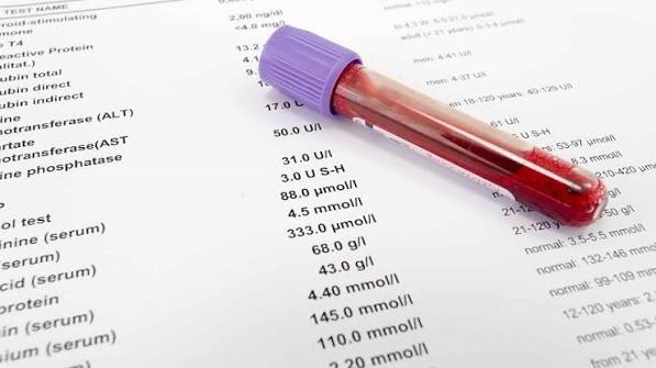
Bilirubin is produced continuously in the body as a byproduct of haem catabolism — predominantly from the breakdown of aged red blood cells (which survive approximately one hundred twenty days in circulation before being removed by the spleen and bone marrow). Haemoglobin released from destroyed red blood cells is broken down into haem and globin; haem is then converted to biliverdin and then to unconjugated (indirect) bilirubin, which is lipid-soluble and cannot be excreted in urine or bile in this form. Unconjugated bilirubin travels in the blood bound to albumin to the liver, where hepatocytes take it up and conjugate it with glucuronic acid using the enzyme UDP-glucuronosyltransferase (UGT1A1) — converting it into water-soluble conjugated (direct) bilirubin that can be excreted into bile. Conjugated bilirubin passes from the liver through the bile ducts into the small intestine, where gut bacteria convert it to urobilinogen — part of which is reabsorbed and excreted in urine (giving urine its pale yellow colour), and the remainder is converted to stercobilin that is excreted in stool (giving stool its brown colour).
This metabolic pathway illuminates why different patterns of bilirubin elevation occur in different conditions. Haemolysis — excessive red blood cell destruction — overwhelms the liver’s conjugation capacity, causing indirect bilirubin to accumulate; direct bilirubin remains low or normal. In Gilbert’s syndrome — the most common benign cause of mild hyperbilirubinaemia, affecting up to ten percent of the population — a genetic variant reducing UGT1A1 activity by thirty to fifty percent impairs conjugation mildly, again producing isolated indirect hyperbilirubinaemia without haemolysis or liver disease. Hepatocellular liver disease (hepatitis, cirrhosis) impairs multiple steps — uptake, conjugation, and excretion — producing mixed hyperbilirubinaemia with both fractions elevated, though the direct fraction typically dominates in significant disease. Biliary obstruction — from common bile duct stones, strictures, or malignancy — prevents the excretion of already-conjugated bilirubin, producing predominantly direct hyperbilirubinaemia; bilirubin then regurgitates into the circulation, appearing in urine (dark urine) while simultaneously failing to reach the bowel (pale or clay-coloured stool). The article on dark urine and its significance in liver disease covers the specific symptom of bilirubin appearing in the urine in detail.
Normal Bilirubin Ranges and What Elevated Levels Mean
The normal range for total bilirubin is generally 3–17 μmol/L (0.2–1.0 mg/dL) in most laboratories, with direct bilirubin below 5 μmol/L (0.3 mg/dL) and the remainder constituting indirect bilirubin. Levels above the upper limit of normal but below the threshold for visible jaundice — typically defined as below 35–50 μmol/L (2–3 mg/dL) — may be reported on blood tests without any visible yellowing. This subclinical hyperbilirubinaemia is important to investigate when persistent or rising, as it may indicate hepatocellular disease, evolving biliary obstruction, or haemolysis that has not yet reached the level producing visible jaundice. Visible jaundice appears when total bilirubin exceeds approximately 35–50 μmol/L (2–3 mg/dL), first visible in the sclera (whites of the eyes, which are particularly sensitive to bilirubin deposition) before becoming apparent in the skin. Deep jaundice — bilirubin above 200 μmol/L (approximately 12 mg/dL) — produces marked yellow-green discolouration and is associated with cholestatic causes including biliary obstruction, primary biliary cholangitis, and severe intrahepatic cholestasis. The article on yellow skin and eyes as signs of liver disease covers the full clinical spectrum of jaundice.
The clinical significance of an elevated bilirubin depends critically on the fractionation pattern and accompanying blood test findings. Isolated indirect hyperbilirubinaemia — elevated total bilirubin with predominantly indirect fraction, normal direct bilirubin, and normal ALT, AST, and ALP — almost always indicates either Gilbert’s syndrome (particularly if it appears in a young adult, fluctuates with illness, fasting, or stress, and has been present chronically) or haemolysis (where a low haemoglobin, elevated reticulocyte count, elevated LDH, and low haptoglobin confirm red blood cell destruction). Both conditions can be evaluated without liver biopsy and both, when correctly identified, spare patients unnecessary anxiety about liver disease. Mixed hyperbilirubinaemia — elevated total bilirubin with both direct and indirect fractions elevated, alongside raised ALT, AST, or ALP — points to hepatocellular or cholestatic liver disease requiring the full investigation pathway. Predominantly direct hyperbilirubinaemia with elevated ALP and GGT, and lower aminotransferases, points specifically to biliary obstruction or cholestatic liver disease. For the clinical significance of pale or clay-coloured stools that accompany obstructive jaundice, see the article on pale stool and bile duct problems.
Gilbert’s Syndrome — The Most Common Benign Cause
Gilbert’s syndrome is the most frequently encountered cause of elevated bilirubin on routine blood testing, present in five to ten percent of the population in Western countries. It is caused by a genetic variant (usually a TA repeat polymorphism in the promoter region of the UGT1A1 gene) that reduces the enzyme activity responsible for bilirubin conjugation by approximately thirty to fifty percent. This reduction does not cause liver disease — the liver is structurally normal, liver function is otherwise preserved, and life expectancy is completely unaffected. Total bilirubin in Gilbert’s syndrome is typically mildly elevated — usually between 17 and 70 μmol/L (1–4 mg/dL) — and fluctuates characteristically: levels rise with prolonged fasting, intercurrent illness, physical exertion, dehydration, menstruation, and stress, and fall with adequate nutrition and hydration. Fasting is the simplest clinical test: bilirubin in Gilbert’s syndrome rises by fifty percent or more after twenty-four hours of fasting, while bilirubin from other causes does not show this fasting-dependent fluctuation.
Gilbert’s syndrome is a diagnosis of exclusion — it is established by demonstrating isolated indirect hyperbilirubinaemia, normal direct bilirubin, normal ALT, AST, ALP, and GGT, normal full blood count (to exclude haemolysis), and a characteristic pattern consistent with known precipitants. Genetic testing (UGT1A1 promoter analysis) is available but rarely necessary for diagnosis when the clinical picture is clear. The practical importance of recognizing Gilbert’s syndrome is to reassure patients that there is no underlying liver disease and no treatment is required — avoiding unnecessary liver biopsies, hepatology referrals, and patient anxiety. It also has pharmaceutical relevance: patients with Gilbert’s syndrome have reduced UGT1A1 activity affecting the metabolism of certain drugs including irinotecan (a chemotherapy agent), nilotinib, and some antiretroviral medications, making pharmacists and prescribers aware of this variant important for medication safety. Understanding that mild bilirubin elevation in the context of Gilbert’s syndrome does not indicate liver pathology allows patients to interpret subsequent blood test results with appropriate context rather than persistent concern. For a comprehensive perspective on how liver-derived symptoms connect, the article on fatigue and liver health provides useful framing.
Frequently Asked Questions About the Bilirubin Test
My total bilirubin is slightly elevated but I don’t look yellow — should I be worried?
Not necessarily — mild elevation in the range of 20–50 μmol/L (1.2–3 mg/dL) without visible jaundice is common and in most cases benign, particularly in young adults. Gilbert’s syndrome is by far the most frequent explanation and requires no treatment. However, a mildly elevated bilirubin that is a new finding (not previously elevated on past blood tests), is accompanied by any elevation in ALT, AST, ALP, or GGT, or is associated with symptoms like pale stool, dark urine, fatigue, or abdominal discomfort warrants further investigation. Your doctor will likely check whether it is predominantly indirect (pointing toward Gilbert’s or haemolysis) or direct (pointing toward liver or biliary pathology), and may request additional tests or liver ultrasound. The key question is whether the elevation is isolated and in keeping with a benign cause, or whether it is part of a broader pattern of abnormal liver function tests — which the fractionation and accompanying panel results help to establish.
What does it mean when both direct and total bilirubin are elevated?
Elevated direct (conjugated) bilirubin is always significant because it indicates that bilirubin has been conjugated by the liver but cannot be excreted normally — either because hepatocytes are releasing it back into the blood due to liver cell damage, or because biliary obstruction is preventing its flow through the bile ducts. Elevated direct bilirubin alongside elevated ALP and GGT points toward cholestatic disease — biliary obstruction (stones, stricture, tumour) or cholestatic liver diseases (primary biliary cholangitis, primary sclerosing cholangitis, intrahepatic cholestasis). Elevated direct bilirubin alongside elevated ALT and AST points toward hepatocellular disease (viral hepatitis, alcoholic hepatitis, drug-induced liver injury, autoimmune hepatitis) where liver cell damage impairs excretion. Imaging — typically liver ultrasound as first-line — is essential to differentiate intrahepatic from extrahepatic causes, as bile duct dilation on ultrasound indicates extrahepatic obstruction requiring urgent cholangiographic investigation (MRCP or ERCP). The combination of jaundice, pale stool, and dark urine is particularly urgent to evaluate because it indicates obstructive jaundice that may be caused by pancreatic head cancer or cholangiocarcinoma — conditions where early diagnosis substantially improves outcomes. This clinical picture is addressed in detail in the article on when to seek urgent medical attention for digestive symptoms.
Sources: NIDDK — Liver Blood Tests · Mayo Clinic — Bilirubin Test · AASLD — Practice Guidelines
Cholestatic Jaundice — Biliary and Intrahepatic Causes
Cholestatic jaundice — characterized by elevated direct bilirubin, raised ALP and GGT, often with only modest ALT and AST elevation, and accompanied clinically by pale stool, dark urine, and sometimes pruritus (itching from bile salt deposition in skin) — is a clinically important pattern requiring systematic investigation to distinguish intrahepatic from extrahepatic causes. Extrahepatic cholestasis — bile duct obstruction occurring outside the liver — is caused by choledocholithiasis (common bile duct stones, the most frequent cause), strictures (from prior surgery, inflammation, or instrumentation), cholangiocarcinoma (bile duct cancer), pancreatic head carcinoma compressing the common bile duct, ampullary carcinoma, and external lymph node compression. Ultrasound demonstrating dilated bile ducts is the key first-line finding that points toward extrahepatic obstruction and triggers urgent MRCP (magnetic resonance cholangiopancreatography) to characterize the level and cause of obstruction, with a view to ERCP (endoscopic retrograde cholangiopancreatography) for therapeutic stone removal or stenting of malignant strictures. The triad of jaundice, pale stool, and dark urine associated with abdominal pain and fever (Charcot’s triad) indicates cholangitis — bacterial infection of the obstructed bile duct — which is a medical emergency requiring immediate antibiotics and urgent biliary drainage.
Intrahepatic cholestasis — impaired bile flow occurring within the liver — produces the same biochemical pattern (elevated direct bilirubin, ALP, GGT, and bilirubin) but without bile duct dilation on ultrasound. Causes include primary biliary cholangitis (PBC, an autoimmune disease predominantly affecting middle-aged women, diagnosed by anti-mitochondrial antibody and characteristic liver histology), primary sclerosing cholangitis (PSC, associated with inflammatory bowel disease, causing multifocal bile duct stricturing visible on MRCP), drug-induced cholestasis (amoxicillin-clavulanate, flucloxacillin, anabolic steroids, oestrogen-containing contraceptives), intrahepatic cholestasis of pregnancy (a pregnancy-specific condition posing fetal risk), total parenteral nutrition (TPN)-associated cholestasis in hospitalized patients, and infiltrative liver diseases (sarcoidosis, lymphoma, amyloid). The cholestatic pattern of bilirubin elevation — particularly when itching accompanies it — warrants the article on itchy skin and liver problems as a companion clinical reference, since pruritus from cholestasis can precede jaundice by weeks.
Haemolytic Causes of Elevated Bilirubin
Elevated bilirubin from haemolysis — excessive red blood cell destruction — is an important non-hepatic cause of hyperbilirubinaemia that requires a fundamentally different investigation and management pathway than liver-related jaundice. In haemolysis, the high bilirubin is predominantly indirect (unconjugated), arising because the rate of red blood cell breakdown exceeds the liver’s conjugation capacity. The liver itself is functioning normally — ALT, AST, ALP, and GGT are normal, and direct bilirubin is not elevated beyond normal. The blood count shows anaemia (low haemoglobin), elevated reticulocyte count (bone marrow working harder to replace destroyed cells), elevated LDH (released from lysed red cells), low haptoglobin (haptoglobin binds free haemoglobin from lysed cells and is cleared from circulation, so low levels indicate intravascular haemolysis), and the peripheral blood film may show abnormal red cell morphology depending on the haemolytic cause. In chronic haemolytic anaemias (hereditary spherocytosis, sickle cell disease, autoimmune haemolytic anaemia), persistent indirect hyperbilirubinaemia can produce pigment gallstones — cholesterol-poor, bilirubin-rich stones that develop at an accelerated rate due to the chronic excess bilirubin load on the biliary system.
Causes of haemolysis presenting with elevated indirect bilirubin in adults include autoimmune haemolytic anaemia (AIHA — warm and cold types, associated with lymphoma, lupus, drugs, and idiopathic), microangiopathic haemolytic anaemia (MAHA — occurring in thrombotic thrombocytopenic purpura/TTP, haemolytic uraemic syndrome/HUS, and disseminated intravascular coagulopathy/DIC), hereditary red cell membrane and enzyme disorders (hereditary spherocytosis, G6PD deficiency — particularly triggered by oxidant drugs, fava beans, or infection), and mechanical haemolysis (from prosthetic heart valves). Malaria is an important cause of haemolytic jaundice globally and should be considered in anyone with recent travel history. The urgency of haemolytic jaundice varies considerably by cause — TTP is a haematological emergency requiring immediate plasma exchange, while hereditary spherocytosis with mild chronic elevation requires management planning for complications rather than immediate escalation. Differentiating liver-origin jaundice from haemolytic jaundice by bilirubin fractionation is therefore one of the most clinically consequential distinctions enabled by this single blood test. The article on easy bruising and liver function provides context on how synthetic markers complement bilirubin in distinguishing liver disease severity.
Bilirubin in Liver Disease Monitoring and Prognosis
In established liver disease, serial bilirubin measurement is one of the most important indicators of disease trajectory and prognosis. Rising bilirubin in a patient with chronic liver disease — whether from cirrhosis, autoimmune hepatitis, primary biliary cholangitis, or viral hepatitis — indicates deteriorating hepatic function and warrants clinical reassessment of the underlying disease activity, potential precipitating factors (infection, medication changes, bleeding), and the current liver disease stage. In cirrhosis, bilirubin above 34 μmol/L (2 mg/dL) contributes significantly to Child-Pugh score calculation (a widely used prognostic scoring system for cirrhosis incorporating bilirubin, albumin, INR, ascites, and encephalopathy), and is one of three variables in the MELD score (Model for End-Stage Liver Disease, along with INR and creatinine) that guides liver transplant listing priority. A rapidly rising bilirubin in a patient with cirrhosis may indicate acute-on-chronic liver failure — a distinct clinical syndrome where an acute precipitant (infection, bleeding, alcohol binge, drug reaction) causes rapid hepatic decompensation superimposed on pre-existing chronic liver disease, carrying a thirty-day mortality that can exceed thirty percent without escalation to specialist care.
In primary biliary cholangitis (PBC), rising bilirubin above 17 μmol/L — despite adequate ursodeoxycholic acid (UDCA) therapy — is a marker of incomplete biochemical response (defined by the Paris criteria and Barcelona criteria as failure to normalize or adequately reduce ALP, AST, and bilirubin after twelve months of UDCA). Incomplete biochemical response identifies patients at higher risk of disease progression to biliary cirrhosis and liver failure, and these patients are candidates for add-on therapy with obeticholic acid or bezafibrate. In hepatitis B and C, bilirubin trends during antiviral therapy provide evidence of treatment response — normalization of bilirubin alongside ALT reduction and viral load suppression confirms biochemical remission. In autoimmune hepatitis, rising bilirubin despite immunosuppression indicates treatment failure or non-compliance and requires urgent reassessment including consideration of second-line agents (mycophenolate mofetil, calcineurin inhibitors) or, in refractory cases, liver transplant evaluation. Understanding bilirubin in the broader clinical context — including symptoms such as abdominal swelling from advanced liver disease — provides the most complete picture of disease severity.
Bilirubin monitoring is also a key component of acute liver failure assessment. In acute liver failure from any cause — acetaminophen toxicity, acute viral hepatitis, Wilson’s disease presenting in acute crisis, ischaemic hepatitis, Budd-Chiari syndrome, or drug-induced acute hepatic necrosis — rapidly rising bilirubin alongside coagulopathy (rising INR), encephalopathy, and creatinine defines the severity of the acute failure and informs the King’s College Criteria assessment for emergency transplant listing. The critical clinical lesson is that in acute liver failure, a falling aminotransferase (ALT and AST) in the context of simultaneously rising bilirubin and INR does not represent improvement — it represents hepatocyte exhaustion and progressive liver failure, requiring immediate escalation rather than reassurance. Serial daily bilirubin measurements in acute liver failure provide the most sensitive window into whether the liver is recovering or deteriorating, making this single test arguably the most important serial marker in the most severe form of liver disease that clinicians encounter.
The bilirubin test — deceptively simple as a single numerical value on a blood panel — encapsulates one of the most diagnostically rich measurements in hepatology when interpreted through the lens of fractionation, accompanying LFT patterns, and clinical context. From benign Gilbert’s syndrome to biliary obstruction requiring urgent endoscopy, from compensated cirrhosis being tracked through the MELD score to acute liver failure demanding emergency transplant evaluation, bilirubin measurement anchors the most consequential clinical decisions in liver disease. Understanding what this test actually measures — not just the number but the fraction, the trend, the accompanying markers, and the clinical picture it fits into — transforms a routine blood result into a reliable guide to what the liver is doing, whether it is coping, and what needs to happen next. The companion articles in this series on ALT and AST, albumin, and alkaline phosphatase complete the framework for interpreting the full liver function panel.
I’ve had mildly elevated bilirubin for years and doctors kept telling me not to worry. After reading the section on Gilbert’s syndrome I finally understand why — apparently I have the genetic variant causing mild indirect bilirubin elevation. It fluctuates when I skip meals which fits perfectly. Wish someone had explained this years ago.
Helen, what you’re describing — mildly elevated bilirubin that rises when you fast, with otherwise normal liver tests — is a textbook presentation of Gilbert’s syndrome. If you haven’t been explicitly told this is Gilbert’s, it’s worth asking your doctor to confirm by checking whether the elevated bilirubin is predominantly indirect (unconjugated) and whether your ALT, AST, ALP, and GGT are all normal. If they are, the diagnosis is essentially established and there is genuinely nothing to treat.
The section explaining that a falling ALT with rising bilirubin and INR means the liver is failing — not improving — is critically important information. I’m a medical student and we don’t always get this counterintuitive concept explained so clearly. Bookmarking this for clinical rotations.