
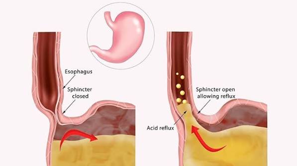
GERD and gastritis are two of the most frequently confused upper gastrointestinal conditions in both clinical practice and everyday conversation. Both cause discomfort in the upper abdomen and chest. Both involve stomach acid. Both may respond to the same medication — a proton pump inhibitor. But they are fundamentally different diseases affecting different parts of the digestive tract, with different underlying causes, different long-term consequences, and different treatments. Confusing them — or treating one as though it were the other — means the actual disease goes unaddressed while the real cause is missed.
What Is GERD?
GERD — gastro-oesophageal reflux disease — is a condition in which gastric contents (acid, pepsin, bile) reflux from the stomach upward into the oesophagus with sufficient frequency or severity to cause troublesome symptoms or mucosal damage. GERD is defined by the presence of troublesome symptoms (heartburn, regurgitation) at least twice a week, by endoscopic evidence of oesophagitis, or by complications such as Barrett’s oesophagus or oesophageal adenocarcinoma.
GERD is primarily a disease of the oesophagus, not the stomach. The stomach produces the acid — but the damage occurs when that acid is abnormally exposed to the oesophageal lining, which is not designed to tolerate it. The stomach lining has specialised acid-resistant epithelium and a thick mucous barrier; the oesophageal squamous epithelium has neither.
The most common mechanism is not a permanently weak lower oesophageal sphincter (LOS) — a common misconception — but rather transient lower oesophageal sphincter relaxations (TLOSRs): episodic, inappropriate relaxations of the LOS that are not associated with swallowing. These allow gastric contents to reflux into the oesophagus. Contributing factors include: hiatus hernia, obesity, large meals, lying supine after eating, alcohol, smoking, and certain foods (fatty foods, coffee, chocolate, mint).
What Is Gastritis?
Gastritis is inflammation of the gastric mucosa — the inner lining of the stomach — confirmed by biopsy. It is a histological diagnosis: symptoms alone cannot establish gastritis. The majority of people with gastritis have no symptoms at all. Gastritis is caused by H. pylori (most common worldwide), NSAIDs, alcohol, autoimmune mechanisms, or bile reflux. For a full explanation of gastritis types and causes, see our article on gastritis: symptoms and causes.
The critical distinction: GERD is acid escaping upward into the oesophagus; gastritis is mucosal inflammation within the stomach itself. GERD does not cause gastritis; gastritis does not cause GERD — though both can coexist.
GERD vs Gastritis: Key Differences
| Feature | GERD | Gastritis |
|---|---|---|
| Organ affected | Oesophagus | Stomach |
| Primary mechanism | Acid refluxing upward | Mucosal inflammation |
| Main symptom | Heartburn, regurgitation | Epigastric pain, nausea (or none) |
| Main causes | TLOSRs, hiatus hernia, obesity | H. pylori, NSAIDs, autoimmune |
| Cancer risk | Oesophageal adenocarcinoma (via Barrett’s) | Gastric adenocarcinoma (via Correa cascade) |
| H. pylori | Inversely associated | Direct causal agent |
| Primary treatment | Lifestyle + PPI for symptom control | Treat cause (eradicate H. pylori, stop NSAIDs) |
Both conditions respond to PPIs — but for different reasons. In GERD, PPIs reduce the acid available to reflux into the oesophagus, healing oesophagitis and relieving heartburn. In gastritis, PPIs reduce the acid irritating an already-inflamed gastric mucosa. But PPIs do not cure the underlying gastritis — only addressing its cause (particularly H. pylori eradication) does.
Symptoms: GERD vs Gastritis Difference
GERD symptoms:
- Heartburn: a burning sensation rising from the lower chest/sternum upward — the cardinal GERD symptom; worse after large meals, bending over, or lying flat
- Regurgitation: effortless return of gastric contents to the throat or mouth — a sour, acidic taste; pathognomonic for GERD
- Chronic cough: reflex cough from proximal acid reaching the upper airway — often nocturnal; may be the dominant symptom without any heartburn
- Hoarseness/voice changes: acid reaching the larynx (laryngopharyngeal reflux, LPR) damaging the vocal cords
- Globus sensation: persistent feeling of a lump or fullness in the throat
- Worsening asthma: through microaspiration and vagally-mediated bronchoconstriction
Gastritis symptoms (when present):
- Epigastric discomfort: upper central abdominal pain — burning, aching, gnawing; stays in the abdomen; does not rise into the chest
- Nausea, bloating, early satiety, belching
- Often asymptomatic — most H. pylori gastritis cases cause no symptoms
The cardinal symptom distinguishing GERD is heartburn — a burning that rises from the sternum — and regurgitation. These do not occur in gastritis. Conversely, the epigastric discomfort of gastritis stays in the upper abdomen and does not characteristically rise.
Causes of GERD vs Causes of Gastritis
GERD risk factors: transient LOS relaxations (primary mechanism); hiatus hernia (found in the majority of severe GERD and Barrett’s cases); obesity (strong, dose-dependent association — weight loss is the most effective single intervention); dietary triggers (fatty foods, coffee, alcohol, chocolate, mint); smoking (reduces LOS pressure); pregnancy; medications that reduce LOS pressure (nitrates, calcium channel blockers, benzodiazepines).
Gastritis causes: H. pylori (most common worldwide — urease, CagA/VacA mechanisms); NSAIDs/aspirin (COX-1 inhibition); alcohol; autoimmune (parietal cell antibodies; pernicious anaemia); bile reflux; rare causes (eosinophilic, granulomatous, CMV). For H. pylori specifically, see our article on H. pylori infection and stomach health.
The H. pylori — GERD inverse relationship: A counterintuitive epidemiological observation is that H. pylori prevalence and GERD/oesophageal adenocarcinoma prevalence show an inverse geographic correlation. The proposed mechanism: H. pylori corpus gastritis impairs parietal cell function, reducing acid production, and thereby reducing the acid available to reflux into the oesophagus. As a consequence, H. pylori eradication in patients with corpus-predominant gastritis may, in some cases, lead to a modest increase or unmasking of GERD symptoms as normal acid secretion is restored. This is not a reason to avoid H. pylori eradication — the cancer prevention and ulcer healing benefits vastly outweigh any modest, manageable effect on GERD symptoms.
Complications
GERD complications:
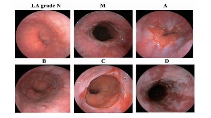
Oesophagitis: acid-induced mucosal erosions of the distal oesophagus, classified by the Los Angeles system: Grade A (mucosal breaks ≤5mm); Grade B (breaks >5mm, not confluent between folds); Grade C (confluent, <75% circumference); Grade D (≥75% of circumference). Grade C and D oesophagitis heals with 8 weeks of high-dose PPI.
Barrett’s oesophagus: metaplastic replacement of normal oesophageal squamous epithelium by columnar intestinal-type epithelium — the most clinically significant GERD complication and the only established precursor to oesophageal adenocarcinoma. Found in approximately 5–15% of patients with chronic GERD symptoms. Annual adenocarcinoma risk approximately 0.3–0.5% (30–40× relative risk vs general population). Requires endoscopic surveillance.
Oesophageal adenocarcinoma: arising from Barrett’s; strongly linked to chronic GERD, obesity, and smoking; incidence has risen sharply in Western countries over the past 40 years.
Peptic stricture: fibrotic narrowing of the distal oesophagus from repeated acid injury; causes progressive dysphagia; treated with endoscopic balloon dilatation.
Gastritis complications:
- Peptic ulcer disease — with risks of bleeding and perforation (see our article on stomach ulcers: what adults should know)
- Gastric adenocarcinoma — via Correa cascade (H. pylori → atrophic gastritis → intestinal metaplasia → dysplasia → cancer)
- Pernicious anaemia — autoimmune gastritis → parietal cell destruction → intrinsic factor loss → B12 malabsorption
- Gastric MALT lymphoma — H. pylori-driven; curable with antibiotic eradication in early-stage disease
Diagnosis
GERD: primarily a clinical diagnosis — typical symptoms and response to a PPI trial is sufficient in most patients without alarm features. Endoscopy is indicated for alarm features, or to assess for Barrett’s oesophagus in high-risk patients. For patients with persistent symptoms despite PPI therapy, ambulatory pH-impedance monitoring (24-hour measurement of acid and non-acid reflux events with symptom correlation) is the most objective test. Oesophageal manometry assesses LOS pressure and oesophageal motility before anti-reflux surgery. According to the NICE CG184 guidance, uninvestigated dyspepsia in adults under 55 without alarm features should be managed with a test-and-treat approach for H. pylori or an empirical PPI trial before referral for endoscopy.
Gastritis: requires endoscopy with biopsy — the Sydney Protocol provides histological diagnosis, H. pylori status, and mucosal staging. Non-invasive H. pylori testing (urea breath test or stool antigen test) supports the test-and-treat approach for uncomplicated cases. Blood tests: FBC, B12, parietal cell antibodies for autoimmune gastritis. Fasting serum gastrin if Zollinger-Ellison syndrome is suspected.
Treatment
GERD treatment:
Lifestyle (first-line): weight loss (most effective single intervention for obese GERD patients); elevate head of bed 15–20cm; avoid large meals and eating within 3 hours of bedtime; avoid trigger foods (fatty/fried, coffee, alcohol, chocolate, mint); quit smoking.
Pharmacological: PPIs — most effective; taken 30–60 minutes before the first meal; used at the lowest dose controlling symptoms; alginate preparations (Gaviscon) for post-meal symptoms; H2 receptor antagonists (famotidine) for breakthrough symptoms.
Anti-reflux surgery: laparoscopic Nissen fundoplication or LINX magnetic sphincter augmentation — appropriate for confirmed GERD patients who cannot tolerate long-term PPIs, prefer surgical management, or have large hiatus hernia; ~80–90% success rates.
Gastritis treatment:
- H. pylori eradication: triple or bismuth quadruple therapy; confirmed with urea breath test
- NSAID cessation: removes causative agent in NSAID-associated gastritis
- PPI therapy: symptomatic relief and mucosal healing
- B12 replacement: lifelong intramuscular injections for autoimmune gastritis
For complete management of peptic ulcer complications, see our article on peptic ulcer disease explained.
Can You Have Both GERD and Gastritis?
Yes — GERD and gastritis are independent conditions that can and frequently do coexist. A patient can have H. pylori-associated gastritis and a hiatus hernia causing GERD-related heartburn simultaneously. They do not cause each other, but share some risk factors (NSAIDs, smoking) and some medication approaches (PPIs).
When both are present, each requires its own management: H. pylori gastritis requires eradication therapy; GERD requires lifestyle modification and PPI therapy. A PPI addresses both simultaneously in terms of acid suppression, but the gastritis also requires eradication. Patients undergoing H. pylori eradication for corpus-predominant gastritis may experience a modest worsening of heartburn after eradication as gastric acid normalises — manageable with standard PPI therapy and not a reason to delay or avoid eradication.
Why GERD and Gastritis Are So Frequently Confused
The confusion between GERD and gastritis is not just a lay misunderstanding — it occurs frequently in clinical settings, particularly in primary care, where both conditions present as upper abdominal discomfort and both respond to the same first-line medication (a proton pump inhibitor). Several factors perpetuate this confusion:
1. The word “gastritis” is clinically overused. The term “gastritis” is applied colloquially to almost any upper abdominal discomfort or nausea, regardless of whether any mucosal inflammation is actually present. In the strict medical sense, gastritis requires biopsy confirmation. Many people who have been told they have “gastritis” by a clinician who has not performed endoscopy actually have functional dyspepsia, GERD, or NSAID-associated gastric irritation that resolves when the NSAID is stopped — none of which is histological gastritis. The loose use of the diagnosis creates a misleading label that can persist in a patient’s medical history for years.
2. Both conditions are common and often coexist. In a population where H. pylori prevalence is 30–40% and GERD prevalence is similar, a significant number of individuals will have both, making it genuinely difficult to attribute a symptom to one condition rather than the other without investigation.
3. Both respond to PPIs. Because both conditions improve with proton pump inhibitor therapy — through different mechanisms — a positive response to a PPI trial does not distinguish between them. It is therefore easy to start a patient on long-term PPI therapy based on a symptom response without ever establishing whether they have GERD, H. pylori gastritis, or functional dyspepsia. This “treat and don’t investigate” approach misses H. pylori, which requires specific antibiotic treatment, and misses Barrett’s oesophagus or gastric intestinal metaplasia, which require surveillance.
4. Symptoms genuinely overlap. Both conditions can cause upper abdominal burning, nausea, and post-prandial discomfort. In patients with atypical presentations — GERD without classic heartburn, or gastritis without H. pylori and without classic epigastric pain — the symptom profile may be non-specifically “dyspeptic” and indistinguishable without investigation.
The practical implication is that while it is reasonable to treat empirically with a PPI in the first instance for typical dyspeptic symptoms, patients with persistent or recurring symptoms, those with risk factors for serious pathology (age over 55, family history of upper GI cancer, longstanding GERD with Barrett’s risk factors), and those in whom H. pylori test-and-treat has not been applied should progress to appropriate investigation rather than remaining on long-term PPIs without a confirmed diagnosis.
GERD and Gastritis: Surveillance and Long-Term Monitoring
GERD surveillance: Not all GERD patients require ongoing endoscopic surveillance. Surveillance is primarily directed at identifying and monitoring Barrett’s oesophagus — the only established GERD complication that confers a meaningful cancer risk. Barrett’s oesophagus is typically found on endoscopy performed for longstanding GERD symptoms, particularly in high-risk groups: male sex, age over 50, obesity, white race, tobacco smoking, and a family history of oesophageal adenocarcinoma or Barrett’s. Once Barrett’s is confirmed, the surveillance interval is determined by the degree of dysplasia found on biopsy: no dysplasia → 3–5 years; low-grade dysplasia → 6–12 months (or endoscopic ablation); high-grade dysplasia → endoscopic mucosal resection or radiofrequency ablation. Patients with typical GERD symptoms but no endoscopic Barrett’s on a prior OGD do not require routine ongoing endoscopic surveillance — PPI therapy and symptom monitoring are sufficient.
Gastritis surveillance: Gastritis surveillance is directed at the gastric cancer risk conferred by atrophic gastritis and intestinal metaplasia, stratified using OLGA/OLGIM staging. Patients at Stage III–IV (high-risk) require endoscopic surveillance every 3 years. Patients with autoimmune gastritis may also require surveillance for gastric neuroendocrine tumours (type 1 ECL-cell tumours arising in the context of achlorhydria-driven hypergastrinaemia). Patients with successfully eradicated H. pylori and no atrophic changes or intestinal metaplasia on staging biopsies generally do not require routine endoscopic surveillance beyond a single post-eradication confirmation. The key message is that gastritis surveillance is stratified by mucosal risk stage, not simply by whether H. pylori was ever present.
For both conditions, shared risk factors such as smoking, obesity, high alcohol intake, and an unhealthy diet worsen outcomes — addressing these comprehensively is as important as pharmacological treatment in the long-term management of upper GI disease.
When to See a Doctor About GERD or Gastritis
Many people with mild GERD symptoms self-manage with over-the-counter antacids or alginate preparations, and uncomplicated GERD does not require immediate specialist referral. However, there are specific circumstances in which professional evaluation is important for both GERD and gastritis, beyond the alarm features already listed above.
For GERD: symptoms that persist or return despite 4–8 weeks of PPI therapy at standard doses; symptoms that require ongoing daily PPI use to remain controlled for more than 4–8 weeks (particularly if you are male, over 50, obese, or a current or former smoker — all risk factors for Barrett’s oesophagus); new symptoms of regurgitation that wake you from sleep; any sensation of food sticking in the oesophagus. For children and adolescents with GERD-like symptoms, specialist paediatric assessment is recommended rather than empirical PPI use, since the causes and management differ from adult GERD.
For gastritis: any patient with persistent upper abdominal symptoms should be tested for H. pylori as part of the diagnostic workup — the test-and-treat approach means that H. pylori can be identified and treated non-invasively without always requiring endoscopy. First-degree relatives of patients with gastric cancer should be offered H. pylori testing and, where available, endoscopic OLGA staging even if asymptomatic. Patients known to take regular NSAIDs who develop new upper abdominal symptoms, particularly any bleeding signs (melaena, haematemesis, iron deficiency anaemia), require prompt evaluation.
Frequently Asked Questions
Heartburn — a burning sensation rising from the lower chest or sternum upward — is the cardinal symptom of GERD, not gastritis. It results from acid irritating the oesophageal lining. Gastritis causes epigastric discomfort (upper central abdominal pain that stays in the abdomen), not the characteristic upward-rising burning of heartburn. The widespread use of the word “heartburn” to describe any upper abdominal burning contributes significantly to the confusion between these two conditions.
GERD does not typically cause gastritis — they have separate mechanisms. GERD is acid refluxing upward into the oesophagus; gastritis is mucosal inflammation within the stomach caused by H. pylori, NSAIDs, or autoimmune mechanisms. Bile reflux (bile flowing upward from the duodenum into the stomach) can cause a chemical gastritis, but this is a specific post-surgical context, not typical GERD. Standard acid reflux into the oesophagus does not cause gastritis.
In a small proportion of patients with corpus-predominant H. pylori gastritis who have had chronically reduced acid secretion, eradicating H. pylori can lead to a modest increase in gastric acid production as the mucosa recovers, potentially unmasking GERD symptoms. This is not a reason to avoid eradication. The cancer prevention and ulcer healing benefits are substantial; any new GERD symptoms arising after eradication are manageable with standard PPI therapy. Most patients with H. pylori gastritis do not notice any change in GERD symptoms after eradication.
Not necessarily for either, initially. GERD in adults under 55 without alarm features can be diagnosed clinically by typical symptoms and confirmed by response to a PPI trial. Gastritis from H. pylori infection can be inferred from a positive urea breath test or stool antigen test using the test-and-treat approach. Endoscopy is indicated for alarm features, for assessing complications (Barrett’s oesophagus, gastritis cancer risk staging), or for initial investigation in patients over 55 with new dyspepsia.
PPIs are the most effective treatment for controlling GERD symptoms and healing oesophagitis, but they do not cure the underlying cause of GERD — they reduce acid to minimise reflux harm rather than preventing the reflux itself. When PPIs are stopped in most GERD patients, symptoms return. For a durable fix, anti-reflux surgery (fundoplication, LINX) addresses the LOS dysfunction. Weight loss in obese patients can reduce reflux frequency sufficiently to allow PPI dose reduction in some cases. PPI therapy is appropriate long-term management for GERD, but it is suppression rather than cure.
Both terms describe metaplastic replacement of normal epithelium by intestinal-type epithelium — but in different organs. Barrett’s oesophagus is intestinal metaplasia of the oesophagus, arising from chronic GERD. Gastric intestinal metaplasia arises in the stomach in the context of H. pylori gastritis and atrophic gastritis. Both are precancerous conditions requiring surveillance, but they predispose to different cancers (oesophageal adenocarcinoma for Barrett’s; gastric adenocarcinoma for gastric intestinal metaplasia) and are managed with different surveillance programmes.
Neither is uniformly more serious. Uncomplicated GERD is a chronic symptom burden but not immediately dangerous; Barrett’s oesophagus is a genuine cancer precursor. Uncomplicated H. pylori gastritis without atrophy carries low short-term risk, but untreated chronic progression to atrophic gastritis over decades represents significant gastric cancer risk. In terms of acute dangerous complications, a bleeding or perforated peptic ulcer from H. pylori gastritis is immediately life-threatening in a way that typical GERD is not. The most useful framing is that both require correct diagnosis, treatment of the underlying cause, and appropriate surveillance — not a ranking of severity.
Medical Disclaimer: This article is for educational purposes only and does not constitute medical advice. Alarm symptoms — haematemesis, dysphagia, unexplained weight loss, black stools, or severe abdominal pain — require urgent medical evaluation.
References
- Katz PO, Gerson LB, Vela MF. Guidelines for the Diagnosis and Management of Gastroesophageal Reflux Disease. Am J Gastroenterol. 2013;108(3):308–328. Available at: PubMed.
- Vakil N et al. The Montreal Definition and Classification of Gastroesophageal Reflux Disease. Am J Gastroenterol. 2006;101(8):1900–20.
- NICE guideline CG184: Dyspepsia and gastro-oesophageal reflux disease. 2014 (updated 2019). Available at: nice.org.uk.
- Malfertheiner P et al. Maastricht V/Florence Consensus Report. Gut. 2017;66(1):6–30. Available at: PubMed.
- NHS. Gastro-oesophageal reflux disease (GERD). Available at: nhs.uk.
I’ve been told at various points over the past five years that I have ‘gastritis’, ‘GERD’, and ‘acid reflux’ — often interchangeably, without endoscopy. I was started on omeprazole after a brief consultation based on my symptoms (upper abdominal burning, occasional nausea), and I’ve been taking it more or less continuously. Reading this article made me realise that I’ve never actually had H. pylori testing, which apparently should have been the first step in evaluating my symptoms. The point about PPI response not distinguishing between GERD and gastritis is exactly what happened in my case — because I responded to omeprazole, the assumption was that the diagnosis was correct. I have an appointment next week and I’ll be specifically asking about the H. pylori test-and-treat approach and whether an endoscopy is warranted given five years of continuous PPI use.
Claire — your situation is unfortunately a common one, and your plan to ask about H. pylori test-and-treat and potential endoscopy is exactly appropriate. The NICE CG184 guidance on dyspepsia recommends that all patients with uninvestigated dyspepsia who are under 55 and without alarm features should be offered H. pylori test-and-treat before a long-term PPI prescription — the test is a urea breath test or stool antigen test (not blood serology, which is less accurate), and if positive, the patient should complete eradication therapy and then reassess symptoms. The fact that your symptoms responded to omeprazole does not confirm GERD and does not exclude H. pylori gastritis — as the article explains, PPIs suppress acid regardless of whether H. pylori is present, so symptom improvement cannot distinguish between the two conditions. After five years of continuous PPI use without a clear underlying diagnosis, your request for H. pylori testing and, if symptoms persist despite eradication (or if H. pylori is negative), an endoscopy is a completely reasonable and justified ask. You would be a candidate for endoscopy on the basis of longstanding symptoms requiring continuous PPI and the need to exclude Barrett’s oesophagus or gastric pathology.
Neil — your understanding of your Barrett’s surveillance situation is accurate and well-reasoned. Short-segment Barrett’s without dysplasia on systematic biopsies is categorised as low-to-intermediate risk under current BSG (British Society of Gastroenterology) and ACG (American College of Gastroenterology) guidelines, and a 3–5 year surveillance interval reflects the low annual progression rate (~0.3% per year) to high-grade dysplasia or adenocarcinoma in this group. The interval shortens significantly if dysplasia is found: low-grade dysplasia now typically prompts either a 6-month repeat biopsy or endoscopic ablation (radiofrequency ablation), since the annual progression rate from LGD to cancer is substantially higher than from Barrett’s without dysplasia. High-grade dysplasia is treated with endoscopic mucosal resection or ablation rather than ongoing surveillance. Your point about Barrett’s versus gastric intestinal metaplasia is an important distinction. They are both intestinal metaplasia — both representing a response to chronic mucosal injury — but the organs affected, the cancer types they predispose to, the cancer risk magnitudes, and the surveillance programmes are entirely separate and should not be conflated.
I was diagnosed with Barrett’s oesophagus two years ago at an endoscopy done for longstanding heartburn — I’d had reflux symptoms for about 12 years without ever being scoped. The Barrett’s was found incidentally; I was told it was short segment with no dysplasia. The table in this article comparing the surveillance intervals for different dysplasia grades is something I hadn’t seen anywhere else — I was told ‘come back in three to five years’ but without any context for why that interval specifically, or what would change it. I now understand that no-dysplasia short-segment Barrett’s puts me in the lower-risk surveillance category (3–5 years) and that if any dysplasia is found on repeat biopsy, that would shorten the interval or lead to ablation. I also found the distinction between Barrett’s oesophagus and gastric intestinal metaplasia clarifying — my wife has gastric intestinal metaplasia from previous H. pylori infection, and we had assumed they were effectively the same condition. The cancer types and surveillance programmes are different.