
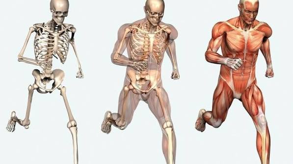
Most adults think of bones, joints, and muscles as separate structures that simply happen to occupy the same body. In practice, they function as a single integrated mechanical system — one where the health of each component depends directly on the others, and where problems in one reliably generate consequences in all three. Understanding how bones, joints, and muscles work together is not an abstract anatomical exercise. It is the basis for understanding why isolated treatments so often produce limited results, why certain injuries predict future ones, and why the most effective strategies for musculoskeletal health address all three components simultaneously rather than each in turn.
The Three Components: A Brief Structural Overview
Before examining how these systems interact, it is useful to understand what each one contributes to the whole.
Bones are the rigid structural framework of the musculoskeletal system. Their primary mechanical roles are to provide lever arms for muscle force, protect vital organs, and maintain the body’s overall structural geometry. Despite their apparent solidity, bones are dynamic living tissue, continuously renewed through a remodeling cycle in which bone-resorbing cells (osteoclasts) remove older bone and bone-forming cells (osteoblasts) deposit new matrix. The balance of this cycle determines bone density and strength, and it is directly influenced by the mechanical forces bones experience — bone that is regularly loaded through weight-bearing activity maintains higher density than bone that is not.
Joints are the articulations between bones that allow controlled movement. The design of each joint type reflects its functional role: the hip and shoulder use ball-and-socket joints that permit wide rotational movement; the knee and elbow are hinge joints optimized for flexion and extension in a single plane; the vertebral joints permit limited, carefully controlled movement in multiple planes. Most joints contain articular cartilage — a smooth, dense connective tissue that reduces friction between bone surfaces — and are enclosed in a joint capsule filled with synovial fluid, which lubricates the joint and delivers nutrients to the avascular cartilage.
Muscles generate and control movement by contracting and relaxing in response to nerve signals. Each muscle is attached to bone at two points via tendons: the origin (typically the more stationary attachment) and the insertion (the attachment on the bone that moves when the muscle contracts). Muscles work in coordinated groups: when one muscle contracts to produce a movement, an opposing muscle (the antagonist) lengthens to control the movement’s speed and prevent joint injury. Muscles also serve as dynamic stabilizers of the joints they cross — providing active support that complements the passive support of ligaments.
The Lever System: How Muscles Move Bones Through Joints
The musculoskeletal system operates as a series of biological levers. In each lever arrangement, the joint acts as the fulcrum, the bone acts as the lever arm, and the muscle provides the force. The specific arrangement — the distances between the joint, the muscle’s attachment point, and the load being moved — determines both the mechanical advantage of the movement and the forces experienced by the joint.
Most musculoskeletal lever systems are designed to prioritize speed and range of motion over force efficiency. The muscle attachment points are typically close to the joint, while the load (the weight of the limb, or an object being held) is further away. This means muscles must generate substantially more force than the actual load being moved — but the trade-off is that a small shortening of the muscle produces a large arc of movement at the hand or foot. The biceps brachii, for example, attaches to the radius close to the elbow joint; it must generate approximately seven times more force than the object being lifted, but this arrangement allows the hand to move quickly through a wide arc with a small muscle contraction.
This mechanical reality has important implications for joint health. The forces transmitted through joints during everyday activities are far greater than the external loads being managed. During level walking, the knee joint experiences compressive forces of approximately 2–3 times body weight. During stair climbing, this rises to 3–4 times body weight. During squatting activities, forces can reach 7–8 times body weight across the patellofemoral joint. Understanding this helps explain why excess body weight has such a disproportionate impact on joint wear — each additional pound translates to several additional pounds of joint force with every step.
Active and Passive Stabilization: How Joints Stay Stable
Joint stability — the ability of a joint to maintain proper alignment under load — comes from two sources that must work together effectively.
Passive stabilization is provided by the static connective tissues: ligaments that connect bone to bone, the joint capsule, and the geometry of the joint surfaces themselves. These structures resist excessive movement through their tensile strength and can withstand significant forces without active muscle engagement. However, passive stabilization alone is insufficient for the dynamic conditions of movement — ligaments and joint capsules cannot respond quickly enough to sudden changes in force direction or unexpected loads.
Active stabilization is provided by the muscles crossing each joint. When muscles contract to stabilize a joint, they increase joint compression (which paradoxically improves stability by pressing joint surfaces together more firmly) and create opposing force couples that prevent unwanted rotation or translation. This active stabilization is continuously adjusted in real time by the nervous system through a process called proprioception — the body’s sense of joint position and movement, provided by sensory receptors in muscles, tendons, and joint capsules.
The practical consequence is that adequate muscle strength around a joint is essential for joint protection. When the muscles crossing a joint are weak, the passive structures — ligaments and cartilage — are exposed to forces and movement patterns they are not designed to manage, accelerating their degradation. This is why muscle strengthening is a cornerstone of both injury prevention and rehabilitation for virtually every joint condition. It is also why joint injuries themselves create a cascade of problems: a ligament injury reduces proprioceptive input, impairing the reflex activation of stabilizing muscles, which in turn exposes the healing ligament to further stress.
The Cartilage Problem: Why Joints Depend on Muscle Health
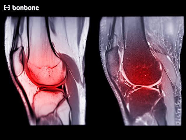
Articular cartilage — the smooth tissue covering the bone ends within joints — presents a fundamental biological challenge: it has no blood supply of its own. Nutrients and oxygen reach cartilage cells through diffusion from the synovial fluid that fills the joint space. This diffusion process is driven by the compression and decompression cycles of normal joint loading — movement literally pumps nutrients into cartilage and waste products out.
This means that appropriate movement and loading is not merely tolerated by cartilage — it is required for cartilage health. Joints that are immobilized or chronically underloaded develop cartilage that is thinner, less resilient, and more vulnerable to damage. This is one reason that complete rest is rarely the appropriate treatment for joint pain from osteoarthritis, and why low-impact exercise is consistently beneficial rather than harmful for most joint conditions.
The role of muscles in cartilage health is therefore twofold. First, strong muscles absorb and distribute joint forces, reducing the peak stress on cartilage with each step or movement. Second, muscles drive the movement and loading cycles that maintain cartilage nutrition. A person who is sedentary due to muscle weakness or pain is simultaneously depriving their cartilage of the mechanical stimulus it requires for ongoing health, while also removing the muscular cushioning that protects it from harmful impact forces.
Research on osteoarthritis outcomes consistently reflects this relationship. Quadriceps weakness precedes and predicts knee osteoarthritis progression. Strengthening the muscles around affected joints reduces pain and improves function. Physical activity that is well-matched to a person’s current capacity consistently outperforms both rest and passive treatments for most forms of degenerative joint disease. Adults managing ongoing joint concerns — as explored in guides to living well with chronic health conditions — benefit from understanding that the active component of treatment, specifically exercise, is not optional in musculoskeletal management.
Bone Remodeling and the Role of Mechanical Loading
One of the most important and underappreciated aspects of bone biology is that bone density is maintained and built in direct response to the mechanical forces bones experience. This relationship — known as Wolff’s Law — means that bone adapts its structure over time to match the loads it regularly bears: bone subjected to higher mechanical stress becomes denser and structurally reinforced in the direction of that stress, while bone that is chronically underloaded loses density.
The practical implication is that weight-bearing exercise is specifically required to maintain bone density — not just any exercise, but exercise that transmits force through the skeleton. Swimming and cycling, while excellent for cardiovascular health and joint-friendly for many musculoskeletal conditions, provide limited osteogenic (bone-building) stimulus because they are not weight-bearing. Walking, running, hiking, dancing, and resistance training all apply mechanical force to bone that stimulates osteoblast activity and density maintenance.
Muscle contractions also directly stimulate bone formation. Every time a muscle contracts forcefully, it exerts tensile stress on the bone at its attachment point. This stress is a powerful driver of bone remodeling at those sites — which is why the bones of the dominant arm in tennis players are measurably denser than those of the non-dominant arm, and why resistance training produces bone density benefits in addition to muscle strengthening.
This mechanobiological relationship helps explain one of the concerning aspects of sarcopenia — age-related muscle loss. As muscles weaken and shrink with aging, the mechanical stimulus they provide to bone is reduced, contributing to bone density loss that parallels the muscle loss. The two processes reinforce each other: weaker muscles → less mechanical bone stimulation → reduced bone density → more fragile bones → greater injury risk from falls → less activity → further muscle loss. Breaking this cycle through consistent resistance training and adequate protein and calcium intake is one of the most important preventive health interventions available to adults in midlife and beyond. A well-structured long-term health plan that encompasses musculoskeletal health alongside metabolic and organ health addresses this interconnected biology systematically.
Proprioception: The Nervous System’s Role in Musculoskeletal Integration
The musculoskeletal system cannot function as an integrated unit without the nervous system as its coordinator. Proprioception — the sensory system that monitors joint position, movement, and force — is the mechanism by which the nervous system continuously updates its model of where the body is in space and adjusts muscle activation accordingly.
Proprioceptive information is gathered by specialized receptors in muscles (muscle spindles, which detect stretch), tendons (Golgi tendon organs, which detect tension), and joint capsules and ligaments (which detect joint position and movement rate). This information streams continuously to the spinal cord and brain, where it is used to coordinate muscle activation — adjusting the stiffness and force of muscles around each joint to maintain stability and produce smooth, controlled movement.
When proprioceptive function is intact, the musculoskeletal system maintains joint stability with impressive precision, even during rapid or unexpected movements. When it is impaired — by joint injury (which damages mechanoreceptors in capsules and ligaments), by pain (which alters motor patterns), by aging (which reduces receptor sensitivity), or by sedentary behavior (which reduces the richness of proprioceptive experience) — joint control deteriorates and injury risk increases.
This is why balance training and proprioceptive exercises are a meaningful component of musculoskeletal health maintenance, not just a consideration after injury. Standing on one leg, walking on uneven surfaces, and exercises that challenge postural control all maintain the neural pathways that protect joints from injury during unexpected movements. Falls in older adults — with their serious consequences for bone health and functional independence — are substantially driven by proprioceptive decline, and balance training consistently reduces fall risk in this population. Annual health monitoring, such as a systematic health checklist approach, can incorporate balance assessment as a standard component of musculoskeletal health evaluation.
When One System Fails: The Cascade of Consequences
Understanding how bones, joints, and muscles work together also helps explain the clinical patterns that healthcare providers see routinely — and why addressing one component in isolation so often provides only partial relief.
Consider the typical progression following a significant joint injury. An anterior cruciate ligament (ACL) tear damages not only the structural ligament but also the proprioceptive receptors within it. The resulting instability and pain reduce load-bearing through the knee. Reduced loading causes quadriceps atrophy — studies document 20–40% quadriceps volume loss within weeks of ACL injury. The combination of instability and weakened muscles exposes the knee cartilage to abnormal loading patterns, accelerating wear at specific cartilage surfaces. Bone remodeling responds to the altered loading, with increased density at some points and decreased density at others. Long-term follow-up studies of ACL-injured individuals show substantially elevated rates of knee osteoarthritis compared to uninjured controls — even after surgical reconstruction and full return to sport.
This cascade is not inevitable when managed comprehensively. Rehabilitation programs that restore proprioception, rebuild quadriceps and hamstring strength, and address movement quality throughout the kinetic chain consistently produce better long-term joint outcomes than those focused narrowly on the injured structure itself. The same principle applies to managing any chronic musculoskeletal condition: an integrated approach that addresses all three systems simultaneously — bones, joints, and muscles — produces outcomes that cannot be achieved by treating each in isolation.
How Do Bones, Joints, and Muscles Work Together? (Quick Summary)
Bones, joints, and muscles form an integrated mechanical system. Bones provide the rigid lever arms; joints are the pivot points that allow controlled movement; muscles generate the forces that move bones through those joints. Muscles also actively stabilize joints, supplementing the passive stability provided by ligaments. Bone density is maintained by the mechanical forces generated through weight-bearing and muscular activity. Joint cartilage health depends on the movement and loading cycles that muscles drive. The nervous system coordinates the entire system through proprioception. Each component depends on the others — which is why problems in one reliably create problems in all three, and why effective musculoskeletal care addresses the whole system rather than any single component.
Practical Implications: What This Means for Your Musculoskeletal Health
The integrated biology of the musculoskeletal system translates into several practical principles for adults managing their health.
Muscle strength protects joints. This is not a metaphor — it is a mechanical reality. Investing in consistent strength training reduces the forces transmitted through cartilage with every step, slows osteoarthritis progression, and reduces fracture risk by both maintaining bone density and reducing fall risk.
Movement is medicine for cartilage. The avascular nature of cartilage means it depends on the pumping action of joint movement for nutrition. Prolonged immobility or inactivity starves cartilage of nutrients; regular, appropriately loaded movement maintains it.
Bone responds to mechanical demand. The skeleton you have in 10 years is substantially shaped by what you ask of your bones today. Weight-bearing and resistance exercise are non-optional components of bone health maintenance — no supplement fully substitutes for the mechanical stimulus that exercise provides.
Proprioception needs training. The neural component of joint stability is trainable and declines with disuse and aging. Balance and coordination exercises should be a regular part of musculoskeletal health maintenance, not only pursued after an injury.
Treat the system, not just the symptom. Pain in one joint is often maintained by weakness or movement problems elsewhere in the kinetic chain. Lower back pain frequently involves hip mobility and core strength. Knee pain is often influenced by foot mechanics and hip muscle function. Understanding the system-level nature of musculoskeletal problems — as healthcare providers increasingly do — produces more durable results than local treatment alone. Adults who track their overall health trajectory over time, building on approaches they may already use for other body systems like the principles of musculoskeletal health or systematic health reviews for health maintenance after age 60, are better positioned to recognize when a pattern of musculoskeletal change requires professional attention.
Frequently Asked Questions
Why does a muscle injury often affect the joint it crosses?
Muscles provide active stabilization of the joints they cross — they are not just movers but also protectors. When a muscle is injured, its ability to contract forcefully and at the right moment is impaired. This reduces the dynamic stability it would normally provide to the adjacent joint, exposing the joint’s passive structures (ligaments, cartilage) to forces they are less equipped to handle alone. This is why hamstring muscle tears increase the load on the knee’s anterior cruciate ligament, and why rotator cuff muscle injuries increase the risk of shoulder joint instability and secondary joint problems.
Does cartilage grow back after it is damaged?
Adult articular cartilage has very limited regenerative capacity because it is avascular (no blood supply) and has few resident stem cells. Minor surface damage may be partially repaired through the production of fibrocartilage — a less specialized repair tissue — but this does not fully restore the mechanical properties of hyaline cartilage. Significant cartilage loss, as in osteoarthritis, does not spontaneously regenerate. Surgical procedures including microfracture, osteochondral transplantation, and cartilage cell transplantation can address focal defects in selected patients, but no technique reliably restores large areas of damaged cartilage to normal function. Research into cartilage regeneration using stem cells and tissue engineering is active but has not yet produced clinically available solutions.
Why do older adults lose muscle faster than younger ones?
Several age-related biological changes accelerate muscle loss (sarcopenia) in older adults. Hormonal changes — declining testosterone in men and estrogen in women — reduce anabolic signaling that promotes muscle protein synthesis. Motor unit remodeling reduces the number and size of fast-twitch muscle fibers (which provide strength and power) preferentially over slow-twitch fibers (which provide endurance). Inflammation associated with aging (termed inflammaging) impairs muscle protein synthesis and promotes muscle protein breakdown. Reduced physical activity, commonly associated with aging, removes the mechanical stimulus needed to maintain muscle mass. Adequate protein intake (particularly leucine-rich protein) and consistent resistance training are the most effective known interventions to slow this process at any age.
Can you damage a joint by exercising too much?
Overuse injuries — damage from repetitive loading that exceeds the tissue’s capacity to repair itself — are real and common in people who increase training load too rapidly or do not allow adequate recovery time between high-intensity sessions. Tendons and bones are particularly susceptible: stress fractures in bone and tendinopathy reflect accumulated microdamage that outpaces remodeling and repair. However, for the vast majority of adults doing moderate recreational exercise, overuse injury is far less of a concern than the substantial harms of inactivity. The principle of progressive loading — gradually increasing exercise intensity and volume to allow tissues to adapt — is the practical answer to balancing exercise benefits against overuse risk.
Does losing weight help joint pain?
Yes, and the effect is substantial. Because joint forces are multiples of body weight during weight-bearing activities, relatively modest weight loss produces disproportionately large reductions in joint loading. A 10-pound weight loss reduces knee joint compressive forces by approximately 40 pounds per step during walking. Clinical studies on weight loss in people with knee osteoarthritis consistently show reductions in pain scores that correlate with the degree of weight loss achieved. The mechanism is primarily mechanical — reduced joint loading — with an additional contribution from reduced systemic inflammation associated with reduced adipose tissue, which also affects joint inflammation through inflammatory cytokine pathways.
How do ligaments differ from tendons, and why does it matter?
Ligaments connect bone to bone, limiting joint movement to safe ranges. Tendons connect muscle to bone, transmitting muscular force to the skeleton. Both are composed primarily of collagen fibers, have limited blood supply, and heal slowly compared to muscle. The clinical significance of the distinction lies in their different responses to loading: tendons are adapted to transmit high tensile forces in the direction of the muscle’s pull and respond well to progressive loading in rehabilitation. Ligaments are adapted for multi-directional restraint and are more sensitive to the direction of applied force. Rehabilitation after ligament injury must address the specific directional instability created by the torn ligament, while tendon rehabilitation focuses on gradually restoring tensile capacity along the line of pull.
What is the kinetic chain, and why is it relevant to joint pain?
The kinetic chain refers to the interconnected sequence of joints, muscles, and bones that work together during movement. In the lower body, the kinetic chain runs from the foot through the ankle, knee, hip, pelvis, and spine — each segment influencing the mechanics of adjacent segments. Restricted ankle mobility, for example, forces the knee and hip into compensatory movement patterns that increase their mechanical loading. Weak hip abductors allow the pelvis to drop during single-leg stance, altering knee alignment and increasing patellofemoral joint stress. Pain or limitation anywhere in the chain typically has upstream and downstream effects throughout it. This is why comprehensive musculoskeletal assessment evaluates movement patterns across the entire relevant kinetic chain, not just the symptomatic joint.
References and Further Reading
- NIH NIAMS — Bones, Muscles, and Joints Overview: Research-based information on musculoskeletal biology and conditions.
- Arthritis Foundation — Joint Protection Principles: Practical guidance on protecting joint structures through daily activities.
- Mayo Clinic — Strength Training: Get Stronger, Leaner, Healthier: Evidence-based overview of resistance training benefits for musculoskeletal health.
- CDC — Physical Activity for Arthritis: Summary of exercise recommendations and evidence for joint health maintenance.
- American Journal of Sports Medicine — Proprioception and Musculoskeletal Injury: Research on the role of proprioceptive training in injury prevention.
- StatPearls — Wolff’s Law and Bone Remodeling: Review of mechanical bone adaptation and its clinical implications.
- NIH MedlinePlus — Muscle Disorders: Authoritative patient resource on muscle health and conditions.
- National Osteoporosis Foundation — Exercise for Bone Health: Guidelines on weight-bearing and resistance exercise for bone density maintenance.
The explanation of why cartilage needs movement to stay healthy was genuinely new information for me. I have moderate osteoarthritis in both hips and my previous understanding was that exercise was something to be cautious about — that moving a damaged joint would cause more wear. Your article’s explanation that cartilage has no blood supply and depends on the pumping action of joint loading for nutrient delivery makes the case for exercise in a way that actually makes biological sense to me rather than just being a recommendation I’m supposed to follow. I’ve since started water aerobics three times per week, which is low impact enough that I don’t have pain during or after, and after two months I notice that my morning hip stiffness has reduced. I think the framing of movement as medicine rather than as a risk to manage would help a lot of people with arthritis who are currently sitting still to protect their joints.
The kinetic chain section at the end explained something I’ve been puzzling over for two years. I had persistent knee pain that multiple imaging studies showed was not coming from the knee joint itself — no significant cartilage loss, no meniscus damage. My physiotherapist eventually identified very tight hip flexors and weak hip abductors from years of desk work as the primary driver. The knee was experiencing abnormal loading because the hip wasn’t controlling alignment during single-leg stance. Six weeks of targeted hip strengthening resolved pain that three years of knee-focused treatment had not. Your article is the first plain-language explanation I’ve seen of why this happens mechanically. I think a lot of people are spending years treating symptoms at the wrong joint because the kinetic chain concept isn’t well communicated in standard care.
Your experience is very consistent with what musculoskeletal research has established about patellofemoral pain and knee pain more broadly. A landmark study by Powers et al. published in the Journal of Orthopaedic and Sports Physical Therapy demonstrated that abnormal hip kinematics — specifically reduced hip abductor and external rotator strength causing dynamic valgus (knee collapse inward) during single-leg loading — is a primary driver of patellofemoral pain in many patients. The knee is experiencing the consequence of a proximal control problem, not a structural knee problem. This pattern is sometimes called ‘the knee as a victim rather than a culprit’ in sports medicine literature. The reason knee-focused treatment fails in these cases is precisely what you experienced: if the underlying hip control deficit is not corrected, the abnormal loading pattern continues regardless of what is done locally at the knee. The clinical challenge is that standard imaging of the knee shows no structural cause, which can lead providers to question the reality of the patient’s pain rather than searching for the functional cause further up the chain. A physiotherapist with a full-body movement assessment approach — rather than one focused on the symptomatic joint — is the most reliable way to identify these patterns.