
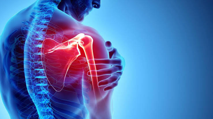
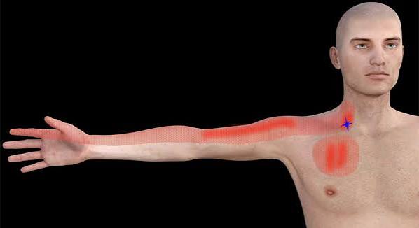
Pain that travels down the arm — radiating from the neck, shoulder, or chest into the upper arm, forearm, or hand — carries a broad differential that includes some of medicine’s most important diagnoses. At one end of the spectrum lies cervical radiculopathy from a compressed disc, which is common, usually resolves with time, and follows a predictable dermatomal pattern. At the other end lies myocardial infarction, in which isolated arm pain — without chest pain — can be the sole presenting symptom. Between these poles sit conditions including thoracic outlet syndrome, shoulder pathology, ulnar nerve entrapment, and, rarely, an apical lung cancer invading the brachial plexus. Accurate assessment depends on identifying the pattern: the quality of the pain, which aspect of the arm and hand is affected, whether it appears at rest or with specific activities, and what associated features are present.
This article covers the major causes of arm pain that radiates distally from the neck or shoulder, with particular attention to the clinical features that distinguish each cause, the red flags that demand immediate assessment, and the investigation pathway. The goal is not to replace clinical evaluation but to give anyone experiencing this symptom the framework to communicate it clearly and to recognise when urgency is required.
What Does Radiating Arm Pain Mean?
Arm pain that appears to travel or radiate from a proximal source is not a single entity. Understanding which mechanism is responsible requires identifying whether the pain is neurological, vascular, cardiac, or referred musculoskeletal in origin:
- Neurological radiating pain (radiculopathy) follows the dermatomal territory of the compressed cervical nerve root — a specific finger or aspect of the forearm and hand. It has a sharp, burning, or electric quality and is typically associated with numbness, tingling, or weakness in the same distribution.
- Referred musculoskeletal pain from the cervical facet joints, shoulder, or neck muscles is dull, aching, and poorly localised. It tends to stay in the shoulder and upper arm, rarely reaching the hand, and has no associated neurological signs.
- Cardiac pain radiating down the arm is typically crushing or heavy in quality, predominantly in the medial aspect of the left arm, and is associated with cardiovascular risk factors — though it can occur in isolation without chest pain, particularly in women and diabetics.
- Thoracic outlet pain involves the lower brachial plexus (C8/T1) and preferentially affects the medial forearm and hand — the ring and little fingers — and is worsened by arm elevation.
Cervical Radiculopathy: The Most Common Neurological Cause
Cervical radiculopathy — compression or irritation of a cervical nerve root — is the most common cause of arm pain that travels in a specific dermatomal distribution. The most frequently affected levels are C6/7 (compressing C7) and C5/6 (compressing C6). The distribution of symptoms is highly predictable and clinically useful:
- C5 (C4/5 disc): lateral arm, deltoid; shoulder abduction weakness; biceps reflex reduced
- C6 (C5/6 disc): thumb and index finger, lateral forearm; biceps and brachioradialis weakness; biceps reflex reduced
- C7 (C6/7 disc, most common): middle finger, posterior forearm; triceps weakness; triceps reflex reduced
- C8 (C7/T1 disc): ring and little finger, medial forearm; intrinsic hand muscle wasting (thenar, hypothenar, interossei)
Neck pain radiating into the arm (brachialgia) is typically present but some patients have arm symptoms with minimal neck pain. Spurling’s test — lateral flexion and extension of the neck toward the symptomatic side combined with gentle axial loading — reproduces the radicular arm pain and is highly specific for cervical radiculopathy. Most episodes improve within 4–12 weeks with physiotherapy, appropriate analgesia, and anti-neuropathic agents (gabapentin, amitriptyline). MRI cervical spine is the investigation of choice when symptoms are severe, persistent, or associated with progressive weakness. Loss of grip strength is a common functional consequence of C8 radiculopathy and cervical myelopathy.
Cervical Myelopathy: When the Cord Itself Is Compressed
Cervical spondylotic myelopathy (CSM) — compression of the spinal cord itself within the cervical canal — is distinct from radiculopathy (nerve root compression) and carries greater clinical significance. Cord compression produces upper motor neurone signs below the level of the lesion: spasticity and hyperreflexia in the legs, upgoing plantar responses (Babinski), and a broad-based spastic gait with poor balance. In the arms, there may be a combination of lower motor neurone signs at the level of compression (root involvement) and upper motor neurone signs below. Lhermitte’s phenomenon (electric shock sensation down the spine on neck flexion) and the Hoffman sign (flicking the middle fingernail causes reflex flexion of the thumb and index finger — an upper motor neurone sign in the hand) are useful clinical findings. Cervical myelopathy is a surgical diagnosis: compression severe enough to produce cord signal change on MRI warrants urgent neurosurgical assessment.
Cardiac Ischaemia: The Diagnosis That Cannot Be Missed
Arm pain that radiates from the chest is a classic feature of myocardial ischaemia — angina and myocardial infarction — mediated through the convergence of cardiac sensory fibres with somatic afferents in the upper thoracic spinal segments. The classic pattern is chest pain or tightness radiating to the left arm, jaw, left shoulder, or upper back. However, several patient groups frequently present with atypical features where arm pain occurs without chest pain: women are more likely than men to present with isolated arm, jaw, or epigastric pain as the primary symptom; diabetic patients with autonomic neuropathy may have reduced pain perception and present with dyspnoea and fatigue rather than chest pain; and older adults may experience only breathlessness or fatigue.
The quality of cardiac arm pain is typically crushing, heavy, or tightening — fundamentally different from the sharp, burning, or electric quality of neurological arm pain. Associated features including diaphoresis (sweating), nausea, breathlessness, and a sense of impending doom raise the clinical probability of a cardiac cause. Any arm pain of uncertain origin in a patient with cardiovascular risk factors (hypertension, hyperlipidaemia, diabetes, smoking, family history of early coronary artery disease) should have a cardiac cause actively excluded with an ECG and troponin measurement before a musculoskeletal cause is assumed. This is a clinical rule that saves lives.
Call 999 immediately if arm pain is accompanied by: chest tightness or heaviness; sweating or clamminess; breathlessness; nausea; a sense of impending doom; or if you have known cardiac disease. Do not wait to see if the pain resolves. An ECG and troponin blood test are the essential first investigations to exclude acute coronary syndrome.
Thoracic Outlet Syndrome
Thoracic outlet syndrome (TOS) results from compression of the brachial plexus, subclavian artery, or subclavian vein as these structures pass between the clavicle and the first rib — the thoracic outlet. The most common form, neurogenic TOS, involves the lower brachial plexus (C8 and T1 nerve roots) because these are the most vulnerable to compression in this space. The clinical picture is medial forearm and hand pain, with numbness and tingling in the ring and little fingers, and weakness and wasting of the intrinsic hand muscles — a distribution that closely mimics C8 radiculopathy or ulnar neuropathy. The key distinguishing feature is that symptoms are worsened by arm elevation: carrying shopping, reaching overhead, and using a computer mouse for extended periods all aggravate neurogenic TOS. Numbness and tingling in the medial hand with arm elevation is a characteristic pattern.
Vascular TOS is less common but more acute in its presentations. Subclavian artery compression can cause intermittent arm pallor, coldness, Raynaud-like colour changes, and in severe cases distal arterial embolism from thrombosis over an atherosclerotic or post-stenotic dilatation in the subclavian artery. Subclavian vein compression produces effort thrombosis — Paget-Schroetter syndrome — characteristically in young athletes who perform repetitive overhead activities (swimmers, volleyball players, weightlifters); the arm becomes acutely swollen and blue after exertion. Management of neurogenic TOS begins with physiotherapy targeting the cervicoscapular muscles; surgical decompression (first rib resection or scalenectomy) is reserved for refractory cases.
Shoulder Pathology: When the Pain Comes from the Joint
Shoulder pathology — rotator cuff impingement, tendinopathy, full-thickness tear, frozen shoulder, or glenohumeral OA — commonly refers pain into the lateral upper arm and can be confused with C5 or C6 radiculopathy. The key distinction is that shoulder pain, even when referred, rarely travels below the elbow. Rotator cuff impingement produces a painful arc (pain at 60–120° of shoulder abduction, improving above and below this range) and may refer into the lateral deltoid region. Full-thickness rotator cuff tears produce weakness of shoulder abduction and external rotation, sometimes acute with an audible or palpable pop. Frozen shoulder (adhesive capsulitis) — most common in middle-aged adults, diabetics, and following shoulder immobilisation — causes severe diffuse shoulder pain that radiates into the upper arm with extreme stiffness; the first movement lost is external rotation. None of these conditions typically produce numbness or tingling in the hand, which is an important clinical sign pointing away from the shoulder and toward the neck or thoracic outlet.
Ulnar Nerve Entrapment
Ulnar neuropathy at the elbow (cubital tunnel syndrome) — the second most common nerve entrapment after carpal tunnel syndrome — compresses the ulnar nerve at the medial epicondyle of the humerus. The ulnar nerve supplies the ring and little fingers (sensory), the hypothenar muscles (little finger opposition and abduction), the interossei (finger abduction/adduction), and the medial two lumbricals. Compression produces tingling and numbness in the ring and little fingers, medial hand, and medial forearm; weakness of grip and pinch; and — in advanced cases — a claw hand deformity (hyperextension at the MCP joints and flexion at the IP joints of the ring and little fingers) due to the loss of the interossei and medial lumbricals that normally flex the MCPs. Froment’s sign — when the patient is asked to hold a piece of paper between the thumb and index finger, the distal joint of the thumb flexes involuntarily because adductor pollicis (ulnar-innervated) is weak and flexor pollicis longus (median-innervated) compensates — confirms adductor pollicis weakness. Nerve conduction studies localise the compression site precisely. Treatment: conservative (avoiding prolonged elbow flexion, padded elbow, splinting) → surgical decompression for persistent or progressive cases.
Pancoast Tumour: The Diagnosis Not to Miss
A Pancoast tumour is an apical lung cancer (most commonly squamous cell carcinoma or adenocarcinoma) that invades the structures at the superior sulcus of the lung: the lower brachial plexus (C8, T1), the sympathetic chain, the subclavian vessels, and sometimes the first and second ribs. The result is a combination of severe medial arm and hand pain (C8/T1 distribution — ring and little fingers, medial forearm) with ipsilateral Horner’s syndrome (ptosis — drooping of the upper eyelid, miosis — constricted pupil, and anhidrosis — absent sweating on the ipsilateral face) from invasion of the cervical sympathetic chain. Hand and intrinsic muscle wasting may develop as the T1 root is progressively compressed.
The diagnosis is frequently delayed by months because the symptoms — arm pain and hand tingling — are attributed to cervical radiculopathy, carpal tunnel syndrome, or shoulder pathology. The combination of lower brachial plexus symptoms plus ipsilateral Horner’s syndrome is pathognomonic and requires urgent chest X-ray and CT chest. Any patient presenting with medial arm pain, hand numbness, and a unilateral drooping eyelid or small pupil should have a chest X-ray on the same day. Early diagnosis substantially improves treatment options (surgery, radiotherapy, and chemotherapy for localised disease).
Referred Pain from the Cervical Spine
The cervical facet joints are synovial joints susceptible to osteoarthritic degeneration and can generate significant pain that refers into the shoulder, scapular region, and upper arm without following a dermatomal territory. C2/3 and C3/4 facet pain can produce occipital headaches and posterior cervical pain. Lower cervical facet pain from C4/5 downward refers to the ipsilateral shoulder and upper arm. The distinguishing features from true radiculopathy are: the pain is dull and aching rather than electric or burning; it does not typically travel below the elbow; there are no neurological signs in the hand or forearm; and Spurling’s test does not reproduce the typical arm pain in its dermatomal distribution. Cervical facet pain is worsened by cervical extension and rotation and improved by flexion — the opposite pattern from cervical disc herniation. Treatment includes physiotherapy, cervical facet joint injections, and medial branch radiofrequency ablation for refractory cases.
Complex Regional Pain Syndrome
Complex regional pain syndrome (CRPS) is a chronic pain condition characterised by severe burning pain, allodynia (pain from light touch), and vasomotor and trophic changes in a limb — disproportionate to any initiating injury. The upper limb is the most commonly affected. CRPS Type 1 (reflex sympathetic dystrophy) occurs without identifiable nerve injury and is most commonly triggered by a distal radius fracture (Colles’ fracture), though it can also develop after MI, stroke, or surgery. The affected hand and forearm show swelling, colour changes (initially warm and red, later cool and cyanotic), sweating abnormalities, and eventual trophic skin and nail changes with osteopenia on X-ray. The Budapest criteria — requiring sensory, vasomotor, sudomotor, and motor/trophic categories — are used for diagnosis. Management is complex: early physiotherapy and pain rehabilitation, with ketamine infusions, sympathetic nerve blocks, and spinal cord stimulation for severe refractory cases.
When Arm Pain Needs Urgent Assessment
- Arm pain + chest tightness/heaviness + sweating/breathlessness → call 999: possible heart attack
- Medial arm pain + ipsilateral drooping eyelid or small pupil (Horner’s) → urgent chest X-ray: possible Pancoast tumour
- Rapidly progressive arm or hand weakness → urgent neurology referral
- Bilateral arm/hand symptoms + legs affected + gait disturbance → cervical myelopathy: urgent MRI
- Acute arm swelling after overhead exertion in an athlete → vascular TOS/effort thrombosis: same-day Doppler
Investigation of Arm Pain
The investigation pathway follows the clinical assessment. For suspected radiculopathy, MRI cervical spine is the gold standard; nerve conduction studies (NCS) are useful to confirm root level when imaging is equivocal or when peripheral neuropathy is in the differential. For cardiac causes, 12-lead ECG and high-sensitivity troponin are mandatory. For suspected Pancoast tumour, chest X-ray followed by CT chest with contrast; PET-CT for staging. For TOS, imaging of the cervicothoracic junction (CT with vascular protocol if vascular TOS is suspected; MRI neurography for neurogenic TOS), Doppler ultrasound for subclavian vein thrombosis. For shoulder pathology, plain X-ray and ultrasound are the first-line investigations; MRI arthrogram for full-thickness rotator cuff tears.
Arm and hand weakness alongside radiating pain narrows the differential and almost always warrants formal neurological assessment. The principle of dermatomal localisation that applies to leg pain applies equally to the arm — identifying which fingers are affected directs the clinical diagnosis.
Key Resources
- NHS: Coronary Artery Disease and Angina
- NHS: Heart Attack — Symptoms and Emergency Action
- NHS: Cervical Spondylosis
Frequently Asked Questions
What causes pain that shoots down the arm?
A shooting, electric, or burning pain that travels down the arm in a specific distribution is most likely caused by cervical radiculopathy — compression of a cervical nerve root by a disc herniation or spondylotic osteophyte. The C6/7 disc level (C7 root) is most commonly affected, producing middle finger, posterior forearm, and triceps symptoms. C5/6 (C6 root) is next most common, producing thumb and index finger symptoms. The pattern of which fingers are affected is the most useful clinical localiser. Thoracic outlet syndrome can produce similar medial arm symptoms by compressing the lower brachial plexus.
Can a heart attack cause arm pain without chest pain?
Yes — this is a clinically important presentation, particularly in women, diabetic patients, and older adults. Cardiac ischaemia can present with isolated left arm pain, jaw pain, or epigastric discomfort without the classic central chest pain. The arm pain in MI is typically heavy, crushing, or aching (not shooting or burning), involves the medial aspect of the left arm, and may be accompanied by sweating, breathlessness, or nausea. Any arm pain of uncertain cause in a person with cardiovascular risk factors should have an ECG and troponin performed to exclude a cardiac cause before a musculoskeletal explanation is assumed.
How is arm pain from the neck different from shoulder pain?
Arm pain from cervical radiculopathy follows a specific dermatomal pattern, usually travels below the elbow to specific fingers, has a neurological quality (burning, shooting, electric), and is associated with numbness or tingling in the same territory. It is worsened by neck movements and Valsalva manoeuvres. Shoulder pathology (rotator cuff, frozen shoulder, OA) produces a more diffuse aching in the shoulder and upper arm, rarely travels below the elbow, and has no associated hand or finger symptoms. Spurling’s test (neck lateral flexion and compression) reproduces radicular arm pain; painful arc testing and shoulder range of motion abnormalities identify shoulder pathology.
What is thoracic outlet syndrome?
Thoracic outlet syndrome (TOS) is compression of the brachial plexus, subclavian artery, or subclavian vein between the clavicle and first rib. Neurogenic TOS — the most common form — involves the lower brachial plexus (C8/T1), producing medial forearm and hand pain and tingling in the ring and little fingers, with symptoms worsened by arm elevation. It is commonly associated with a cervical rib (a congenital extra rib from C7), poor posture, or muscular hypertrophy. Physiotherapy targeting the cervicoscapular region is first-line treatment; first rib resection is curative for refractory neurogenic TOS.
What does Horner’s syndrome mean when combined with arm pain?
Horner’s syndrome — unilateral drooping of the upper eyelid (ptosis), a constricted pupil (miosis), and absent sweating (anhidrosis) on the same side — combined with medial arm and hand pain is a red flag combination for Pancoast tumour: an apical lung cancer invading the lower brachial plexus (C8/T1) and the cervical sympathetic chain. This combination requires urgent chest X-ray and CT chest on the same day. The diagnosis is frequently delayed because the arm symptoms are attributed to cervical disc disease — awareness of this pattern can lead to earlier diagnosis when treatment options are better.
Why do I get arm pain at night?
Nocturnal arm pain has several possible causes depending on the pattern. Cervical radiculopathy is often worse at night because lying flat reduces cervical lordosis and may increase pressure on a compressed root. Carpal tunnel syndrome (median nerve compression at the wrist) classically causes nocturnal paraesthesiae — waking with tingling and numbness in the first three and a half fingers, often relieved by shaking the hand. Thoracic outlet syndrome may be aggravated by certain sleeping positions. Shoulder pathology (particularly frozen shoulder) is notoriously painful at night, especially when lying on the affected shoulder. Cardiac ischaemia can cause resting arm pain (unstable angina) — this should always be considered when arm pain at rest has a crushing quality and is associated with cardiovascular risk factors.
When should I see a doctor urgently about arm pain?
Seek emergency care immediately if arm pain is accompanied by chest tightness, breathlessness, sweating, or nausea — particularly if you have cardiovascular risk factors. These may indicate a heart attack. Seek same-day urgent assessment if you notice a drooping eyelid or unequal pupils alongside arm and hand pain — this combination requires a chest X-ray to exclude a Pancoast tumour. Book an urgent appointment (within days) for rapidly worsening hand or arm weakness, bilateral arm symptoms with any leg involvement, or arm pain that has not improved after four to six weeks of conservative management.
References
- Caridi JM, et al. Cervical radiculopathy: a review. HSS J. 2011;7(3):265–272.
- Panagopoulos J, et al. Natural history of radiculopathy. Spine. 2017;42(2):91–99.
- Sanders RJ, et al. Thoracic outlet syndrome: a review. Neurologist. 2008;14(6):365–373.
- Fehrenbacher JW. Pancoast tumour. Ann Thorac Surg. 2018;106(1):313–316.
- Moseley GL, Flor H. Targeting cortical representations in the treatment of chronic pain. Neurorehabil Neural Repair. 2012;26(6):646–652.
- NHS. Heart attack. nhs.uk. Updated 2022.
- Chung TT, et al. Complex regional pain syndrome. Curr Pain Headache Rep. 2014;18(4):411.
- Waldman SD. Cervical spondylosis. In: Pain Management. WB Saunders; 2011.
The section on distinguishing shoulder pain from cervical radiculopathy is exactly what I find myself explaining to patients. I had a frozen shoulder diagnosis for eight months before an MRI of my cervical spine showed a C6/C7 disc herniation — the radiating pain to the thumb and index finger with reduced bicep reflex was the clue the physiotherapist picked up when the shoulder treatment wasn’t working. The key difference described here — that shoulder pain is localised to the deltoid and reproduced by shoulder movements, while radiculopathy radiates distally and is reproduced by neck movements — should be better known.
Fiona raises an important clinical point — the overlap between shoulder pathology and cervical radiculopathy is one of the most common diagnostic challenges in musculoskeletal medicine. The examination findings that distinguish them are reliable: cervical radiculopathy reproduces arm symptoms with the Spurling manoeuvre (lateral neck flexion toward the affected side with compression), and shoulder pathology reproduces pain with rotator cuff provocative tests. When both are present — cervicogenic shoulder pain — the pattern can be particularly confusing. The C6 level (thumb and index finger, reduced bicep reflex) and C7 level (middle finger, reduced tricep reflex) are the most commonly affected levels in disc herniation. Martin, your case illustrates the critical principle that any arm pain reproduced by exertion must be evaluated for cardiac cause before a musculoskeletal explanation is accepted — exertional reproduction, relief with rest, and absence of positional or movement-dependent features point strongly to an ischaemic origin.
I had arm pain for about three months that my GP initially thought was a trapped nerve in the neck. When I mentioned it came on with exertion and resolved with rest, I was sent for an urgent cardiology referral. The angiogram showed a significant LAD stenosis. I had a stent placed and the arm pain resolved completely. The article correctly flags this as a presentation that must not be missed — exertional arm pain that comes and goes is cardiac until proven otherwise.