
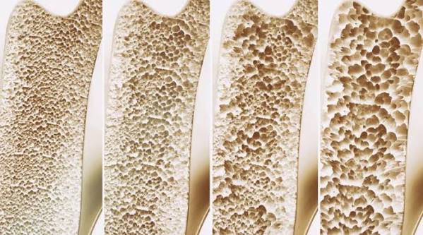
Bone density — the amount of mineral (primarily calcium and phosphorus) packed into a given volume of bone — is the most important measurable predictor of fracture risk in adults. When a clinician requests a bone density test, or when a DEXA scan report comes back with T-scores and Z-scores, understanding what those numbers mean, why they matter, and how bone density changes over a lifetime gives patients the context they need to act on that information effectively. Bone density is not a fixed biological constant — it peaks in early adulthood, declines gradually with age, and is modified — for better or worse — by diet, exercise, hormones, and medications throughout life.
What Is Bone Density?
Bone consists of a protein scaffold — primarily collagen — that is mineralised with calcium phosphate crystals (hydroxyapatite). The density of this mineralised matrix, measured in grams per square centimetre (g/cm²) by DEXA scanning, is what clinicians mean by bone mineral density (BMD). A higher BMD means more mineral per unit area, which translates directly into greater bone strength and resistance to fracture under mechanical load.
Bone is not a static material. It is continuously remodelled throughout life by two cell types working in opposing directions:
- Osteoclasts — dissolve (resorb) old or damaged bone, releasing calcium and phosphorus back into the bloodstream
- Osteoblasts — form new bone by laying down collagen and mineralising it with calcium phosphate
When osteoclast activity exceeds osteoblast activity — as happens progressively with age, and sharply at the menopause — net bone loss results. This is the fundamental mechanism of osteoporosis and osteopenia. The balance of these two cell populations is regulated by hormones (oestrogen, testosterone, parathyroid hormone, calcitonin), mechanical loading (exercise), and nutritional factors (calcium, vitamin D).
Peak Bone Mass
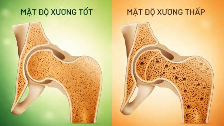
Peak bone mass is the maximum bone density an individual achieves, typically between the ages of 25 and 30. It is the single most important determinant of bone health in later life: the higher the peak, the more bone loss can be sustained before the osteoporosis threshold is crossed. Genetics account for an estimated 60–80% of peak bone mass variation between individuals, explaining why a family history of osteoporosis or hip fracture is itself a clinical risk factor. The remaining 20–40% is determined by factors that can be influenced during childhood and early adulthood:
- Calcium intake during childhood and adolescence — particularly the pubertal growth spurt when bone mineral is being deposited rapidly
- Vitamin D status — essential for calcium absorption and bone mineralisation
- Physical activity — weight-bearing exercise during growth stimulates osteoblast activity and directly builds bone mass
- Hormonal environment — sex hormone levels during puberty, thyroid function, and growth hormone all influence the peak achieved
- Absence of conditions that reduce bone mass — eating disorders, amenorrhoea (in athletes or with malnutrition), and malabsorption during the growth period can permanently reduce peak bone mass
How Bone Density Changes With Age
After peak bone mass is reached, gradual bone loss begins — in both men and women — from the early thirties. In men, this loss is slow and relatively linear: approximately 0.3–0.5% of bone mass per year throughout adult life. In women, the trajectory is different in two important respects:
- Premenopausal bone loss is similar to men at approximately 0.3–0.5% per year
- At the menopause, loss accelerates dramatically — to 2–3% per year in the first five years post-menopause — driven by the loss of oestrogen’s suppression of osteoclast activity
- After the acute menopausal phase, loss returns to approximately 1% per year
This is why postmenopausal women have significantly higher osteoporosis prevalence than men of the same age — not because men are not affected, but because the female trajectory includes a period of very rapid loss at a time when women are often not being monitored for bone health. The clinical implication is that DEXA screening becomes relevant at an earlier age for women than for men, and earlier still for women with early or surgical menopause.
How Is Bone Density Measured?
DEXA scanning (dual-energy X-ray absorptiometry) is the gold standard measurement for bone mineral density. It uses two low-energy X-ray beams at different energy levels to distinguish bone from soft tissue, calculates BMD at the hip (total hip and femoral neck) and lumbar spine (L1–L4), and produces T-scores and Z-scores. DEXA is accurate, reproducible, quick (10–15 minutes), and delivers a minimal radiation dose. Full detail on what the scan involves and what to expect is in our guide to DEXA scan: what adults should know.
Other measurement methods include:
- Quantitative CT (QCT): three-dimensional measurement of trabecular and cortical bone separately; higher radiation dose; used in research and some specialist centres
- Peripheral DEXA (pDXA) and quantitative ultrasound (QUS): measure bone density at peripheral sites (heel, forearm); lower cost and accessible, but less clinically validated for fracture prediction than central DEXA; used for screening in some settings but not for diagnosis
- Plain radiography: insensitive for early bone loss — approximately 30% of bone density must be lost before it is visible on plain X-ray
Understanding T-Scores and Z-Scores
DEXA results are expressed in two ways:
The T-score compares your measured BMD to the average BMD of a healthy young adult at peak bone mass (the reference population). It is expressed in standard deviations: a T-score of 0 means exactly average peak bone mass; −1.0 means one standard deviation below; −2.5 means two and a half standard deviations below. The WHO classification thresholds — normal (T ≥ −1.0), osteopenia (−1.0 to −2.5), and osteoporosis (T ≤ −2.5) — are based on this comparison. The T-score is used for postmenopausal women and men over 50.
The Z-score compares your BMD to an age-matched and sex-matched peer group — people of your own age and sex. A Z-score of −2.0 or below means bone density is significantly lower than expected for your age, which is clinically important and should prompt investigation for a secondary cause. The Z-score is used in premenopausal women, men under 50, and children. Using the T-score in these groups would be misleading — it would classify most normal adults as having osteopenia simply because they are no longer at their twenties peak. More on the DEXA classification system is in our guide to bone density test: a simple guide.
Factors That Affect Bone Density
Bone density at any point in adult life reflects the cumulative result of peak bone mass achieved, the rate of subsequent loss, and the modifying effect of lifestyle and medical factors. The most important modifiable influences on bone density throughout adult life are:
- Weight-bearing and resistance exercise: mechanical loading of bone through ground-reaction forces (walking, jogging, dancing, tennis) and muscle tension (resistance training) is the primary anabolic stimulus for osteoblast activity. Sedentary individuals lose bone more rapidly. More detail is in our guide to osteoporosis: symptoms, causes, and prevention.
- Calcium: the primary structural mineral in bone. Inadequate intake increases parathyroid hormone secretion, which increases osteoclast activity to maintain serum calcium at the expense of skeletal calcium. Full detail on dietary calcium is in our guide to calcium and bone health.
- Vitamin D: regulates intestinal calcium absorption; deficiency — very common in UK adults — impairs mineralisation and increases parathyroid hormone-driven bone resorption. See our guide to vitamin D and bone health.
- Oestrogen and testosterone: sex hormones are the most powerful regulators of osteoclast activity; their loss at menopause (oestrogen) and gradually with age (both sexes) is the primary driver of age-related bone loss
- Glucocorticoids (steroids): prednisolone and other corticosteroids at therapeutic doses directly inhibit osteoblast function; long-term use is the most common secondary cause of osteoporosis
- Smoking: impairs osteoblast function and accelerates bone loss at all ages; one of the most modifiable risk factors for bone density decline
- Body weight: low body weight (BMI below 19) is associated with lower bone density; mechanical loading from body weight and adipose-derived oestrogen both contribute to bone maintenance
Medical Conditions That Reduce Bone Density
Beyond the lifestyle and hormonal factors discussed above, a number of medical conditions independently reduce bone density — often considerably. Identifying and treating these secondary causes is an important part of managing anyone diagnosed with osteopenia or osteoporosis, particularly when low bone density is unexpected for a patient’s age or sex (indicated by a Z-score of −2.0 or below).
Malabsorption conditions: Coeliac disease is one of the most significant secondary causes — impaired small intestinal absorption of calcium and vitamin D means the skeleton is chronically under-mineralised even in people with seemingly adequate dietary intake. Both diagnosed and undiagnosed coeliac disease are associated with significantly lower bone density; the effect is partly reversible with strict gluten-free diet and supplementation. Inflammatory bowel disease (Crohn’s disease and ulcerative colitis) similarly impairs absorption, particularly when active disease involves the small bowel. Bariatric surgery — particularly Roux-en-Y gastric bypass — bypasses the segment of intestine where calcium is most efficiently absorbed, and bone density loss after bariatric surgery is a well-recognised complication that requires proactive management.
Endocrine conditions: Hyperparathyroidism (elevated parathyroid hormone, usually from a benign adenoma) drives osteoclast activity and mobilises calcium from bone, preferentially reducing cortical bone density at the forearm and hip. Hyperthyroidism — whether primary or caused by excess thyroid hormone treatment — accelerates bone turnover. Diabetes, particularly type 1, is associated with lower bone density; the mechanism involves impaired insulin-like growth factor signalling on osteoblasts. Cushing’s syndrome (excess cortisol from any cause) produces the same effect as therapeutic glucocorticoids — osteoblast suppression with significant bone loss.
Chronic kidney disease (CKD): Advanced kidney disease significantly disrupts bone and mineral metabolism through multiple mechanisms: reduced activation of vitamin D (which occurs in the kidney), impaired calcium and phosphate handling, elevated parathyroid hormone, and the direct effect of uraemia on bone cells. Renal osteodystrophy — the collective term for CKD-related bone disease — requires specialist management and is often underdiagnosed until bone density is severely reduced.
Rheumatoid arthritis: both the inflammatory process itself and the glucocorticoids used to manage flares contribute to accelerated bone loss, particularly at sites adjacent to inflamed joints (periarticular osteoporosis) as well as systemically. Patients with rheumatoid arthritis have approximately twice the fracture risk of age-matched controls and should be regularly assessed with DEXA and considered for bone protection therapy.
Investigating for secondary causes when bone density is unexpectedly low — with a standard blood panel including calcium, phosphate, ALP, thyroid function, full blood count, and renal and liver function — is standard practice and can identify reversible contributors to bone loss that, when treated, may prevent further deterioration.
Why Bone Density Matters Beyond Osteoporosis
Bone density exists on a continuous spectrum — fracture risk increases progressively as density falls, not only below the osteoporosis threshold. This is clinically important because it means that even T-scores in the osteopenia range carry meaningfully elevated fracture risk compared with normal bone density, and that bone density considerations apply to any adult with below-average T-scores rather than only those who have crossed the −2.5 line.
Importantly, a significant proportion of fragility fractures in absolute terms occur in people with osteopenia rather than osteoporosis — precisely because osteopenia is much more prevalent in the population. In a typical UK cohort of women over 50, more fragility fractures occur annually in women with osteopenia than in women with osteoporosis, because the proportion of women with osteopenia is several times larger, even though individual fracture risk per person is lower. This is the statistical logic behind the FRAX tool, which uses continuous T-score values (not category thresholds) alongside clinical risk factors to estimate 10-year fracture probability, enabling treatment decisions to be based on overall fracture probability rather than diagnostic category alone. Osteopenia-specific guidance is in our guide to osteopenia: what adults should know.
Frequently Asked Questions
Can bone density be improved?
Bone density can be modestly improved in younger adults through consistent weight-bearing and resistance exercise combined with adequate calcium and vitamin D. In older adults — particularly postmenopausal women — pharmacological treatment (bisphosphonates, denosumab, teriparatide) produces measurable T-score improvements. Teriparatide, as an anabolic agent that stimulates new bone formation rather than simply slowing resorption, produces the largest density gains — typically 8–12% at the lumbar spine over two years — and is used for severe osteoporosis or after antiresorptive treatment failure. For most adults with osteopenia or early osteoporosis, stabilisation of bone density (preventing further loss) through lifestyle measures and, where indicated, medication is the realistic and clinically meaningful goal.
What T-score indicates osteoporosis?
A T-score of −2.5 or below at the hip or lumbar spine on DEXA scanning defines osteoporosis, according to the WHO classification. Severe osteoporosis is defined as a T-score of −2.5 or below plus a prior fragility fracture. The T-score threshold was established based on the observed relationship between bone density and fracture probability in large population studies of postmenopausal women, and the −2.5 value was chosen to approximate the lifetime fracture risk that was considered clinically significant. It is a pragmatic threshold for communication and treatment decisions — not a biological boundary, as fracture risk increases continuously across the entire T-score range.
Does bone density change after a fracture?
A fragility fracture is the most powerful predictor of a subsequent fracture — this is an independent effect of the fracture event itself, beyond its association with low bone density. The fracture itself does not directly reduce bone density at other sites, but it is a signal that fracture risk is higher than the DEXA T-score alone might indicate, possibly because bone quality factors (collagen structure, microarchitecture) that are not captured by BMD measurement also play a role. For this reason, NICE guidance recommends that any person who sustains a fragility fracture is assessed for osteoporosis and offered treatment if appropriate — regardless of their T-score at the time.
How quickly does bone density change?
Bone density changes slowly under most circumstances — this is why DEXA monitoring intervals are measured in years, not months. In untreated postmenopausal women, annual bone loss averages approximately 1% per year after the early menopausal phase. With bisphosphonate treatment, this loss is halted and T-scores typically improve by 3–5% at the lumbar spine over three years. With teriparatide, gains of 8–12% at the lumbar spine over two years have been documented. The most rapid bone density losses occur in acute situations: immobilisation (e.g., prolonged bed rest), high-dose glucocorticoid initiation, and the acute menopausal transition (2–3% per year). DEXA scanning more frequently than every two years provides little additional information in most clinical contexts because the measurement precision error of DEXA means that changes below approximately 3% are within the error range of the test.
Is bone density the same as bone strength?
Bone density is the most important measurable contributor to bone strength, but it is not the only one. Bone quality — which encompasses the microarchitecture of trabeculae, the degree of collagen crosslinking, the accumulation of microdamage, and the mineralisation pattern — also contributes to how much force a bone can withstand before fracturing. This is why some people with T-scores in the osteopenia range sustain fragility fractures, while others with osteoporosis T-scores do not. BMD explains approximately 60–70% of the variance in bone strength across populations. The clinical relevance is that fracture prediction based on BMD alone is imperfect, and clinical risk factors for fracture (age, prior fracture, family history, glucocorticoid use) add meaningful predictive information beyond the T-score — which is the basis for the FRAX tool’s combined approach.
What is bone density measured in?
Bone mineral density is measured in grams per square centimetre (g/cm²) by DEXA scanning — the mass of mineral in a two-dimensional projected area of bone. This areal measurement (as opposed to a volumetric measurement) is a practical simplification that works well in population studies but can be influenced by bone size, which is why very tall individuals or those with large vertebrae may have slightly higher areal BMD than their true volumetric density warrants. Quantitative CT (QCT) measures volumetric BMD in mg/cm³ and can separate trabecular from cortical compartments, but it is used primarily in research rather than routine clinical practice due to higher cost and radiation dose.
At what age does bone density start to decline?
Bone density begins to decline gradually from the early thirties in both men and women — after peak bone mass is reached between approximately 25 and 30 years of age. In the decades before menopause, the rate of loss is slow at around 0.3–0.5% per year. At the menopause, loss accelerates sharply to 2–3% per year in women for approximately five to ten years. After this acute phase, loss reverts to approximately 1% per year for the rest of life. In men, the decline is gradual and linear throughout adult life at approximately 0.3–0.5% per year, with no equivalent of the acute menopausal phase, though testosterone decline in later decades contributes to accelerated loss in some men.
Summary
Bone density — measured as bone mineral density (BMD) in g/cm² by DEXA scanning — reflects the amount of mineral packed into the bone matrix and is the most important measurable predictor of fracture risk. It peaks in the late twenties, shaped primarily by genetics, diet, and exercise during growth, then declines gradually through adult life — with an accelerated phase at the menopause in women. The T-score classifies BMD relative to young-adult peak: normal (≥ −1.0), osteopenia (−1.0 to −2.5), and osteoporosis (≤ −2.5). Key modifiable influences on bone density throughout life include weight-bearing and resistance exercise, calcium and vitamin D intake, avoidance of smoking and excess alcohol, and management of medical conditions and medications that accelerate bone loss. Secondary causes — including coeliac disease, hyperparathyroidism, CKD, rheumatoid arthritis, and glucocorticoids — should be investigated and addressed when bone density is lower than expected for age. Because fracture risk increases continuously across the T-score spectrum, bone density matters not only for those meeting the formal osteoporosis diagnosis but for all adults with below-normal density. Related topics are covered in depth in our guides to osteoporosis: symptoms, causes, and prevention, osteopenia, and the DEXA scan.
Medical disclaimer: This article is for general educational purposes and does not constitute medical advice. Consult a qualified healthcare professional for personalised bone health assessment.
References:
NHS. Osteoporosis. NHS. 2023.
NICE CG146. Osteoporosis: assessing the risk of fragility fracture. NICE. 2023.
Kanis JA, et al. The diagnosis of osteoporosis. J Bone Miner Res. 1994;9(8):1137–1141.
Bonjour JP, et al. Peak bone mass. Osteoporos Int. 2009;20(Suppl 2):S3–S14.
International Osteoporosis Foundation. Bone density testing. iofbonehealth.org. 2023.
I was diagnosed with coeliac disease at age 48 and my first DEXA scan three months after diagnosis showed a Z-score of −2.3 — well below what would be expected for my age. My gastroenterologist explained that undiagnosed coeliac disease for years had been impairing my calcium absorption, and that my bones had been under-mineralised throughout that period without any obvious symptoms. I am now two years post-diagnosis, strictly gluten-free, on calcium and vitamin D supplementation, and my follow-up DEXA showed improvement to −1.6 — still below age-expected but much better. The article’s section on secondary causes is accurate and something I wish I had known earlier — coeliac disease is not immediately thought of as a bone disease, but the link with low bone density is direct and significant.
Frances, your case is a clear illustration of the clinical significance of Z-scores. A Z-score of −2.3 at age 48 — well below the −2.0 threshold that triggers investigation for secondary causes — correctly directed the diagnostic workup toward coeliac disease as the driver of unexpectedly low bone density for your age. The improvement from −2.3 to −1.6 over two years on strict gluten-free diet with calcium and vitamin D supplementation is a meaningful response, reflecting restored intestinal calcium absorption working through normal osteoblast activity. Stabilising and gradually improving the Z-score on strict dietary adherence is the realistic goal; your trajectory is encouraging. David, your savings account analogy is a good teaching tool because it correctly frames peak bone mass as a finite resource that is built once (in youth) and then drawn down. The key clinical message it needs to carry is the asymmetry: the building phase is time-limited (ends by approximately age 30), while the drawing-down phase runs for the rest of life. Interventions during the building phase — impact exercise, calcium, vitamin D, weight — have a permanent effect on the peak that cannot be replicated later. On swimming and cycling: these are non-weight-bearing activities that load the skeleton differently — muscles contract against water or pedals but the skeleton is not subjected to ground-reaction forces through the axial skeleton, which is the osteogenic stimulus. Patients active in these sports need weight-bearing exercise specifically added to their programme for bone health benefit.
I’m 32 and a physiotherapist, so I’ve always been interested in bone biology. When I explain to patients why it’s important to do weight-bearing exercise in their 20s specifically — not just from middle age onwards — I often struggle to get the point across clearly. The section on peak bone mass in this article captures the argument very well: the higher the peak you build by age 30, the more bone loss you can sustain later in life before crossing the osteopenia or osteoporosis threshold. The analogy I use with patients is a savings account — building a larger balance in your twenties means you can make more withdrawals later before you run out. The point about swimming and cycling being excellent cardiovascular exercise but irrelevant for bone density is particularly worth communicating to patients who assume any exercise is bone-protective.