
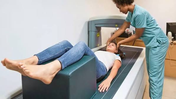
A bone density test is the only reliable way to assess the strength of your bones before a fracture occurs. It measures bone mineral density — the amount of mineral packed into your bones — and uses that measurement to estimate fracture risk and guide decisions about treatment and prevention. The most common and accurate bone density test is the DEXA scan (dual-energy X-ray absorptiometry), which takes approximately 10–15 minutes, involves no injection or special preparation, and delivers a very low radiation dose. Understanding what the test involves, who needs it, how to interpret the results, and what to do with those results is the foundation of any informed approach to bone health.
What Is a Bone Density Test?
A bone density test measures bone mineral density (BMD) — the concentration of calcium and other minerals in bone tissue, expressed as a density (grams per square centimetre, g/cm²). Higher bone mineral density means stronger bones that are more resistant to fracture; lower density means weaker, more fragile bones with increased fracture risk. The test identifies osteoporosis and osteopenia before a fracture occurs, enabling preventive or protective treatment to be started. It is the equivalent of a blood test for cholesterol — it identifies risk before the event (fracture in the case of bone density; heart attack in the case of cholesterol) at a stage when intervention can make a meaningful difference.
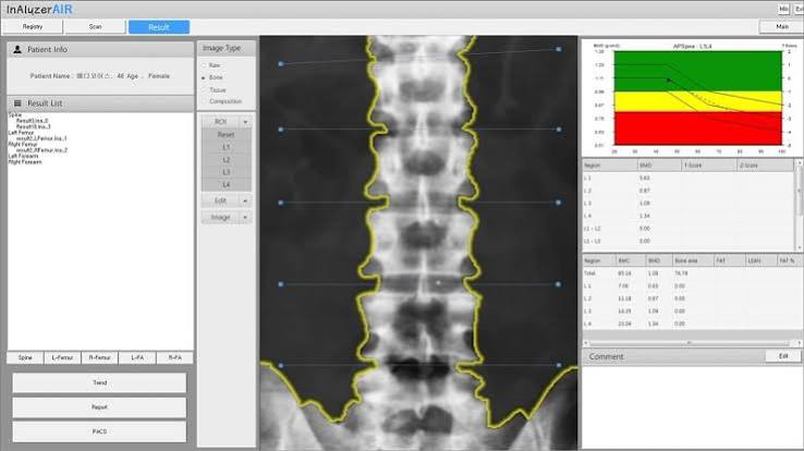
The standard test used in clinical practice is the DEXA scan. DEXA stands for dual-energy X-ray absorptiometry — it uses two X-ray beams at different energy levels to distinguish bone from surrounding soft tissue and calculates the density of the bone being imaged. The clinical standard is to measure BMD at the lumbar spine (L1–L4) and at the hip (femoral neck and total hip), since these are the two sites where osteoporotic fractures are most clinically significant and where measurement is most standardised. The lowest T-score across these sites is used for diagnosis.
Who Should Have a Bone Density Test?
NICE guidance identifies the following groups as appropriate for bone density testing:
- Women aged 65 and over — routine assessment recommended regardless of other risk factors
- Women aged 50–64 with one or more clinical risk factors: early menopause (before 45), surgical menopause (oophorectomy), prior fragility fracture, family history of hip fracture, low BMI (below 19 kg/m²), glucocorticoid use, secondary causes (coeliac disease, inflammatory bowel disease, rheumatoid arthritis, hyperthyroidism)
- Men aged 70 and over
- Men aged 50–69 with significant risk factors, particularly secondary causes or prior fragility fracture
- Any adult who has sustained a fragility fracture — a fracture from a fall from standing height or less
- Anyone starting long-term glucocorticoid therapy (prednisolone ≥5 mg/day for ≥3 months)
The FRAX tool (developed by the University of Sheffield for the WHO) can be used before DEXA to calculate 10-year fracture probability from clinical risk factors alone. If the FRAX score is above the intervention threshold even without BMD input, treatment may be recommended without DEXA. If the FRAX score is below the threshold for assessment, scanning may not change management. FRAX is most useful in the intermediate range, where the DEXA result will tip the decision. More on BMD and what T-scores mean is in our guide to bone density: what it means.
What Happens During a DEXA Scan?
The DEXA scan is one of the most straightforward imaging investigations in clinical practice. Preparation is minimal:
- No fasting or dietary restriction is required
- Calcium supplements should not be taken for 24 hours before the scan, as residual calcium in the gut can affect measurements
- Remove metal jewellery, belts, and clothing with metal zips or buttons near the spine and hip
- Wear comfortable clothing that can be adjusted; a hospital gown is not always necessary
- Inform the radiographer if you have had a recent barium examination, CT scan with contrast, or nuclear medicine scan — some contrast agents can interfere with DEXA measurements
- Inform the radiographer if there is any possibility of pregnancy
During the scan, you lie flat on a padded table. The scanner arm passes over the hip and then the lumbar spine while you remain still. The procedure involves no injection, no enclosed tunnel, and no discomfort. The scan typically takes 10–15 minutes in total. The radiation dose is extremely low — approximately 1–10 microsieverts (μSv), compared with approximately 10 μSv for a chest X-ray and approximately 8,000 μSv for a CT scan of the abdomen. The annual background radiation exposure in the UK is approximately 2,700 μSv, giving a sense of scale. Full detail on the DEXA scan is in our guide to DEXA scan: what adults should know.
Understanding the Results
DEXA results are reported as two scores at each measurement site (hip and lumbar spine):
The T-score compares your measured bone mineral density to the average density of a healthy young adult at peak bone mass (the reference population). Each unit represents one standard deviation. The WHO classification based on T-score is:
- Normal: T-score at or above −1.0
- Osteopenia: T-score between −1.0 and −2.5
- Osteoporosis: T-score at or below −2.5
- Severe osteoporosis: T-score at or below −2.5 plus a fragility fracture
The Z-score compares your bone density to an age-matched and sex-matched reference group. A Z-score of −2.0 or below means your bone density is significantly lower than expected for someone of your age and sex — which should trigger investigation for a secondary cause. The Z-score is used for premenopausal women and men under 50, where the T-score comparison to young adults is misleading. In postmenopausal women both scores are usually reported; clinical decisions use the T-score.
The report will typically also include:
- The absolute BMD value (g/cm²) at each site — important for monitoring change over time on the same machine
- A comparison graph showing where your density falls on the age-matched population curve
- The site with the lowest T-score — which drives the diagnosis
Accuracy and Limitations of DEXA
DEXA is highly accurate and reproducible, but it has limitations that are worth understanding. The test measures areal bone mineral density — a two-dimensional projection — rather than true volumetric density. This means that larger bones, or vertebrae affected by osteophytes (bony outgrowths of osteoarthritis), can produce artificially elevated lumbar spine T-scores. When there is significant lumbar osteoarthritis in an older patient, the lumbar spine BMD may appear better than it actually is, making the hip result the more reliable measurement.
DEXA measures density, not bone quality — the microarchitecture, collagen cross-linking, and mineralisation pattern that also contribute to fracture resistance. This is why BMD explains approximately 60–70% of the variation in bone strength across populations, with the remainder attributable to quality factors that DEXA cannot capture. In practice, this means that some people with osteopenia fracture, and some with osteoporosis T-scores do not — and why clinical risk factors alongside the T-score (via FRAX) give a more complete fracture probability estimate than the T-score alone.
The minimum interval for meaningful repeat DEXA is generally two years — bone density changes slowly and DEXA’s measurement precision error (approximately 1–2%) means that changes below approximately 3% fall within the test’s error range and are not clinically interpretable. Scanning more frequently than every two years in stable clinical situations does not add useful information.
What Happens After the Test?
The results will be reviewed by a clinician — either the specialist who ordered the scan or your GP through the fracture liaison service pathway — and interpreted alongside your clinical risk factors using FRAX. Based on this combined assessment, the management pathway falls into one of three categories:
- Normal T-score, low overall fracture risk: lifestyle advice (calcium, vitamin D, weight-bearing exercise), repeat scan in five to ten years if ongoing risk factors
- Osteopenia with low-to-moderate fracture risk: optimise lifestyle, calcium and vitamin D, address modifiable risk factors, repeat DEXA in two to three years
- Osteoporosis or high fracture risk: pharmacological treatment (usually bisphosphonate first-line), calcium and vitamin D co-prescription, repeat DEXA after three years of treatment
Osteoporosis management following a positive DEXA result is detailed in our guide to osteoporosis: symptoms, causes, and prevention. Osteopenia management without immediate pharmacological treatment is covered in our guide to osteopenia: what adults should know.
Monitoring Treatment With DEXA
For patients on pharmacological treatment for osteoporosis, repeat DEXA scanning is used to assess treatment response over time. Understanding how DEXA is used in this monitoring role — and what constitutes a meaningful change — helps patients know what to expect from their follow-up scans.
Bisphosphonate monitoring: NICE guidance recommends repeat DEXA at the three-year point for patients on oral bisphosphonates (alendronate, risedronate) and at three years for those receiving annual intravenous zoledronate. A meaningful treatment response is defined as stable or improving T-scores — neither of which alone indicates fracture prevention, but a declining T-score while on treatment warrants review for compliance, calcium and vitamin D adequacy, and secondary causes. Most patients on bisphosphonates will show T-score improvement of approximately 3–5% at the lumbar spine over three years; the hip typically shows smaller changes because it is predominantly cortical bone, which is slower to respond.
Denosumab monitoring: Patients on denosumab (six-monthly subcutaneous injection) are typically rescanned annually or every two years, given that denosumab produces more rapid BMD changes than bisphosphonates — lumbar spine gains of 5–8% over two years are documented in trials. An important monitoring point specific to denosumab is ensuring the six-monthly injection is not delayed or missed: a gap in treatment triggers a rebound increase in bone resorption that can result in rapid bone loss and, in some cases, multiple vertebral fractures. DEXA results should always be reviewed alongside injection compliance.
Teriparatide monitoring: Teriparatide (the anabolic parathyroid hormone analogue) produces the most rapid BMD changes — typically 8–12% at the lumbar spine over two years — and is the agent most likely to show clear T-score improvement at the annual monitoring DEXA. The BMD gains on teriparatide are not retained without a subsequent antiresorptive agent; transitioning to bisphosphonate or denosumab after the two-year teriparatide course is essential, and the post-teriparatide DEXA establishes the new baseline from which monitoring continues.
Interpreting monitoring scans: When comparing serial DEXA results, always ensure comparisons are made on the same machine and using the same anatomical positioning — a change in machine or site-of-measurement protocol introduces systematic measurement differences that are not clinically meaningful. A T-score change of less than 3% between scans may fall within the measurement precision error of DEXA and should be interpreted cautiously. A decline in T-score of more than 3–5% while on established treatment warrants clinical review — possibilities include non-compliance with medication, inadequate calcium or vitamin D co-supplementation, a newly active secondary cause, or a condition requiring a change of treatment agent.
Peripheral Tests and Their Role
In addition to central DEXA (hip and spine), peripheral DEXA (pDXA) and quantitative ultrasound (QUS) can measure bone density at peripheral sites — most commonly the heel, forearm, or finger. These devices are smaller, lower cost, and sometimes used in community screening settings or pharmacies. They provide an indication of bone density status and can identify individuals who may benefit from central DEXA referral, but they are not validated for diagnosis in the same way as central hip and spine DEXA, and treatment decisions should not be made on the basis of peripheral test results alone.
A low result on a heel ultrasound or peripheral DEXA should prompt referral for formal central DEXA at hip and spine rather than direct treatment initiation. This is important because the correlation between peripheral and central BMD is imperfect — a peripheral result can be falsely reassuring or falsely alarming relative to the central hip and spine measurement that actually drives diagnosis and treatment decisions. Results from different machines or different anatomical sites are not directly comparable, so serial monitoring must be done on the same machine and at the same site to be interpretable. If you receive a peripheral screening result from a community device, treat it as a prompt to seek GP referral for central DEXA rather than as a definitive diagnostic result.
Frequently Asked Questions
Does a bone density test hurt?
No. A DEXA bone density test is entirely painless and non-invasive. There is no injection, no dye, no enclosed space, and no discomfort. You lie on a padded examination table while the scanner arm passes over the hip and spine area. The procedure takes approximately 10–15 minutes and is one of the most straightforward imaging investigations available. The only requirement is to lie reasonably still during the scan, which most people have no difficulty with. The radiation dose is minimal — equivalent to a fraction of a chest X-ray — and there are no known risks associated with the procedure.
How often should I have a bone density test?
The appropriate interval depends on the result and clinical context. For adults with normal T-scores and few risk factors, repeat scanning every five to ten years is generally sufficient. For those with osteopenia and active risk factors, two to three years is appropriate. For those on treatment for osteoporosis, the standard is to rescan after three years of bisphosphonate therapy or annually on teriparatide. Annual scanning in stable osteopenia is not recommended — the measurement precision of DEXA means changes below approximately 3% are within the test’s error range and do not provide actionable information. More frequent scanning adds cost without clinical benefit in most cases.
Can I eat before a bone density test?
Yes — there is no dietary restriction before a standard DEXA bone density test. The one important exception is calcium supplements: these should not be taken for 24 hours before the scan, as residual calcium in the gut can theoretically interfere with the measurement. Regular food and other medications can be taken as normal. If you take calcium supplements daily, simply skip the dose on the day before and the morning of the scan. Water and normal meals are fine throughout.
Is a bone density test the same as a bone scan?
No — these are two completely different investigations. A bone density test (DEXA scan) measures bone mineral density to assess osteoporosis risk. A bone scan (radionuclide bone scintigraphy) is a nuclear medicine test that uses a radioactive tracer to show areas of abnormal bone activity — it is used to detect bone metastases (cancer spread to bone), fractures, infection, and other structural bone disease. The two investigations use different technology, serve different clinical purposes, require different preparation, and should not be confused. If your doctor has requested a bone density test, it is a DEXA to measure osteoporosis risk — not a cancer investigation.
Can I get a bone density test on the NHS?
Yes. DEXA scanning is available on the NHS and is funded for patients who meet the clinical criteria set by NICE guidance. Referral is typically made by a GP or specialist (orthopaedics, rheumatology, endocrinology) for patients who meet the age and risk factor criteria. The GP can use the online FRAX tool to calculate fracture risk before referring, to ensure the scan will contribute usefully to management. DEXA scanning is not currently offered as a universal population screening programme in the UK — it is targeted to individuals at elevated fracture risk. Some pharmacies offer peripheral heel ultrasound screening for a fee; a positive result from these should prompt NHS DEXA referral rather than treatment.
What is the FRAX score and how does it relate to a bone density test?
FRAX is a web-based risk calculator (developed by the University of Sheffield for the WHO) that estimates an individual’s 10-year probability of sustaining a major osteoporotic fracture (hip, spine, wrist, or humerus) and hip fracture specifically. It takes clinical risk factors as inputs — age, sex, BMI, prior fracture, family history, smoking, alcohol, steroid use, rheumatoid arthritis, and secondary causes — and can be used with or without the DEXA T-score. When the T-score is included, the fracture probability estimate is more accurate. The FRAX score helps clinicians decide whether to recommend treatment based on overall fracture risk rather than the T-score category alone, which is particularly useful in the osteopenia range where T-scores are below normal but the appropriateness of medication depends on overall risk.
What if my T-score is different at the spine and hip?
It is common for T-scores to differ between the lumbar spine and hip, and the lower of the two scores drives the diagnosis. Lumbar spine T-scores may be falsely elevated in older patients due to lumbar osteoarthritis, which causes osteophyte formation and increases the apparent bone density on DEXA. In patients over 65, the hip T-score is generally considered more reliable when there is discordance between sites. The pattern also has some diagnostic value: vertebral T-scores more negative than hip T-scores are characteristic of early postmenopausal bone loss (which preferentially affects the trabecular bone of the spine), while the reverse pattern (hip worse than spine) is more typical of age-related cortical bone loss or secondary causes.
Summary
A bone density test — most commonly a DEXA scan measuring bone mineral density at the hip and lumbar spine — is the primary tool for diagnosing osteoporosis and osteopenia and estimating fracture risk before a fracture occurs. The test is painless, quick, involves minimal radiation, and requires no injection or fasting. T-scores from normal (≥ −1.0) through osteopenia (−1.0 to −2.5) to osteoporosis (≤ −2.5) classify BMD and inform treatment decisions, with the FRAX tool adding clinical risk factors for a more complete fracture probability estimate. Who needs the test is determined by age and clinical risk factors according to NICE guidance, with women over 65 and both sexes with risk factors or prior fracture as the primary indications. Results should be interpreted alongside clinical context, and management — from lifestyle optimisation through to pharmacological treatment — should follow accordingly. Related guides to bone health are available on bone density: what it means, osteoporosis: symptoms, causes, and prevention, and the DEXA scan itself.
Medical disclaimer: This article is for general educational purposes and does not constitute medical advice. Consult a qualified healthcare professional for personalised bone health assessment and DEXA referral.
References:
NHS. DEXA scan. NHS. 2023.
NICE CG146. Osteoporosis: assessing the risk of fragility fracture. NICE. 2023.
Royal Osteoporosis Society. Bone density scanning. theros.org.uk. 2023.
WHO FRAX. Fracture Risk Assessment Tool. University of Sheffield.
Kanis JA, et al. The diagnosis of osteoporosis. J Bone Miner Res. 1994;9(8):1137–1141.
I had my first DEXA scan at 65 after my GP suggested it as a routine check. I was quite nervous beforehand — I’d had an MRI a few years ago and found the enclosed space difficult, so I assumed a DEXA would be similar. In fact it was completely different. I lay on an open padded table with nothing over me, the scanner arm just passed slowly over my hip and then they repositioned me for the spine, and it was done in about 12 minutes. Not claustrophobic at all. My results showed osteopenia at the spine (T-score −1.8) and normal at the hip (T-score −0.9). My GP explained that the spine result can be slightly affected by mild arthritis and that the overall picture was low-to-moderate risk, so she recommended calcium and vitamin D plus a follow-up DEXA in two years. This article is a very accurate description of the experience and I think it would reassure anyone who is apprehensive about the procedure.
Susan, your experience captures a very common pre-procedure anxiety that comes from conflating DEXA with MRI — the enclosed gantry, the noise, the prolonged positioning. DEXA is categorically different: open table, short duration, no enclosed space, no injection. The discordance you describe between your spine and hip T-scores (−1.8 and −0.9 respectively) is also very common and clinically recognisable. Lumbar spine T-scores in women over 65 are frequently influenced by mild degenerative osteophytes from osteoarthritis, which increase the apparent areal BMD on DEXA and produce a more favourable lumbar result than true bone quality warrants. Your GP’s approach — treating the worse of the two scores as more informative but recognising the overall picture as low-to-moderate risk — is entirely appropriate. Anthony, your three-year monitoring T-score trajectory (−2.7 to −2.3, an approximately 7% improvement at the lumbar spine) is above the expected average for alendronate at three years (typically 3–5%), indicating a good treatment response. On the denosumab question: the rebound resorption phenomenon on denosumab discontinuation is one of the most important practical distinctions between bisphosphonates and denosumab — bisphosphonates remain in bone mineral for years after stopping and continue to have residual antiresorptive effect, while denosumab effect is entirely reversible within months of a missed dose. This does not mean denosumab is a worse treatment, but it does mean that once started it should either be continued indefinitely or transitioned carefully to a bisphosphonate rather than simply stopped.
I have been on alendronate for three years for osteoporosis diagnosed after a wrist fracture (T-score −2.7 at the lumbar spine). My monitoring DEXA at three years showed improvement to −2.3, which my bone health nurse said was a good response to treatment and indicated I should continue for another two years before reassessment. What I found helpful about this article is the explanation of what a ‘good response’ actually means numerically — I hadn’t understood that a 3–5% improvement over three years was the expected benefit, or that treatment works primarily by stabilising rather than dramatically reversing bone loss. The section on denosumab rebound is something I wasn’t aware of — I had considered asking my GP to switch to injections as an alternative to weekly tablets, and understanding that missing a denosumab dose can cause rapid bone loss is something I would want to factor into that decision.