

Blood in the urine — whether visible to the naked eye or found on a routine laboratory test — is one of the symptoms that warrants medical evaluation in all adults, without exception. Seeing red or pink in the toilet is alarming, and that alarm is appropriate: blood in the urine has a wide range of causes, but none of them should be left uninvestigated. In some cases the cause is benign and resolves quickly. In others, it is the first visible signal of a serious condition — bladder cancer, kidney cancer, or a progressive kidney disease — where early detection makes a real difference. This article covers what causes blood in the urine, how it is evaluated, and when urgent care is needed.
Gross vs. Microscopic Hematuria
Blood in the urine — medically called hematuria — falls into two categories based on whether it is visible.
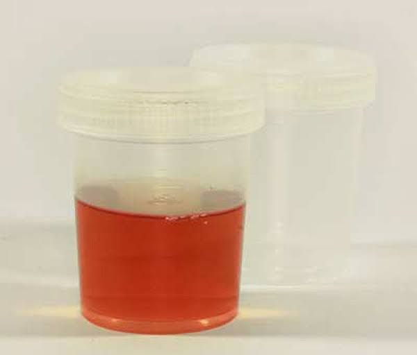
Gross hematuria is blood that is visible to the naked eye. It can turn urine pink, bright red, or a dark tea or cola color depending on the quantity and source of the bleeding. Even a tiny amount — one milliliter in a liter of urine — is enough to produce visible discoloration. Gross hematuria can be frightening when it appears, but the color alone does not indicate severity — what matters is the underlying cause.
Microscopic hematuria is blood that is invisible but detectable on laboratory testing. It is defined as three or more red blood cells per high-power field on microscopic examination of a properly processed urine sample. It is often found incidentally during a routine urinalysis — the person had no idea blood was present. Microscopic hematuria appears in approximately 4 percent of urine dipstick screens in the general adult population. (AUA, 2020)
Both types require medical evaluation. Gross hematuria demands more urgent attention; microscopic hematuria discovered on a routine test should be followed up within 2 to 4 weeks.
Pseudohematuria — Mimics of Blood in Urine
Red or dark urine is not always caused by blood. Eating beets or rhubarb can turn urine red or pink through the excretion of betanin pigment — a condition called beturia. Medications including phenazopyridine (urinary analgesic), rifampin (antibiotic), and some antipsychotics discolor urine in distinctive ways. Menstrual blood contamination in women can produce a false positive.
Myoglobinuria — dark, tea-colored urine from severe muscle breakdown (rhabdomyolysis) — tests positive on a blood dipstick but shows no red blood cells on microscopy. The distinction matters: rhabdomyolysis requires its own urgent treatment (IV hydration to protect the kidneys) and a different diagnostic pathway.
A positive dipstick for blood should always be confirmed with microscopic urinalysis before any clinical decision is made. A dipstick positive that shows no RBCs on microscopy means hemoglobin or myoglobin is present — not red blood cells — which changes the next steps entirely.
What Causes Blood in the Urine?
Urinary Tract Infections
UTIs are the most common cause of hematuria in women of reproductive age. Bacterial inflammation damages the bladder and urethral lining, releasing red blood cells into the urine. UTI-related hematuria is almost always accompanied by painful urination, urgency, and frequency — which distinguishes it from the painless hematuria that raises the most concern for malignancy. A positive urine culture confirms the diagnosis; hematuria typically resolves with antibiotic treatment within days.
In older adults and men, even when a UTI is present, underlying pathology should still be considered — bladder cancer and structural abnormalities can coexist with UTI and should not be dismissed because an infection was found.
Kidney Stones
Kidney stones cause hematuria in approximately 85 percent of cases. As the stone moves through the ureter, it damages the urothelium (the lining of the ureter), releasing red blood cells into the urine. The combination of blood in the urine and sudden, severe flank or groin pain is the classic kidney stone presentation. Kidney stones affect approximately 1 in 11 Americans. (NIDDK, 2023) The article on painful urination covers kidney stone management in detail.
Bladder Cancer
Bladder cancer is the cause of hematuria that requires the most urgent attention. Its classic presentation is painless gross hematuria — blood in the urine with no associated pain, urgency, or infection. This combination is clinically critical: because the hematuria causes no discomfort, it is easily dismissed or attributed to dehydration — but it is precisely this pattern that is most suggestive of malignancy.
Bladder cancer is the sixth most common cancer in the United States, with approximately 83,000 new cases diagnosed annually. (ACS, 2024) It is significantly more common in men than women, and the single most important modifiable risk factor is cigarette smoking — smokers face two to three times the bladder cancer risk of non-smokers. Other risk factors include occupational exposure to aromatic amines (dye, rubber, and leather industries), prior pelvic radiation, and chronic bladder irritation.
The AUA’s 2020 Microhematuria Guidelines are clear: all adults with confirmed gross hematuria — even a single episode — should have a complete urologic evaluation including cystoscopy and upper tract imaging. (AUA, 2020) Approximately 15 to 20 percent of adults with painless gross hematuria are found to have bladder cancer on evaluation. The hematuria can be intermittent — appearing then seeming to resolve on its own. This apparent resolution is never a reason to delay evaluation.
Kidney Disease and Glomerulonephritis
Several kidney diseases cause hematuria by allowing red blood cells to pass through the damaged glomerular filtration membrane. These conditions are distinguished from urologic causes by the presence of red blood cell casts on microscopy, accompanying proteinuria, and declining eGFR.
IgA nephropathy — the most common primary glomerulonephritis worldwide — typically presents as episodic gross hematuria producing dark cola-colored urine, triggered by an upper respiratory or gastrointestinal infection. Between episodes, persistent microscopic hematuria is usually present. IgA nephropathy progresses to end-stage kidney disease in approximately 20 to 25 percent of patients over 20 years without treatment. KDIGO 2023 guidelines now include sparsentan and intestinal-release budesonide (Nefecon) alongside the traditional ACE/ARB backbone. (KDIGO, 2023) Tracking kidney health numbers — eGFR and UACR — is essential for monitoring IgA nephropathy progression.
Alport syndrome is a hereditary nephritis caused by COL4A gene mutations affecting the glomerular basement membrane. X-linked Alport syndrome — the most common form, accounting for approximately 80 percent of cases — causes persistent microscopic hematuria in all affected males from childhood, often with episodic gross hematuria. Progressive CKD, sensorineural hearing loss, and characteristic eye abnormalities complete the syndrome. A family history of kidney disease alongside hematuria and hearing loss should always prompt consideration of Alport syndrome. The article on family history and kidney disease risk discusses hereditary nephropathies in detail.
Lupus nephritis presents as hematuria, proteinuria, and declining eGFR — often with red blood cell casts on microscopy. Active lupus nephritis is a medical urgency; without treatment, it can progress to chronic kidney disease and kidney failure within months.
Anti-GBM disease and ANCA vasculitis are rare but serious causes of rapidly progressive glomerulonephritis — hematuria with rapidly declining eGFR over days to weeks. Both are medical emergencies requiring immediate specialist nephrology evaluation.
Benign Prostatic Hyperplasia (Men)
In older men with BPH, dilated veins on the surface of the prostate and bladder base can bleed — particularly with straining or physical activity. The hematuria is typically self-limiting. 5-alpha reductase inhibitors (finasteride, dutasteride) reduce prostate vascularity over 3 to 6 months and are effective at reducing BPH-related hematuria recurrence. BPH is a diagnosis of exclusion for hematuria — bladder cancer must be excluded first.
Renal Cell Carcinoma (Kidney Cancer)
Renal cell carcinoma was traditionally characterized by the classic triad of hematuria, flank pain, and a palpable mass. Today, most RCC is detected incidentally on imaging before symptoms develop — the classic triad, when present, often suggests advanced disease. Approximately 80,000 new RCC cases are diagnosed annually in the United States. (ACS, 2024) Localized RCC is treated surgically; metastatic RCC is managed with targeted therapy and immune checkpoint inhibitors.
Other Causes
Exercise-induced hematuria produces transient gross or microscopic hematuria following intense physical exercise — particularly long-distance running. It resolves completely within 72 hours of rest and requires no specific treatment. If it persists beyond 72 hours, other causes must be excluded.
Anticoagulant therapy (warfarin, apixaban, rivaroxaban) increases bleeding throughout the body. However, hematuria in an anticoagulated patient should never be attributed solely to the anticoagulant — the AUA guidelines are explicit that anticoagulation unmasks an underlying lesion rather than causing hematuria on its own. Full evaluation is still required.
Sickle cell trait can cause painless hematuria, typically from the left kidney, due to papillary necrosis — ischemic damage to the renal papillae from sickling of red blood cells in the medulla. This can occur in otherwise healthy carriers of the sickle cell gene.
Timing Within the Void — What It Can Suggest
The timing of blood within the urinary stream can provide a clue to its anatomical source:
- Initial hematuria (blood at the very beginning, clearing as voiding continues): suggests urethral or prostatic source
- Terminal hematuria (blood at the end of the stream): suggests bladder neck or trigone
- Total hematuria (blood mixed throughout the entire void): suggests bladder, ureter, or kidney
When Blood in the Urine Is a Medical Emergency
Go to the emergency department if: gross hematuria with blood clots making urination difficult or impossible; severe flank or groin pain suggesting an obstructing kidney stone; gross hematuria with fever and chills (possible pyelonephritis or obstructed infected kidney).
Seek care the same day or next day if: gross hematuria appears without pain. Painless gross hematuria must be evaluated promptly — do not wait to see if it recurs. Even a single episode warrants cystoscopy and upper tract imaging.
Schedule within 2–4 weeks if: microscopic hematuria is found incidentally on a routine urinalysis with no current symptoms. Not an emergency, but should not be indefinitely deferred.
How Blood in the Urine Is Evaluated
Urinalysis with microscopy confirms true hematuria (RBCs present), distinguishes it from hemoglobinuria/myoglobinuria (positive dipstick, no RBCs), identifies RBC casts (glomerular disease), detects protein (kidney disease), and identifies bacteria (infection). (NIDDK, 2023)
eGFR and UACR are checked in all patients. Hematuria combined with significant proteinuria and declining eGFR suggests a glomerular disease rather than a urologic source.
CT urogram (CTU) — CT with contrast in a urographic protocol — is the gold standard for upper tract imaging. It detects kidney masses, ureteral tumors, stones, and structural abnormalities with high sensitivity.
Cystoscopy is mandatory for all adults with unexplained gross hematuria. A thin scope passed through the urethra directly visualizes the bladder interior — detecting tumors, stones, and mucosal lesions.
Urine cytology examines shed urothelial cells for malignant features. A useful adjunct to cystoscopy in higher-risk patients.
Kidney biopsy is performed when evaluation points toward glomerular disease — hematuria with significant proteinuria, declining eGFR, red blood cell casts, or a history suggesting autoimmune kidney disease.
AUA Risk Stratification for Microscopic Hematuria
The 2020 AUA Microhematuria Guidelines introduced risk categories to guide workup intensity:
Low risk (women under 50, no smoking, no risk factors, no prior hematuria): observation with repeat urinalysis in 6 months may be an acceptable initial approach.
Intermediate risk (adults aged 50–59, limited smoking history, no gross hematuria): cystoscopy plus upper tract imaging recommended; renal ultrasound acceptable in preference to CT.
High risk (adults over 60, heavy smoking — more than 30 pack-years — occupational chemical exposure, or prior gross hematuria): full evaluation with cystoscopy plus CT urogram recommended promptly. (AUA, 2020)
Treatment Depends on the Cause
UTI: Antibiotics appropriate to the identified pathogen.
Kidney stones: Hydration, pain management, tamsulosin; ureteroscopy with laser lithotripsy for larger stones.
Bladder cancer: TURBT for non-muscle-invasive disease, with intravesical BCG immunotherapy for high-risk cases. Radical cystectomy for muscle-invasive disease. (ACS, 2024)
IgA nephropathy and glomerulonephritis: ACE/ARB backbone; sparsentan and budesonide (Nefecon) per KDIGO 2023 for IgA nephropathy; immunosuppression for rapidly progressive forms.
Renal cell carcinoma: Surgical resection for localized disease; targeted therapy and immune checkpoint inhibitors for metastatic disease.
BPH-related hematuria: 5-alpha reductase inhibitors (finasteride, dutasteride) reduce prostate vascularity and hematuria recurrence within 3 to 6 months.
Exercise-induced hematuria: Rest for 72 hours and confirm resolution. No specific treatment required.
Frequently Asked Questions
Is blood in urine always serious?
Not always, but it always warrants evaluation. A UTI or exercise-induced hematuria is benign and resolves with treatment or rest. However, blood in the urine can also be the first sign of bladder cancer, kidney cancer, or glomerulonephritis — conditions where early detection dramatically improves outcomes. There is no reliable way to tell benign from serious based on the color or amount of blood alone. A complete evaluation is the only way to know.
Can a UTI cause blood in urine?
Yes. UTI is the most common cause of hematuria in women of reproductive age. The hematuria is almost always accompanied by painful urination, urgency, and frequency. In younger, low-risk women, hematuria that resolves completely with antibiotic treatment and does not recur is likely UTI-related. In adults over 50 or anyone with risk factors for bladder cancer, follow-up evaluation after UTI treatment is still appropriate.
What color is blood in urine?
Blood causes a range of colors depending on the amount, source, and how long it has been in the urine. Fresh blood from a lower source produces pink or bright red urine. Blood from the kidneys or that has been in the urine longer produces a darker tea, cola, or brownish color. Any visible color change should be evaluated regardless of hue.
Should I go to the ER for blood in my urine?
Go to the ER if gross hematuria is accompanied by blood clots making urination difficult, severe flank pain suggesting an obstructing kidney stone, or fever and chills suggesting an infected obstructed kidney. For painless gross hematuria without these severe accompanying symptoms, seek care the same day or next day — urgent, but not necessarily an ER visit. Microscopic hematuria discovered on a routine test warrants evaluation within 2 to 4 weeks.
Can blood in urine go away on its own?
The visible blood can appear to resolve — hematuria from UTIs clears with treatment, and bladder cancer often produces intermittent hematuria that seems to go away between episodes. This apparent resolution is not reassurance that the cause has resolved. A confirmed episode of gross hematuria requires full evaluation even if the blood is no longer visible. (Mayo Clinic, 2024)
Blood in the urine is never a symptom to dismiss as coincidence and then forget. Even a single episode of visible hematuria warrants a complete evaluation. The workup — urinalysis, cystoscopy, upper tract imaging — is straightforward, and the payoff is significant: finding bladder cancer at a localized, surgically treatable stage rather than an advanced one, or identifying a kidney disease while effective intervention is still possible. Most people who undergo hematuria evaluation do not have cancer. But for those who do, that evaluation is what makes the difference.
Hematuria in Special Populations
Hematuria in Women
Women are diagnosed with UTIs far more frequently than men, and UTI-related hematuria is the most common cause of blood in the urine in premenopausal women. However, this higher UTI frequency creates a clinical risk: hematuria in women is more often attributed to infection — sometimes without a full workup — which can delay the diagnosis of bladder cancer or upper tract pathology. The AUA guidelines specifically note that women have historically been under-evaluated for hematuria compared to men.
Bladder cancer, while less common in women than men, is diagnosed at a more advanced stage in women on average — likely because early symptoms are dismissed as recurrent UTIs. Any woman with hematuria that does not fully resolve after appropriate UTI treatment, or hematuria without a confirmed bacterial infection, should receive the same full evaluation as any other adult. Postmenopausal women with hematuria have a substantially higher pre-test probability for malignancy and should be evaluated promptly.
Hematuria in Men
Men have a lower lifetime rate of UTIs but a significantly higher rate of bladder cancer. In men, even a single episode of gross hematuria should prompt urologic evaluation without delay. BPH is common in older men and can cause hematuria, but it is a diagnosis of exclusion — bladder cancer and renal cell carcinoma must be ruled out before BPH is accepted as the explanation. Men with a long smoking history and hematuria are in the high-risk category per AUA guidelines and should receive full evaluation including CT urogram and cystoscopy.
Hematuria in Children and Young Adults
In children and young adults, the differential for hematuria shifts markedly toward glomerular disease and congenital conditions. IgA nephropathy commonly presents in teenagers and young adults with episodic gross hematuria following a respiratory infection. Alport syndrome often becomes clinically apparent in childhood. Thin basement membrane nephropathy — the most common cause of persistent microscopic hematuria in children — is generally benign but requires monitoring. In any child with hematuria, a blood pressure measurement and urinalysis for protein are the critical first steps; hypertension and proteinuria alongside hematuria point strongly toward glomerular disease requiring nephrology referral.
What to Expect From a Hematuria Evaluation
Understanding what a hematuria workup involves can reduce anxiety and help people seek care promptly rather than avoiding evaluation out of uncertainty about what it entails.
The initial visit typically includes a detailed history (smoking history, medications, occupational exposures, family history of kidney disease or cancer), physical examination, urinalysis with microscopy, and urine culture if infection is suspected. Blood tests — including eGFR, creatinine, and UACR — assess kidney function and screen for glomerular disease.
Cystoscopy — if recommended — is performed as an outpatient procedure, typically under local anesthetic. A thin, flexible scope is passed through the urethra into the bladder. The procedure takes 5 to 15 minutes. Most people experience mild discomfort and a burning sensation during urination for 24 to 48 hours afterward. Cystoscopy directly visualizes the bladder lining, which no imaging test can fully replicate. For most adults with gross hematuria, this procedure is the most important single component of the workup.
CT urogram is a CT scan with IV contrast given in a urographic (urine-phase) protocol. It takes 20 to 30 minutes and provides detailed imaging of the kidneys, ureters, and bladder — detecting masses, stones, strictures, and anatomical abnormalities. The main consideration is the use of IV contrast; patients with reduced kidney function or contrast allergy require modified protocols.
Timeline: For high-risk patients with painless gross hematuria, evaluation should be completed as quickly as possible — ideally within 2 to 4 weeks of initial presentation. For low-risk patients with incidental microscopic hematuria, completing the workup within 6 to 8 weeks is appropriate. Most evaluations result in a benign explanation; the minority that identify serious pathology benefit enormously from the early detection.
Monitoring After Treatment
After a cause of hematuria has been identified and treated, follow-up depends on the underlying diagnosis.
For UTI-related hematuria, a repeat urinalysis 4 to 6 weeks after completing antibiotics confirms resolution. Persistent microscopic hematuria after confirmed successful UTI treatment requires further evaluation — it should not be assumed to still be infection-related.
For bladder cancer, cystoscopic surveillance is performed every 3 months for the first 2 years after TURBT, then every 6 months for years 2 through 4, then annually if no recurrence is found. Bladder cancer recurs in approximately 50 to 70 percent of cases after initial TURBT — surveillance is not optional. A new episode of hematuria in a bladder cancer survivor should be evaluated immediately rather than assumed to be benign.
For IgA nephropathy and glomerular diseases, ongoing monitoring of eGFR, UACR, and blood pressure is the backbone of long-term management. People with IgA nephropathy who have significant proteinuria (UACR >0.5 g/g), hypertension, or reduced eGFR at diagnosis are at the highest risk of progression and require the most intensive monitoring and treatment. The article on kidney health numbers covers how to track these values over time.
For kidney stones, dietary and metabolic evaluation identifies the stone type (calcium oxalate, uric acid, struvite, cystine) and drives prevention — increased fluid intake, dietary modifications, and medications where indicated. Recurrence rates for untreated kidney stone disease are high — approximately 50 percent within 10 years — making post-episode evaluation and prevention counseling worthwhile.
Sources: AUA Microhematuria Guidelines 2020 (auanet.org), American Cancer Society (cancer.org), NIDDK (niddk.nih.gov), KDIGO 2023 (kdigo.org), Mayo Clinic (mayoclinic.org)
Bookmarked this article on blood in urine: possible causes and immediately — going to reference it regularly. It is refreshing to see an article that acknowledges individual variation rather than one-size-fits-all advice. I wish I had found this article earlier — would have saved a lot of confusion.
This is one of the clearest explanations of blood in urine: possible causes and I have found. The connection between lifestyle choices and long-term outcomes is explained clearly here. Appreciate the effort that went into researching and writing this — it shows.
Came across this while researching blood in urine: possible causes and for a family member. The connection between lifestyle choices and long-term outcomes is explained clearly here. Thank you for making complex medical information accessible without dumbing it down.