
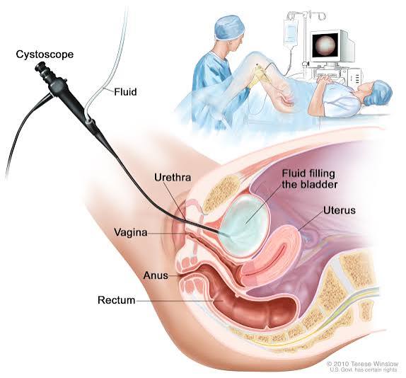
Most urinary symptoms — blood in the urine, recurrent urinary tract infections, or difficulty emptying the bladder — can often be evaluated with urine tests, blood tests, and imaging. But when a urologist needs to see inside the bladder directly, there is only one way to do it: cystoscopy. A thin tube with a camera and light is introduced through the urethra and into the bladder, allowing direct visual inspection of the bladder wall, ureteral openings, and urethra.
The word cystoscopy often causes anxiety because it sounds invasive, but flexible cystoscopy — the form used for most outpatient diagnostic purposes — is performed in a clinic setting with only a local anesthetic gel applied to the urethra. Most patients complete the procedure in 5 to 10 minutes. Understanding why cystoscopy is ordered, what the urologist is looking for, and what to expect before and after the procedure can help patients approach the test with significantly less anxiety.
What Is Cystoscopy
Cystoscopy is a procedure in which a cystoscope — a thin tube equipped with a lens, a light source, and a channel for irrigation fluid — is passed through the urethra and into the bladder. The urologist watches a live video monitor while advancing the scope, allowing direct inspection of the urethra, bladder neck, and the entire bladder interior. Sterile water or saline is slowly introduced through the irrigation channel to gently distend the bladder, which allows the walls to open up and become fully visible.
There are two main types of cystoscopes:
- Flexible cystoscope: A thin, bendable scope (typically 3 to 5 mm in diameter) that follows the natural curves of the male or female urethra without requiring the patient to be repositioned. Flexible cystoscopy is performed in a clinic or procedure room using only topical lidocaine gel applied to the urethra. Most patients do not require oral sedation or intravenous medication. The procedure typically takes 5 to 10 minutes.
- Rigid cystoscope: A straight, inflexible scope that provides a wider channel for surgical instruments. Rigid cystoscopy is performed in an operating room under regional (spinal) or general anesthesia. It is used when the procedure involves not just looking but also doing — taking a biopsy, removing a stone, resecting a bladder tumor, or placing a ureteral stent.
Why Cystoscopy Is Recommended
Cystoscopy is ordered for a variety of urological conditions, but the most common indication is blood in the urine — either visible to the eye (gross hematuria) or detected only on a urine test (microscopic hematuria).
Blood in the urine (hematuria): The American Urological Association (AUA) 2020 microhematuria guidelines recommend cystoscopy for all adults 35 years and older with any episode of visible blood in the urine, regardless of other factors. For microscopic hematuria, the AUA uses a risk stratification system based on age, smoking history, prior radiation to the pelvis, and other factors. Intermediate- and high-risk patients are directed to cystoscopy plus upper tract imaging. More information on hematuria evaluation is available in our guide on blood in urine test results.
Bladder cancer surveillance: After a diagnosis of bladder cancer, cystoscopy is the primary tool for monitoring whether the cancer has recurred. Surveillance intervals depend on the tumor grade and stage: patients with high-grade or muscle-invasive disease typically undergo cystoscopy every 3 months for the first 2 years after treatment, then every 6 months for the next 2 years, and then annually thereafter. Bladder cancer has one of the highest recurrence rates of any cancer, which makes ongoing cystoscopic surveillance essential.
Recurrent urinary tract infections: Women who experience three or more UTIs per year, or whose infections are associated with unusual features (such as blood in the urine, resistant organisms, or failure to respond to standard treatment), may be referred for cystoscopy to evaluate for an underlying bladder abnormality. Related: our article on urine culture testing explains how UTIs are diagnosed and characterized.
Lower urinary tract symptoms not responding to treatment: Men with significant urinary symptoms that do not improve with medication may undergo cystoscopy to evaluate for a urethral stricture, bladder neck obstruction, or intraluminal lesion. Women with urgency, frequency, and pelvic pain that suggests interstitial cystitis may undergo cystoscopy under anesthesia with hydrodistension, which both confirms the diagnosis and may provide temporary symptom relief.
Ureteral stent placement and removal: Ureteral stents are placed and removed cystoscopically. Stent placement is required after ureteroscopy for kidney stone management, after ureteral surgery, or to relieve ureteral obstruction. Stent removal is performed in the office with a flexible cystoscope; no anesthesia is typically required for uncomplicated stent removal.
What Cystoscopy Can Find
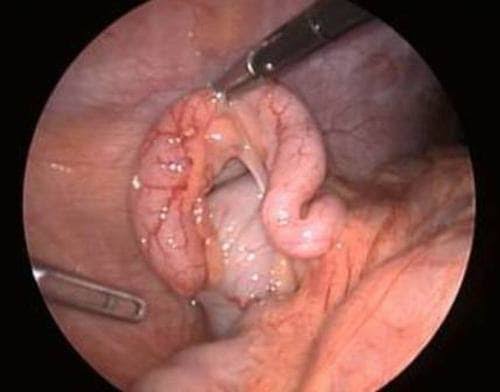
A skilled urologist examining the bladder interior can identify a wide range of findings that would be invisible to any imaging modality.
Normal findings: A healthy bladder has smooth, pale pink mucosa. The two ureteral orifices — the openings where urine enters the bladder from each ureter — are visible on the posterior wall. Normal efflux is observed as clear jets of urine periodically emerging from each orifice approximately every 15 to 30 seconds.
Bladder tumors: The two main visual presentations are papillary lesions and flat lesions. Papillary tumors have a frond-like appearance — small finger-like projections extending into the lumen. They are typically low-grade and non-muscle-invasive, though biopsy is required to confirm. Flat lesions (carcinoma in situ, or CIS) appear as a reddened, velvety patch on the bladder wall and may be subtle or nearly invisible under standard white light. CIS is high-grade with significant malignant potential, making it the most dangerous finding to miss.
Bladder stones: Stones within the bladder are immediately visible cystoscopically and can be fragmented and removed in the same session using laser or mechanical lithotripsy through a rigid cystoscope.
Ureteral efflux assessment: Observing whether urine is flowing normally from each ureteral orifice provides direct information about kidney function on each side. Bloody efflux from one orifice immediately localizes a bleeding source to the upper urinary tract on that side. An absent or sluggish efflux from one orifice suggests ureteral obstruction on that side.
Other findings: Bladder diverticula (pouches in the bladder wall that can harbor stones or tumors), foreign bodies (including eroded mesh from pelvic floor repair surgery), urethral strictures, and signs of trabeculation from chronic outlet obstruction are all visible cystoscopically. For context on imaging tests that complement cystoscopy, see our article on kidney ultrasound.
Flexible vs. Rigid Cystoscopy and Blue-Light Technology
Flexible cystoscopy is appropriate for diagnostic evaluation, bladder cancer surveillance, and ureteral stent removal. It is well-tolerated in clinic with local anesthetic. Women typically experience minimal discomfort. Men may experience more discomfort as the scope traverses the prostate.
Rigid cystoscopy under anesthesia is required for transurethral resection of bladder tumor (TURBT), bladder biopsy, cystoscopy in patients who cannot tolerate flexible scope, bladder stone lithotripsy, ureteral stent placement requiring fluoroscopic guidance, and hydrodistension for interstitial cystitis evaluation.
Blue-light cystoscopy (photodynamic diagnosis, or PDD) represents a significant advance in detecting flat CIS. One hour before cystoscopy, a solution containing hexaminolevulinate (sold as Cysview or Hexvix) is instilled into the bladder through a catheter. Hexaminolevulinate is preferentially absorbed by malignant urothelial cells. When the cystoscope switches to blue light, malignant cells fluoresce pink-red, making flat CIS lesions that are invisible under standard white light suddenly visible. Clinical studies have shown blue-light cystoscopy detects CIS approximately 20 to 30% more often than white-light cystoscopy alone. The FDA has approved hexaminolevulinate for use at initial TURBT and at surveillance cystoscopy for patients with prior high-risk disease.
How to Prepare for Cystoscopy
- Urine culture before the procedure: If a urinary tract infection is present, cystoscopy will typically be postponed until the infection is treated.
- Medications: Patients taking blood-thinning medications should discuss with their urologist whether these need to be temporarily held. Diagnostic flexible cystoscopy without biopsy typically does not require stopping anticoagulants.
- Antibiotic prophylaxis: Current AUA guidance does not recommend routine prophylactic antibiotics for all patients undergoing flexible cystoscopy. Prophylaxis is recommended for high-risk patients: immunocompromised patients, those with prior recurrent UTIs, indwelling catheters, structurally abnormal urinary tracts, or prosthetic cardiac valves.
- Transportation: Patients who receive only topical lidocaine gel can drive themselves home. Patients who receive IV sedation or oral anxiolytic medication need a driver.
What Happens During the Procedure
The genital area is cleaned with an antiseptic solution. For men, approximately 10 mL of lidocaine gel is instilled directly into the urethra and held in place for 2 to 5 minutes to allow the anesthetic to take effect. Women receive a smaller volume applied to the urethral opening.
The urologist then gently introduces the flexible cystoscope through the urethra, advancing it slowly while watching the monitor. Sterile fluid flows through the scope to distend the bladder and clear any debris. The urologist systematically examines the bladder in a methodical pattern — the dome, lateral walls, posterior wall, trigone, both ureteral orifices, and bladder neck — then examines the urethra during withdrawal.
Most patients report a sensation of pressure or urge to urinate rather than significant pain. Men typically experience more discomfort than women due to the longer, curved male urethra and prostate. The discomfort generally peaks as the scope passes through the prostatic urethra and resolves once it is in the bladder.
What to Expect After Cystoscopy
After a diagnostic flexible cystoscopy, most patients return to normal activities within a few hours. The following are expected and temporary:
- Dysuria (burning with urination): Typically present for 24 to 48 hours.
- Urinary urgency and frequency: May persist for 1 to 2 days.
- Mild blood in the urine: Pink-tinged or faintly bloody urine is expected for 24 to 48 hours.
- Increased fluid intake: Drinking 2 or more liters of water in the first 24 to 48 hours helps flush the urethra and reduces infection risk.
Contact your provider immediately if you experience fever above 38°C (100.4°F), significant bleeding (dark red blood or passing blood clots), inability to urinate, worsening pain beyond 48 hours, or shaking, chills, or feeling severely unwell.
Cystoscopy and Bladder Cancer: Staging and Surveillance
Bladder cancer staging depends critically on how deeply the tumor has invaded the bladder wall. Non-muscle-invasive disease: Stage Ta (confined to the mucosa, papillary), Stage T1 (invading the lamina propria but not the muscle), and Tis (carcinoma in situ — flat, high-grade). Muscle-invasive disease (T2 and beyond) requires aggressive treatment — radical cystectomy with or without neoadjuvant chemotherapy, or definitive chemoradiation.
After a diagnosis of non-muscle-invasive bladder cancer, surveillance cystoscopy is essential because urothelial carcinoma has an exceptionally high recurrence rate — up to 50 to 70% within 5 years for high-grade disease. The standard post-treatment surveillance schedule for high-risk patients includes cystoscopy and urine cytology every 3 months for the first 2 years, then every 6 months for the next 2 years, then annually. Urine cytology complements cystoscopy: it is highly sensitive for detecting high-grade or CIS disease. Low-grade, low-risk Ta tumors follow less intensive intervals: cystoscopy at 3 months post-resection, then annually for 5 years if no recurrence is detected. Information on urine tests used alongside cystoscopy is available in our article on urinalysis.
Risks and Complications of Cystoscopy
Diagnostic flexible cystoscopy is a very safe procedure with a low overall complication rate.
Urinary tract infection: The most clinically significant risk, occurring in approximately 0.5% to 2% of cases. Risk is higher in patients with urinary stasis, indwelling catheters, immunosuppression, or a structurally abnormal urinary tract. Fever developing within 48 hours requires prompt evaluation and antibiotic treatment.
Urethral injury or false passage: Advancing the cystoscope through a narrow or previously instrumented urethra can create a false passage in the urethral wall. This is uncommon with experienced operators using flexible scopes. Patients with prior urethral surgery or known stricture should inform their urologist before the procedure.
Temporary urinary retention: After cystoscopy — particularly after procedures performed under anesthesia with bladder distension — some patients are temporarily unable to urinate. If retention occurs, a temporary urethral catheter is placed for 12 to 24 hours.
Bladder perforation: Extremely rare in diagnostic flexible cystoscopy; a recognized risk of TURBT. Small perforations are managed conservatively; large perforations may require surgical repair.
Frequently Asked Questions
Does cystoscopy hurt? Most patients experience pressure or the urge to urinate rather than sharp pain during flexible cystoscopy. Men typically find the procedure more uncomfortable than women. Lidocaine gel significantly reduces urethral discomfort. The procedure is brief — typically 5 to 10 minutes — which is reassuring for patients who are anxious about sustained discomfort.
How soon will I know the results? For a purely visual diagnostic cystoscopy with no biopsy, the urologist can typically share findings immediately after the procedure while the patient is still in the clinic. If a suspicious area requires biopsy, the biopsy sample is sent to pathology and results are usually available within 5 to 7 business days.
How often do I need cystoscopy after bladder cancer? Surveillance intervals depend on the tumor’s grade and stage. High-risk bladder cancer typically requires cystoscopy every 3 months for the first 2 years, every 6 months for the following 2 years, then annually. Low-risk disease follows annual cystoscopy after an initial negative 3-month scope. Your urologist will personalize the schedule based on your specific tumor characteristics.
Cystoscopy in Special Clinical Situations
Beyond the standard indications, cystoscopy plays a specific and sometimes critical role in several clinical scenarios that patients and providers encounter:
Evaluation before pelvic radiation therapy: Patients who are about to undergo radiation therapy for cervical cancer, prostate cancer, or rectal cancer sometimes undergo baseline cystoscopy before treatment to document the condition of the bladder mucosa and confirm the absence of pre-existing lesions. This baseline is important because radiation to the pelvis can cause radiation cystitis — inflammation and fragility of the bladder wall — months to years after treatment is completed. Having a pre-treatment cystoscopy establishes whether any mucosal abnormality detected later was pre-existing or radiation-induced.
Evaluation of radiation cystitis: Radiation cystitis presents with hematuria (often gross), urinary frequency, urgency, and pain in patients who have received prior pelvic radiation. The bladder wall becomes friable (easily bleeding) due to loss of normal mucosal blood supply and the development of abnormal dilated vessels called telangiectasias. Cystoscopy directly visualizes these telangiectasias, confirms the diagnosis, and allows treatment using fulguration (electrocautery) or laser coagulation of bleeding vessels through the scope’s working channel. In severe cases, cystoscopy with hydrodistension may temporarily compress bleeding vessels and provide relief.
Evaluation after pelvic mesh complications: Synthetic mesh used in pelvic floor repair surgery (for stress urinary incontinence or pelvic organ prolapse) can rarely erode through the vaginal wall into the bladder or urethra. Patients present with recurrent UTIs, pain, or hematuria. Cystoscopy directly visualizes the eroded mesh as a hard white or rough irregular material protruding into the bladder lumen. Identification of mesh erosion on cystoscopy directs the surgical approach for mesh removal, which is typically required to resolve symptoms.
Cystoscopy in children: While most cystoscopy is performed in adults, pediatric urology uses rigid cystoscopy (under general anesthesia) to evaluate congenital urinary tract anomalies. The most common pediatric cystoscopic procedures include evaluation of vesicoureteral reflux (VUR) — abnormal backflow of urine from the bladder into the ureter — and injection of bulking material around the ureteral orifice to correct VUR (endoscopic treatment with Deflux). Posterior urethral valves — a congenital obstruction in the male urethra — are diagnosed and treated cystoscopically under general anesthesia.
Interstitial cystitis (IC) / bladder pain syndrome (BPS): IC/BPS is a chronic bladder condition causing pelvic pain, pressure, and urinary urgency and frequency without a bacterial infection. The diagnosis is one of exclusion — other causes of bladder symptoms must be ruled out first. Cystoscopy under anesthesia with hydrodistension (filling the bladder under anesthesia to its maximum capacity) is used both diagnostically (the urologist assesses for glomerulations — pinpoint hemorrhages on the bladder wall that appear after distension — and Hunner lesions, a specific ulcerated area highly predictive of IC) and therapeutically (hydrodistension itself may temporarily improve symptoms in some patients, presumably by disrupting abnormal neural signaling from the bladder wall). Patients with Hunner lesions benefit from direct treatment of the lesions by fulguration or corticosteroid injection through the scope.
Navigating the Cystoscopy Result: What Happens Next
Understanding what comes after a cystoscopy result — whether normal or abnormal — helps patients engage constructively with their care team.
Normal cystoscopy in a patient with hematuria: A normal cystoscopy in a patient being evaluated for hematuria does not definitively rule out all causes of bleeding. Bladder cancer was not found, which is reassuring, but upper tract sources (kidney or ureter) are evaluated with separate imaging. In a low-risk patient with microscopic hematuria and a normal cystoscopy, the AUA recommends repeat urinalysis at 12 months. If hematuria persists at that follow-up, upper tract imaging and potentially repeat cystoscopy are warranted. In a high-risk patient, a normal cystoscopy does not eliminate the need for upper tract CT urogram, which must be completed as part of the full hematuria workup.
Abnormal finding referred for biopsy or TURBT: When cystoscopy identifies a bladder lesion, the urologist will schedule a separate procedure — rigid cystoscopy under anesthesia — for biopsy or TURBT, depending on the lesion’s appearance. Patients should ask the urologist about the estimated time to scheduling this procedure (typically within 2 to 4 weeks of the diagnostic cystoscopy), what to expect from the TURBT recovery (catheter, irrigation, restrictions), and what pathology results will determine in terms of further treatment (intravesical therapy, radical cystectomy, surveillance schedule).
Recurrence identified at surveillance cystoscopy: When a recurrent bladder tumor is found at a surveillance cystoscopy, the urologist will determine based on its appearance and prior history whether it can be managed with a repeat office fulguration (burning of a small papillary recurrence) or requires return to the operating room for formal TURBT. A recurrence does not automatically mean the cancer is progressing to a more serious stage — low-grade tumors frequently recur as low-grade tumors — but high-grade recurrence at any stage warrants discussion of intensified intravesical therapy (BCG instillation) and possibly more aggressive surgical options.
Practical Questions to Ask Your Urologist Before Cystoscopy
Coming to a cystoscopy appointment with prepared questions helps patients get the most useful information before, during, and after the procedure. The following are questions patients commonly find valuable:
- What are you specifically looking for in my bladder? Knowing whether the urologist is primarily evaluating for a tumor, a stone, a stricture, or an infection-related finding helps contextualize the procedure’s purpose and what a normal vs. abnormal result would mean.
- Will I need a biopsy or any treatment during this cystoscopy? Understanding in advance whether the appointment is purely diagnostic or may include therapeutic steps helps patients prepare emotionally and logistically (arranging transportation, taking the afternoon off work).
- If you find a suspicious area, when can the biopsy be scheduled? Ask this before the cystoscopy so you understand the next-step timeline rather than learning about a possible scheduling delay only after the procedure is done.
- How often will I need cystoscopy going forward? Whether the surveillance is for bladder cancer, recurrent hematuria, or interstitial cystitis, having a clear schedule for future procedures allows patients to plan their year accordingly.
- What should I do if I cannot tolerate the procedure? If the patient has significant anxiety or a history of urethral stricture that could make the procedure painful, discussing anxiolytic premedication, a smaller-caliber scope, or scheduling a procedure under anesthesia prevents an uncomfortable or aborted exam.
Cystoscopy is one of the most direct and clinically informative procedures in urology. When performed for the right indication and interpreted by an experienced urologist, it provides information that cannot be obtained by any other means — allowing accurate diagnosis, appropriate staging, and timely treatment of conditions ranging from benign bladder abnormalities to early-stage bladder cancer.
Sources: AUA Microhematuria Guidelines | Urology Care Foundation — Cystoscopy | American Cancer Society — Bladder Cancer | NIDDK Urologic Diseases | Related: Blood in Urine Test Results | Urinalysis: What It Can Show | Kidney Ultrasound: What It Shows | Urine Culture Test Explained
Really well-written article on cystoscopy: what patients should know. The connection between lifestyle choices and long-term outcomes is explained clearly here. Shared this with three friends who are dealing with related issues. Very useful resource.
Came across this while researching cystoscopy: what patients should know for a family member. The connection between lifestyle choices and long-term outcomes is explained clearly here. Thank you for making complex medical information accessible without dumbing it down.
My doctor recommended I look into cystoscopy: what patients should know and this article covered it perfectly. The article answered questions I didn’t even know I had until I started reading. Thank you for making complex medical information accessible without dumbing it down.