
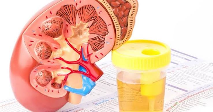
Protein in urine — a condition called proteinuria — is one of the most important early warning signs that the kidneys are under stress. Under normal circumstances, the kidneys act as a highly selective filter: they allow water, small molecules, and waste products to pass into the urine while retaining proteins in the bloodstream. When protein appears in the urine in significant amounts, it means the filter has been damaged or overwhelmed. Finding and addressing this damage early is one of the most effective ways to slow the progression of kidney disease.
Proteinuria is often silent — most people with protein in their urine have no symptoms until the levels are very high. This is why routine screening is essential for people with diabetes, hypertension, or a family history of kidney disease. This guide explains how protein ends up in urine, what different types and levels mean, what diseases cause it, and what treatments effectively reduce it.
How Protein Ends Up in Urine
The kidney’s filtration system is built to be selective. The glomerular filtration barrier consists of three layers: the fenestrated endothelium, the glomerular basement membrane, and the podocytes — specialized cells with foot processes that interdigitate to form filtration slits. Together, these layers form a barrier that effectively prevents proteins larger than approximately 67 kilodaltons from entering the filtrate.
Albumin, the most abundant plasma protein, has a molecular weight of about 67 kilodaltons — right at the exclusion threshold — and carries a negative charge that is repelled by the similarly negatively charged glomerular basement membrane. The small amount of albumin that does filter through is largely reabsorbed by the proximal tubule cells before reaching the final urine. In a healthy person, total urinary protein is less than 150 milligrams per day, and albumin excretion is less than 30 milligrams per gram of creatinine.
When the glomerular filtration barrier is damaged — by diabetic nephropathy, hypertensive nephrosclerosis, lupus nephritis, or focal segmental glomerulosclerosis — its selectivity breaks down. Albumin begins leaking into the filtrate in increasing amounts, and as damage progresses, larger proteins follow. The presence and quantity of protein in the urine is therefore a direct measurement of glomerular barrier integrity.
Types of Proteinuria — Not All Are the Same
Glomerular proteinuria is the most common and clinically important type in adults. It results from damage to the glomerular filtration barrier, and albumin is the dominant protein lost. The ACR is the appropriate test, and the KDIGO albuminuria classification (A1/A2/A3) applies directly. Causes range from diabetic nephropathy and hypertensive nephrosclerosis to the full spectrum of primary and secondary glomerulonephritis.
Tubular proteinuria results from the tubules’ failure to reabsorb low-molecular-weight proteins — such as beta-2 microglobulin and retinol-binding protein — that normally filter freely through an intact glomerulus. The albumin-specific ACR may be normal while total urine protein is elevated. Causes include Fanconi syndrome, heavy metal toxicity, and tenofovir-associated kidney toxicity in HIV patients. Urine protein electrophoresis (UPEP) is needed for full characterization.
Overflow proteinuria occurs when a protein is produced in quantities that overwhelm reabsorption capacity. The classic example is multiple myeloma, where abnormal plasma cells produce massive amounts of immunoglobulin light chains (Bence Jones proteins). These proteins are not albumin, so the standard urine dipstick may be falsely negative — UPEP is required to detect them.
Transient or functional proteinuria is benign and does not indicate underlying kidney disease. Fever, vigorous exercise, acute stress, and decompensated heart failure can all cause temporary protein leakage that resolves when the triggering condition resolves. This is why a single elevated ACR or positive dipstick should always be confirmed with repeat testing before attributing it to kidney disease.
Orthostatic proteinuria occurs almost exclusively in young adults under 30. Protein appears when the person is upright but not when lying down. It is diagnosed by comparing a first morning void (after lying supine overnight, which is normal) with a daytime sample (which shows elevated protein). No treatment is needed; it typically resolves on its own.
How Much Protein Is Too Much?
The KDIGO albuminuria classification uses the ACR:
- A1 (less than 30 mg/g): Normal to mildly increased. No treatment indicated solely for albuminuria; monitoring recommended in high-risk groups.
- A2 (30 to 300 mg/g): Moderately increased (formerly “microalbuminuria”). The earliest sign of diabetic nephropathy. Treatment with ACEi or ARB is indicated in diabetes or CKD.
- A3 (greater than 300 mg/g): Severely increased (formerly “macroalbuminuria”). High risk of CKD progression and cardiovascular events. Full treatment strongly indicated.
Normal total urine protein is less than 150 mg per day. The nephrotic range begins at more than 3.5 grams per day — enough protein loss to cause edema, hypoalbuminemia, hyperlipidemia, and elevated thrombosis risk.
A critical limitation: the dipstick protein test detects only albumin in the A3 range and completely misses the A1 and A2 ranges. A patient with an ACR of 150 mg/g — well into the A2 range with established early diabetic nephropathy — will have a completely negative dipstick. ACR testing, not dipstick, is the appropriate screening tool for proteinuria in high-risk patients. See the guide on the urine albumin test for full ACR detail.
Nephrotic vs. Nephritic Syndrome
Nephrotic syndrome is defined by massive proteinuria (more than 3.5 grams per day) combined with hypoalbuminemia (serum albumin below 3.5 g/dL), edema, hyperlipidemia, and lipiduria. The low plasma albumin reduces oncotic pressure, causing fluid to leak into tissues: periorbital edema worst in the morning, pitting edema of the ankles and legs, and in severe cases, ascites and pleural effusions. The urine often appears foamy, and oval fat bodies (showing as Maltese crosses under polarized light) are seen on microscopy. Causes in adults include minimal change disease, membranous nephropathy, FSGS, diabetic nephropathy, and amyloidosis.
Nephritic syndrome has inflammation as its dominant feature: hematuria (often gross, tea-colored or cola-colored), hypertension, oliguria, and fluid retention — with proteinuria that is present but less massive than in nephrotic syndrome. Red blood cell casts on urinalysis are the diagnostic signature, confirming that glomerular inflammation is shedding RBCs into the tubular space. Causes include IgA nephropathy (the most common glomerulonephritis worldwide), post-infectious GN (classically following streptococcal infection), lupus nephritis, ANCA-associated vasculitis, and anti-GBM disease (Goodpasture’s syndrome).
The distinction between nephrotic and nephritic presentations guides the initial differential diagnosis, the urgency of evaluation, and the subsequent workup including serological tests and kidney biopsy.
Causes of Protein in Urine
Diabetes mellitus is the most common cause of proteinuria in adults worldwide. A2 albuminuria (30–300 mg/g ACR) is the first detectable laboratory sign of kidney involvement — appearing years before creatinine rises or eGFR falls. This is the window where aggressive treatment with ACE inhibitors, ARBs, and SGLT-2 inhibitors can substantially slow or reverse kidney damage. See the guide on chronic kidney disease for the full progression context.
Hypertension causes a gradual rise in ACR through the A2 range as elevated systemic blood pressure transmits to the glomerular capillaries, causing hyperfiltration injury and progressive glomerulosclerosis. ACR typically rises slowly in hypertensive nephrosclerosis rather than reaching nephrotic levels, unless hypertension is severely uncontrolled.
Glomerulonephritis encompasses immune-mediated kidney diseases including IgA nephropathy (IgA immune complex deposition; most common GN worldwide), lupus nephritis (immune complex deposition from circulating anti-dsDNA), FSGS (podocyte injury; associated with obesity, HIV, heroin use), membranous nephropathy (subepithelial immune deposits; most common cause of nephrotic syndrome in non-diabetic adults; primary form identified by anti-PLA2R antibody), and minimal change disease (most common nephrotic syndrome in children).
Infections: hepatitis B causes membranous nephropathy; hepatitis C is associated with membranoproliferative GN and cryoglobulinemic vasculitis; HIV causes collapsing FSGS (HIV-associated nephropathy, HIVAN). Medications: NSAIDs can cause minimal change disease or membranous nephropathy; heroin is associated with FSGS; lithium causes tubular toxicity and occasionally FSGS.
Preeclampsia is defined by new-onset hypertension after 20 weeks of gestation with proteinuria greater than 300 mg per 24 hours, requiring urgent management to protect both mother and fetus.
Symptoms and When to Suspect Proteinuria
Most proteinuria is asymptomatic at A1 and A2 levels. The first symptom many patients notice is foamy urine — persistent foam that remains in the toilet after voiding, resembling detergent foam. This occurs because protein reduces the surface tension of urine. Not all foam is pathological, but foam that persists for more than a minute is worth reporting to a provider.
At nephrotic-range levels, edema becomes visible: puffiness around the eyes in the morning, swelling of the feet and ankles by the end of the day, tightness of rings and shoes, and in severe cases, abdominal swelling from ascites. In high-risk populations (diabetes, hypertension), proteinuria is almost always detected on screening rather than from symptoms — reinforcing why annual ACR testing matters.
How Proteinuria Is Measured and Monitored
Urine ACR (albumin-to-creatinine ratio) is the recommended test for screening and routine monitoring. Serial ACR values track disease progression and treatment response — a 30% or greater reduction from baseline indicates a clinically meaningful response. See the companion article on the albumin-to-creatinine ratio for full interpretation guidance.
24-hour urine protein collection provides precise total protein measurement and is used in nephrotic syndrome monitoring or when ACR is difficult to interpret. UPEP (urine protein electrophoresis) is required when overflow proteinuria from myeloma is suspected.
Treatment — What Lowers Protein in Urine
ACE inhibitors and ARBs are first-line therapy for proteinuria — they dilate the efferent arteriole, reducing intraglomerular pressure and albumin leakage. This antiproteinuric effect is partially independent of blood pressure lowering. SGLT-2 inhibitors provide an additional 20–40% ACR reduction on top of RAAS blockade (CREDENCE, DAPA-CKD, EMPA-KIDNEY trials) and are now guideline-recommended for CKD with proteinuria regardless of diabetes status.
Finerenone (FIDELIO-DKD trial) reduces proteinuria by approximately 30% in diabetic kidney disease patients on background RAAS blockade — additive, not a replacement. Blood pressure control to below 130/80 mmHg and a low-sodium diet (less than 2.3 g sodium/day) further enhance antiproteinuric efficacy.
Conclusion
Protein in urine is a signal that deserves attention — not alarm, but prompt evaluation. When found in the A2 or A3 range on a confirmed ACR test, it indicates that the kidney’s filtration barrier has been damaged and that kidney-protective treatment is warranted. The earlier proteinuria is identified and treated, the more kidney function can be preserved over the long term.
For the full picture of kidney laboratory testing, see the guides on the urinalysis, urine albumin test, and kidney function tests.
Special Populations: Proteinuria in Different Clinical Scenarios
While the principles of proteinuria evaluation apply broadly, certain patient populations require additional context when interpreting ACR results or deciding on the pace of workup.
Children with proteinuria have a different differential from adults. Minimal change disease is by far the most common cause of nephrotic syndrome in children aged 1 to 8, presenting with sudden-onset massive edema after a viral illness or vaccination. It responds well to corticosteroids in most cases. IgA nephropathy is the most common cause of persistent microscopic hematuria with mild proteinuria in older children and adolescents. Post-infectious GN — particularly post-streptococcal — is a common cause of nephritic syndrome in school-age children, typically resolving without specific treatment if renal function is preserved at presentation.
Older adults may have mild A2 albuminuria from age-related glomerulosclerosis rather than from treatable kidney disease. A rising ACR in an older adult is still significant, but the threshold for aggressive workup should account for the baseline age-related changes in glomerular structure. In contrast, heavy proteinuria (A3 or nephrotic range) in an older adult is not expected from aging alone and should prompt evaluation for myeloma (serum and urine protein electrophoresis), membranous nephropathy, amyloidosis, or other conditions more common in this age group.
Patients with diabetes and proteinuria deserve particular emphasis on treatment combination. The current evidence-based approach for a patient with type 2 diabetes, CKD, and A2 or A3 albuminuria involves three synergistic treatments: an ACE inhibitor or ARB (RAAS blockade), an SGLT-2 inhibitor, and finerenone — in addition to optimal glycemic control (target HbA1c 6.5–8% depending on individual risk) and blood pressure below 130/80 mmHg. This triple combination addresses three distinct pathways through which diabetes damages the glomerular barrier and, in clinical trials, substantially reduces the rates of kidney failure and major adverse cardiovascular events.
Patients with lupus require proteinuria monitoring as part of disease activity assessment. The WHO/ISN classification of lupus nephritis distinguishes six classes based on kidney biopsy findings, and the presence of proteinuria — its quantity, its trend, and whether it is accompanied by active urinary sediment (RBC casts, WBC casts) — is used alongside serum complement levels, anti-dsDNA titers, and creatinine to determine whether a patient is in remission or relapse. Proteinuria greater than 500 mg/day in a patient with known lupus that was previously in remission is a signal for urgent re-evaluation, including consideration of repeat kidney biopsy.
Patients post-transplant who develop de novo proteinuria — new albumin excretion exceeding 300 mg/day in a previously normal allograft — face a different differential than native kidney disease. In the transplant context, proteinuria may reflect chronic allograft nephropathy (a combination of immune injury, calcineurin inhibitor toxicity, and ischemic changes), recurrence of the original disease (particularly FSGS, which has a 20–30% recurrence rate in the allograft), or de novo glomerulonephritis in the transplant. Because each of these causes has a different treatment, kidney biopsy of the transplant is often necessary to guide management.
What to Expect at a Nephrology Evaluation for Proteinuria
When a primary care provider refers a patient for proteinuria, the nephrology evaluation typically follows a structured approach designed to establish the type, degree, and cause of proteinuria, and to determine whether kidney biopsy is needed.
The evaluation begins with a detailed medical history: duration of diabetes or hypertension, any family history of kidney disease, prior episodes of kidney stones or UTI, medication list (including OTC NSAIDs and supplements), alcohol and substance use history, and any systemic symptoms suggesting autoimmune disease (rash, joint pain, oral ulcers, photosensitivity) or hematological malignancy (bone pain, fatigue, hypercalcemia).
Blood tests ordered at the nephrology visit typically include a comprehensive metabolic panel (creatinine, BUN, electrolytes, bicarbonate, glucose), a complete blood count, serum albumin, serum protein electrophoresis (SPEP), complement levels (C3, C4), ANA, ANCA, anti-GBM antibody, hepatitis B and C serology, and HIV testing if not recently performed. The combination of which tests are positive helps direct the differential diagnosis toward specific glomerular diseases before considering biopsy.
Kidney biopsy is recommended in adults with proteinuria in the A3 range that is unexplained by a clear clinical diagnosis (such as long-standing diabetes with gradual ACR progression), or in any patient with nephrotic syndrome of unclear cause, or with a nephritic picture (hematuria, RBC casts) that warrants histological classification for treatment. The biopsy result — which identifies the specific pattern of glomerular injury using light microscopy, immunofluorescence, and electron microscopy — is the definitive step that guides the choice of immunosuppressive therapy, determines prognosis, and informs discussions about the likelihood of treatment response.
Living with Proteinuria: Practical Steps for Patients
For patients who have been told they have protein in their urine, the diagnosis often raises practical questions that go beyond what was covered in the clinic visit. The following guidance addresses the most common concerns.
Will my proteinuria go away? This depends on the cause. Transient proteinuria from fever, exercise, or orthostatic causes is expected to resolve completely. Proteinuria from treated UTIs or other acute conditions typically clears within weeks after resolution. Glomerular proteinuria from diabetic nephropathy, hypertension, or glomerulonephritis may improve substantially with treatment — in some cases returning to the A1 range — but in established kidney disease, complete resolution is less common. The goal in most cases is reduction of 30% or more from baseline and stabilization, which slows progression even if proteinuria does not disappear entirely.
Can diet affect protein in urine? Diet influences proteinuria through several pathways. A high-sodium diet raises systemic blood pressure and intraglomerular pressure, worsening protein leakage and blunting the effect of ACE inhibitors and ARBs — reducing sodium intake to below 2.3 grams per day is one of the most underutilized interventions in proteinuric kidney disease. A very high protein diet increases the glomerular filtration rate and may temporarily increase the filtered protein load, though moderate dietary protein (0.8–1.0 g/kg/day) is not restricted in most CKD guidelines; severe restriction is only considered in advanced CKD under dietitian supervision. Weight loss in obese patients with glomerular hyperfiltration — a known mechanism of obesity-related FSGS — reduces ACR by reducing the hemodynamic stress on the glomerulus.
Should I avoid NSAIDs? Yes, in most cases of proteinuric kidney disease, NSAIDs should be avoided. They reduce renal prostaglandin synthesis, which normally dilates the afferent arteriole to maintain glomerular blood flow; by blocking this effect, NSAIDs can acutely reduce GFR and elevate creatinine, particularly in patients already on ACE inhibitors or ARBs (a combination that reduces glomerular perfusion pressure from both sides of the arteriolar system). Long-term NSAID use can also directly cause kidney damage through minimal change disease or membranous nephropathy. Acetaminophen in appropriate doses is the preferred over-the-counter pain reliever for most patients with proteinuric kidney disease.
What symptoms should prompt an urgent call to a provider? Patients with established proteinuria should contact their provider promptly if they notice: sudden marked increase in leg swelling, difficulty breathing (which may indicate pleural effusion or pulmonary edema from severe hypoalbuminemia), sudden darkening or reddening of the urine (suggesting a new episode of hematuria from an acute flare of glomerulonephritis), significant decrease in urine output over 24 to 48 hours, or a concurrent illness that prevents them from taking their kidney medications and maintaining adequate hydration. These symptoms may indicate rapidly progressive kidney disease or a complication of the nephrotic state (such as thrombosis) that requires urgent evaluation.
How often will my proteinuria be checked? Monitoring frequency depends on the current ACR level, the underlying condition, and the treatment response. For a patient with stable A2 albuminuria on ACE inhibitor therapy with controlled blood pressure and blood sugar, annual ACR testing may be sufficient once stability is established. For a patient with A3 albuminuria who has recently started treatment or who is being evaluated for a new cause, ACR testing every 3 months is appropriate to assess the treatment response and catch any progression early. For patients with nephrotic-range proteinuria under specialist care, more frequent monitoring — monthly or every 2 to 3 months — may be appropriate during active treatment phases.
Sources: National Kidney Foundation — Proteinuria | NIDDK — CKD Tests & Diagnosis | Mayo Clinic — Protein in Urine
This is one of the clearest explanations of protein in urine: causes and meaning I have found. The practical tips made this immediately actionable, not just theoretical. Shared this with three friends who are dealing with related issues. Very useful resource.
Bookmarked this article on protein in urine: causes and meaning immediately — going to reference it regularly. The specific numbers and thresholds mentioned are exactly what I needed to understand my results. This is exactly why I prefer this website over generic health platforms.
This is one of the clearest explanations of protein in urine: causes and meaning I have found. The connection between lifestyle choices and long-term outcomes is explained clearly here. Exactly the kind of evidence-based information that is hard to find in one place.