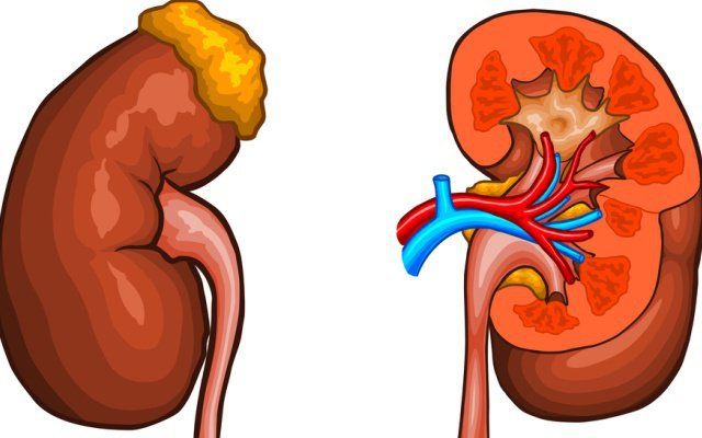
When most people hear “kidney cancer,” they are hearing about renal cell carcinoma (RCC) — the malignancy that accounts for 90 to 95 percent of all primary kidney cancers in adults. But RCC is not a single disease. It is a family of distinct tumors, each arising from different cell types within the kidney, each with its own molecular biology, clinical behavior, metastatic pattern, and response to treatment. The distinction between subtypes matters profoundly: the same stage IV tumor in the kidney carries a very different prognosis and requires a very different treatment strategy depending on whether it is clear cell, papillary type 1, papillary type 2, or chromophobe histology.
Clear Cell Renal Cell Carcinoma
Clear cell RCC (ccRCC) is the most common RCC subtype, representing 70 to 75 percent of all cases. The name comes from the microscopic appearance of the tumor cells — filled with lipid and glycogen that washes out during tissue processing, leaving the cells appearing clear. Grossly, clear cell RCC has a characteristic golden-yellow cut surface, often with areas of hemorrhage, necrosis, and cystic change.
The molecular hallmark is inactivation of the VHL (von Hippel-Lindau) tumor suppressor gene, occurring in approximately 90 percent of sporadic ccRCC cases. In normal cells, the VHL protein targets the hypoxia-inducible factors HIF-1α and HIF-2α for degradation when oxygen is adequate. When VHL is lost, HIF-1α and HIF-2α accumulate constitutively, driving expression of VEGF and other growth factors that promote tumor angiogenesis. This molecular dependency on the VHL-HIF-VEGF axis is precisely why anti-VEGF targeted therapies and the newer HIF-2α direct inhibitor belzutifan are so effective in ccRCC — the tumor has built its survival on a single molecular pathway, and disrupting that pathway cuts its lifeline.
Beyond VHL, additional mutations in chromatin remodeling genes (PBRM1, BAP1, SETD2) further shape the biology of ccRCC. BAP1 mutations are associated with higher nuclear grade and worse prognosis. PBRM1 mutations predict response to immune checkpoint therapy. The WHO-ISUP nuclear grade (1–4) — ranging from grade 1 (small round nuclei, inconspicuous nucleoli) to grade 4 (bizarre giant cells) — is a key prognostic factor and influences adjuvant therapy recommendations after resection.
Papillary Renal Cell Carcinoma
Papillary RCC (pRCC) is the second most common subtype at 15 to 20 percent of cases. It is not a single disease but two biologically and clinically distinct tumors that share a histological pattern of papillary (finger-like) projections.
Type 1 papillary RCC consists of single-layer papillae covered by cells with small, pale cytoplasm and low nuclear grade. It is strongly associated with mutations in the MET proto-oncogene. In hereditary papillary RCC (HPRC) syndrome, germline MET mutations cause bilateral, multifocal type 1 pRCC tumors. Type 1 pRCC has a favorable prognosis and often follows an indolent course.
Type 2 papillary RCC consists of papillae with large, eosinophilic cells and high nuclear grade. It has significantly worse prognosis and is associated with fumarate hydratase (FH) gene mutations in hereditary leiomyomatosis and RCC (HLRCC) syndrome. Because FH loss drives a highly aggressive metabolic phenotype, tumors can spread even when small — patients with HLRCC-associated type 2 pRCC should not be managed with active surveillance regardless of tumor size.
Clear cell RCC: responds well to anti-VEGF TKIs + checkpoint immunotherapy. Papillary RCC: less VEGF-dependent; MET inhibitors (cabozantinib) + immunotherapy preferred. Chromophobe RCC: largely indolent; limited systemic data; surgery is primary treatment. Sarcomatoid (any subtype): responds preferentially to dual checkpoint blockade (nivo+ipi).
Chromophobe Renal Cell Carcinoma
Chromophobe RCC (chRCC) accounts for approximately 5 percent of RCC cases and arises from the intercalated cells of the cortical collecting duct. Chromophobe tumors show losses of multiple chromosomes (1, 2, 6, 10, 13, 17, 21) without VHL mutations — a completely different genomic profile from ccRCC. The clinical behavior is markedly more indolent than other subtypes: stage for stage, chromophobe RCC has the best prognosis of any RCC histology. Local recurrence after resection is uncommon, and distant metastasis is relatively rare.
Birt-Hogg-Dubé syndrome (FLCN gene mutations) predisposes to chromophobe RCC, clear cell RCC, and oncocytoma-chromophobe hybrid tumors, as well as pulmonary cysts and fibrofolliculomas. Renal tumors in BHD syndrome are often multifocal and bilateral but tend to follow an indolent course.
Rare RCC Subtypes
Collecting duct carcinoma (Bellini duct carcinoma) arises from medullary collecting duct cells; accounts for <1% of renal tumors; highly aggressive (median survival <1 year metastatic); treated with platinum-based chemotherapy like urothelial cancer rather than standard RCC regimens.
Medullary carcinoma occurs almost exclusively in patients with sickle cell trait or disease; characterized by SMARCB1 (INI1) loss; typically affects young patients (median age mid-20s); almost uniformly metastatic at presentation; median survival measured in months.
Translocation RCC involves TFE3 or TFEB gene fusions; disproportionately affects children and young adults; may be identified by immunohistochemistry (TFE3 nuclear staining) when conventional markers are atypical.
SDH-deficient RCC involves mutations in the succinate dehydrogenase complex (SDHB/C/D/A) in the setting of SDH deficiency syndrome, which also causes paragangliomas and gastrointestinal stromal tumors. Genetic counseling and family member surveillance are essential.
Sarcomatoid Differentiation
Sarcomatoid differentiation is not a separate subtype but a dedifferentiated transformation that can occur within any conventional RCC subtype. When sarcomatoid features are present, tumor cells acquire a spindle-cell morphology with dramatically more aggressive behavior. Historically carrying a very poor prognosis with poor response to VEGF-targeted therapies, the discovery that sarcomatoid tumors show high PD-L1 expression and immune cell infiltration transformed their treatment: the CheckMate 214 trial found that nivolumab + ipilimumab produced a 56 percent objective response rate in sarcomatoid ccRCC, compared to 18 percent for sunitinib. Sarcomatoid differentiation, once a dire prognostic marker, now identifies a population that preferentially benefits from immunotherapy.
Diagnosis and Imaging
CT urogram with contrast is the primary diagnostic tool — the key imaging feature is contrast enhancement (typically >15–20 Hounsfield units) confirming solid tumor vascularity. Clear cell RCC is particularly hypervascular; papillary and chromophobe RCC show more modest enhancement. MRI is superior for IVC tumor thrombus characterization. Renal mass biopsy is increasingly used for 1–3 cm masses to confirm histological subtype before choosing between active surveillance and intervention.
Treatment of Metastatic RCC
Current first-line standard-of-care combinations for metastatic ccRCC include nivolumab + ipilimumab (CheckMate 214; 5-year OS 43% vs. 27% for sunitinib), pembrolizumab + axitinib (KEYNOTE-426; all IMDC risk groups), lenvatinib + pembrolizumab (CLEAR trial; highest ORR at ~71%), and cabozantinib + nivolumab (CheckMate 9ER). The HIF-2α inhibitor belzutifan (LITESPARK-005) is approved for previously treated metastatic ccRCC and for VHL disease-related RCC, representing the first therapy to directly target the VHL-HIF axis downstream. Adjuvant pembrolizumab (KEYNOTE-564) is now standard for high-risk resected RCC.
Selecting the right treatment depends critically on knowing the histological subtype, IMDC risk category, and prior treatment history. A multidisciplinary genitourinary oncology team — including medical oncology, urology, interventional radiology, and pathology — provides the most informed treatment selection. For the full overview of kidney cancer types and staging, see our guide to kidney cancer. For symptom recognition, see our article on kidney cancer symptoms, and for hematuria evaluation, our guide to kidney cancer blood in urine.
Sources: National Cancer Institute — Kidney Cancer | American Cancer Society — Kidney Cancer | National Kidney Foundation
IMDC Risk Stratification for Metastatic RCC
For patients with metastatic RCC, the IMDC (International Metastatic RCC Database Consortium) risk score is the standard prognostic tool used to select first-line therapy and predict overall survival. It assigns one adverse point each for six factors: Karnofsky performance status less than 80 percent, time from diagnosis to systemic therapy less than one year, hemoglobin below the lower limit of normal, corrected calcium above the upper limit of normal, neutrophil count above the upper limit of normal, and platelet count above the upper limit of normal.
Patients with zero adverse factors are classified as favorable risk; one to two factors as intermediate risk; three to six factors as poor risk. IMDC risk category directly influences first-line treatment selection. Favorable-risk patients may be offered pembrolizumab + axitinib or lenvatinib + pembrolizumab. Intermediate- and poor-risk patients derive particular benefit from nivolumab + ipilimumab. The IMDC score also serves as the standard stratification variable in RCC clinical trials, allowing comparison of results across studies.
How Tumor Biology Informs Drug Selection in ccRCC
The molecular dependency of clear cell RCC on the VHL-HIF-VEGF axis explains the drug landscape that has emerged over two decades. Anti-VEGF therapies (sunitinib, pazopanib, axitinib, cabozantinib) inhibit VEGF receptor signaling, starving the tumor of the growth factor signals that drive angiogenesis. These drugs produce objective responses in 20 to 40 percent of patients but do not eliminate the disease — tumor cells eventually develop resistance by activating alternative survival pathways.
Checkpoint immunotherapy (nivolumab, pembrolizumab, ipilimumab) works through a completely different mechanism: releasing the immunological brakes that tumors use to suppress T-cell killing. In ccRCC, the tumor microenvironment is richly infiltrated by immune cells, and VHL loss upregulates immunosuppressive signals (PD-L1, CD8+ T-cell exhaustion). Checkpoint blockade reverses this immune suppression and can produce durable complete responses — meaning the tumor disappears entirely and does not return even after therapy is stopped. Approximately 10 to 15 percent of patients with metastatic ccRCC achieve complete responses on nivolumab + ipilimumab, and some remain in remission years after discontinuing treatment.
The combination of checkpoint immunotherapy with a VEGF-targeted TKI (pembrolizumab + axitinib; lenvatinib + pembrolizumab) is hypothesized to provide complementary benefits: the TKI produces rapid tumor shrinkage while simultaneously modifying the tumor microenvironment to be more amenable to immune attack, and the checkpoint inhibitor provides the durable immune-mediated control. This hypothesis underpins the remarkable objective response rates (>70%) seen with lenvatinib + pembrolizumab in the CLEAR trial — higher than either drug alone.
Adjuvant Therapy After Nephrectomy
For decades, adjuvant systemic therapy after curative-intent nephrectomy showed no benefit in randomized trials — sunitinib, sorafenib, pazopanib, and even early checkpoint trials produced negative or marginal results. This changed with the KEYNOTE-564 trial of adjuvant pembrolizumab, which enrolled patients with high-risk resected clear cell RCC (pT2 grade 4 or higher, pT3 or higher, pN1, or M1 with no evidence of disease post-resection or metastasectomy) and showed a significant disease-free survival benefit compared to placebo.
Pembrolizumab is now recommended as adjuvant therapy for patients with clear cell RCC at high risk of recurrence after surgery. The benefit appears greatest in patients with T3–T4 or node-positive disease. Whether the DFS benefit translates into an overall survival benefit — and which patients can safely omit adjuvant therapy — remains under investigation in ongoing follow-up data.
Managing Side Effects of RCC Systemic Therapies
Understanding and managing treatment-related toxicities is as important as selecting the right drug. The two main toxicity profiles encountered in RCC treatment are immune-related adverse events (from checkpoint inhibitors) and VEGF-pathway toxicities (from TKIs).
Checkpoint immunotherapy toxicities (immune-related adverse events, irAEs) arise because PD-1/PD-L1 and CTLA-4 blockade can activate immune responses against normal tissues in addition to cancer cells. Common irAEs in RCC treatment include colitis (diarrhea, cramping), hepatitis (elevated liver enzymes), thyroid dysfunction (hypothyroidism more common than hyperthyroidism), skin rash, and pneumonitis (cough, dyspnea). Most irAEs respond to corticosteroids when treated promptly. Patients must report new symptoms immediately — delaying treatment of irAEs allows immune-mediated organ damage to progress.
TKI toxicities are related to VEGF signaling inhibition in normal tissues: hypertension (managed with antihypertensive medications; do not simply stop TKI without oncologist guidance), hand-foot syndrome (painful redness and peeling of palms and soles; urea-based creams, dose reduction), fatigue, mucositis, diarrhea, and hypothyroidism (TSH should be monitored routinely).
Emerging Therapies and Clinical Trials
The pace of RCC drug development remains rapid. Belzutifan — the first FDA-approved HIF-2α inhibitor — targets the HIF-2α transcription factor directly, downstream of VHL, without the vascular side effects of VEGF pathway inhibitors. Approved in 2021 for VHL disease-related RCC and in 2023 for previously treated metastatic ccRCC, belzutifan produces durable responses with a distinct side effect profile dominated by anemia (from suppression of EPO production) and hypoxia (from suppression of erythropoietic EPO).
Triplet combinations — adding a third agent to the current dual-agent standard — are being evaluated in Phase 3 trials. The COSMIC-313 trial examined cabozantinib + nivolumab + ipilimumab versus nivolumab + ipilimumab + placebo, showing improved PFS in intermediate/poor-risk patients. Whether triplet therapy becomes standard depends on continued OS follow-up.
For non-clear cell RCC subtypes (papillary, chromophobe, rare histologies), dedicated clinical trials are critically needed. These subtypes have been historically underrepresented in trials that enroll primarily clear cell RCC, leaving treatment largely empirical. Patients with non-ccRCC histology should be strongly encouraged to enroll in trials specifically designed for their subtype.
Active Surveillance for Small RCC
Not all RCC requires immediate treatment. For small renal masses (T1a, typically ≤3 cm) in elderly patients or those with significant comorbidities, active surveillance — monitoring with serial imaging without immediate intervention — is a guideline-endorsed option. The median growth rate of small RCC under surveillance is approximately 0.28 cm per year, and the risk of metastatic progression during surveillance is low for most masses. Active surveillance is also appropriate while awaiting biopsy results or while patients complete a shared decision-making process about treatment options. Surveillance typically involves CT or MRI at 3–6 months after initial imaging, then annually if stable.
The decision between active surveillance, ablation, and surgery for a small RCC depends on the patient’s age, comorbidities, remaining renal function, histological subtype (if biopsy has been performed), tumor growth rate, and patient preferences. This decision should be made in consultation with an experienced urological oncologist who can individualize the approach based on all available clinical information.
Genetic Counseling and Hereditary RCC
Approximately 3 to 5 percent of all RCC cases are caused by inherited germline mutations in one of several cancer predisposition genes. The clinical features that suggest hereditary RCC include: diagnosis before age 46, bilateral or multifocal kidney tumors, a positive family history of kidney cancer (particularly if early-onset or bilateral), or the presence of extrarenal features characteristic of a specific hereditary syndrome. Identifying a hereditary syndrome has major implications for the patient — in some syndromes (HLRCC), it changes the threshold for surgical intervention even for small tumors — and for family members who may benefit from genetic testing and early surveillance.
The major hereditary RCC syndromes include VHL syndrome (bilateral clear cell RCC plus hemangioblastomas and pheochromocytomas), hereditary papillary RCC (bilateral type 1 papillary RCC from germline MET mutations), Birt-Hogg-Dubé syndrome (multifocal chromophobe and hybrid oncocytoma from FLCN mutations), and hereditary leiomyomatosis and RCC (HLRCC; aggressive type 2 papillary RCC from FH mutations). All patients with clinical features suggesting a hereditary syndrome should be referred for formal genetic counseling and germline testing. Genetic counselors can guide which genes to test, interpret results in the context of the patient’s personal and family history, and coordinate cascade testing of at-risk relatives.
Quality of Life During RCC Treatment
Living with renal cell carcinoma — particularly metastatic RCC on long-term systemic therapy — involves managing both cancer and treatment-related effects on quality of life. Fatigue is the most universally reported symptom, present in 60 to 70 percent of patients on checkpoint immunotherapy or TKIs. Exercise — even moderate, regular walking — has been shown in multiple oncology studies to reduce cancer-related fatigue and improve functional status, and it is safe and recommended for most RCC patients during treatment.
Diet plays a supporting role: maintaining adequate protein intake (to counter muscle catabolism from both cancer and steroids used to treat irAEs), controlling sodium intake to manage TKI-related hypertension, and avoiding nephrotoxic foods and supplements to protect remaining renal function are all practical considerations for patients managing RCC. A registered dietitian with oncology experience can provide individualized guidance.
Mental health support is an underutilized resource in oncology. Patients with advanced RCC face the psychological burden of a chronic, incurable-but-treatable disease — a disease they may live with for many years, through multiple lines of therapy, with ongoing uncertainty about the next scan result. Anxiety, depression, and fear of recurrence are common and underdiagnosed. Screening for psychological distress at oncology follow-up visits, and referral to oncology-specialized psychologists or social workers, is part of comprehensive RCC care.
The Role of Cytoreductive Nephrectomy in the Immunotherapy Era
Cytoreductive nephrectomy — removal of the primary kidney tumor in patients with metastatic disease, performed to reduce overall tumor burden before systemic therapy — was a standard of care in the targeted therapy era, based on two randomized trials (SWOG 8949, EORTC 30947) that showed a survival benefit for nephrectomy plus interferon-alpha over interferon alone. These trials were conducted before the era of effective systemic therapy, and their findings may not generalize to the modern immunotherapy landscape.
The CARMENA trial (2018) challenged cytoreductive nephrectomy in the sunitinib era, showing that sunitinib alone was non-inferior to nephrectomy + sunitinib in intermediate- and poor-risk patients. Whether cytoreductive nephrectomy adds benefit in the checkpoint immunotherapy era is being studied prospectively (PROBE trial). Current NCCN guidelines recommend cytoreductive nephrectomy for favorable-risk patients and selected intermediate-risk patients, with individualized decision-making in the context of systemic therapy timing and patient performance status.
For patients considering all aspects of their kidney cancer care — from the initial diagnosis through systemic therapy decisions — our guide to kidney cancer overview provides a comprehensive starting point. Patients should also review our articles on kidney cancer symptoms and blood in urine from kidney cancer to understand the full clinical picture of this disease.
Post-Treatment Surveillance
After curative-intent surgery for localized RCC, ongoing surveillance is essential. Low-risk tumors (T1a, Fuhrman grade 1–2) require CT at 6 months post-surgery and then annually for 3 years. Intermediate- and high-risk tumors (T2 or higher, or T1 with grade 3–4 features, or sarcomatoid differentiation) require CT at 3–6 months, then every 6 months for 2 years, then annually for at least 5 years. Chest CT is obtained at the same intervals to detect pulmonary metastases — the most common site of RCC recurrence, typically appearing as round nodules bilaterally. Patients who develop any new symptoms during the surveillance interval — bone pain, cough, headaches, neurological changes, or new hematuria — should undergo unscheduled imaging rather than waiting for the next scheduled scan. Early detection of recurrence, when the metastatic burden is limited, opens the possibility of metastasectomy — surgical removal of isolated metastases — which can achieve durable complete remissions in carefully selected patients with oligometastatic RCC recurrence.