
Kidney cancer has a paradox at its core: the organ most capable of harboring a growing tumor for years without causing a single noticeable symptom. The kidneys sit deep in the retroperitoneum — surrounded by fat and protected by the lower ribs — with enough reserve function that a tumor can grow to substantial size before displacing enough tissue to cause pain, producing enough bleeding to color the urine, or creating enough bulk to be felt from the outside. This is why approximately 50 percent of all kidney cancers are discovered incidentally, found on a CT scan or ultrasound performed for an entirely unrelated reason.
But symptoms do occur, particularly as tumors grow larger or begin to spread. Knowing what kidney cancer symptoms look like — and understanding that blood in the urine is never something to dismiss as a probable infection or minor irritation — can make the difference between a stage I diagnosis with a 93 percent five-year survival and a stage IV diagnosis with a dramatically different prognosis. Recognizing the symptoms is an act of advocacy for your own health.
The Classic Triad — and Why It Usually Means Advanced Disease
Medical students learn the “classic triad” of kidney cancer: hematuria (blood in the urine), flank pain, and a palpable abdominal or flank mass. In textbooks, this triad neatly summarizes the defining features of the disease. In clinical practice, it is a warning rather than a teaching point — because when all three are present simultaneously, the tumor has usually grown large enough to declare itself in multiple ways, and the cancer is rarely localized.
The classic triad occurs in only 6 to 10 percent of kidney cancer presentations and is associated with advanced disease, including large primary tumors and a higher likelihood of regional or distant spread. The majority of kidney cancers are found before this triad develops — most through incidental imaging, and some through individual symptoms that appear before all three elements coexist. Understanding each component separately is therefore more clinically useful than waiting for the full triad to appear.
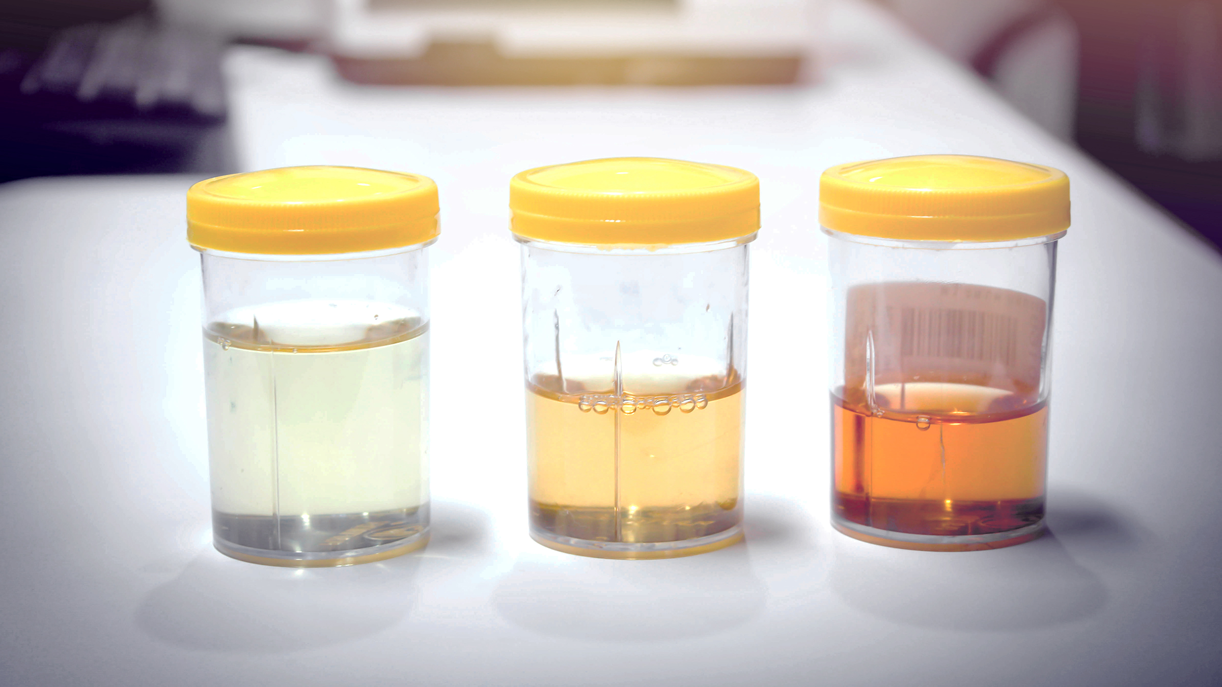
Blood in the Urine (Hematuria) — The Most Important Warning Sign
Hematuria — the presence of blood in the urine — is the single most common kidney cancer symptom and the one most likely to prompt patients to seek evaluation early enough to make a curative difference. Hematuria in kidney cancer has several important characteristics:
It is often painless. Unlike the hematuria caused by kidney stones, which is typically accompanied by severe colicky pain, kidney cancer often produces blood in the urine without any pain at all. Painless hematuria is a red flag that demands investigation — it should never be assumed to be a urinary tract infection without proper evaluation.
It may be intermittent. Tumor bleeding in kidney cancer is not constant. A patient may notice red or cola-colored urine on one occasion and then see nothing abnormal for weeks or months before it recurs. This intermittency creates false reassurance — the bleeding stopped on its own, so it must not be serious. In fact, the intermittent nature of tumor-related hematuria makes it more important to investigate the first episode rather than waiting to see if it happens again.
It can be gross or microscopic. Gross hematuria is visible to the naked eye — urine appears pink, red, brown, or cola-colored. Microscopic hematuria is invisible to the eye but detected on urinalysis when red blood cells are found under the microscope. Both types warrant evaluation. Microscopic hematuria found incidentally on a routine urine test should trigger the same investigative pathway as gross hematuria when no benign cause accounts for it.
Any amount matters. There is no safe threshold of blood in the urine. Even a single episode of gross hematuria — even if it resolved within hours — is sufficient indication for a urological evaluation that includes upper urinary tract imaging.
Any blood in the urine — even one episode, even painless, even if it resolved — requires a CT urogram and urological evaluation. Do not wait for a second episode. Do not assume it is a UTI without imaging. One test can catch a kidney cancer before it spreads.
Flank Pain — What It Feels Like and What to Do
Flank pain in kidney cancer is typically a dull, persistent ache or pressure sensation on one side of the back, located below the lower ribs and above the pelvis. Unlike the severe, cramping pain of a kidney stone, kidney cancer-related flank pain tends to be chronic and lower in intensity — a constant background discomfort rather than an acute crisis. The pain arises from the tumor expanding within the kidney and stretching the renal capsule, the fibrous membrane surrounding the kidney that contains pain-sensing nerve fibers.
An important exception: when a kidney tumor bleeds into the collecting system and a clot forms, the clot can travel down the ureter and cause acute, colicky pain similar to a kidney stone — sometimes called clot colic. This typically occurs simultaneously with visible blood in the urine and warrants same-day evaluation.
Back pain is extraordinarily common, and the vast majority of people with back pain do not have kidney cancer. Features of flank pain that raise suspicion for a renal cause: one-sided location below the ribs, persistent duration beyond 2 weeks without mechanical explanation, accompanying urinary symptoms, constitutional symptoms like unexplained weight loss, or absence of pain relief with typical musculoskeletal treatments like NSAIDs and rest.
Unexplained Weight Loss, Fatigue, and Anemia
As kidney cancer grows, it produces inflammatory cytokines — TNF-alpha, IL-6, and IL-1 — that drive systemic metabolic effects: loss of appetite, accelerated muscle breakdown (cancer cachexia), and chronic fatigue. These are not specific to kidney cancer, but they are important signals that something systemic is occurring.
Unintentional weight loss of more than 5 to 10 pounds over a few months, without dietary change or deliberate effort, is a red flag symptom for any malignancy. It is particularly significant when combined with any other kidney cancer symptom.
Fatigue is multifactorial: inflammatory cytokines create a state of chronic exhaustion, and anemia — present in up to 70 percent of patients with advanced kidney cancer — reduces oxygen delivery to tissues, compounding the sense of exhaustion. The anemia of kidney cancer is typically the normocytic, normochromic anemia of chronic disease, distinct from iron deficiency anemia, and may not respond to iron supplementation.
Paraneoplastic Syndromes — When the Tumor Speaks Through Hormones
Renal cell carcinoma produces hormones, enzymes, and cytokines that cause systemic effects entirely separate from the direct mechanical presence of the tumor — paraneoplastic syndromes. RCC produces them more frequently than almost any other solid tumor.
Hypercalcemia (elevated blood calcium) is caused by PTHrP (parathyroid hormone-related protein) secreted by RCC cells. Symptoms follow the classic mnemonic — “bones, groans, moans, and stones”: bone pain, abdominal pain, neurological effects like confusion and depression, and kidney stones from elevated calcium in the urine. Severe hypercalcemia causes cardiac arrhythmias and can be life-threatening. A newly elevated calcium level in a patient without parathyroid disease should prompt imaging to evaluate for malignancy.
Polycythemia (erythrocytosis) is caused by ectopic erythropoietin production by RCC cells, driving abnormally elevated red blood cell production. Patients may appear ruddy or flushed and experience headaches, dizziness, or itching after bathing. This is the opposite of the anemia that most kidney cancer patients experience — representing a subset of tumors with high EPO output.
Stauffer syndrome is a distinctive RCC paraneoplastic effect: non-metastatic hepatic dysfunction manifesting as elevated liver enzymes and alkaline phosphatase, without any actual liver metastasis. It is caused by cytokines produced by the tumor that impair hepatocyte function and resolves completely after successful nephrectomy. Its importance: it can mimic liver metastasis on imaging, leading to inappropriate upstaging.
Fever and night sweats without infection occur in approximately 20 percent of kidney cancer patients, mediated by IL-1, IL-6, and TNF-alpha. Patients may be evaluated extensively for infection before kidney cancer is considered.
New-onset hypertension in a patient without prior hypertension can be caused by tumor renin production or by compression of the renal artery by a large mass, activating the renin-angiotensin system.
Symptoms of Metastatic Kidney Cancer
When kidney cancer has spread beyond the kidney, the symptoms depend on which organs are involved:
- Lung metastases (most common): persistent cough, coughing up blood (hemoptysis), progressive shortness of breath, pleuritic chest pain
- Bone metastases (second most common): deep, constant, progressive pain in the spine, pelvis, or long bones — worse at night. Pathological fracture can occur with minimal trauma. Vertebral metastases causing spinal cord compression are a medical emergency requiring urgent imaging and intervention
- Brain metastases: new morning headaches, nausea, seizures, personality changes, focal neurological deficits (limb weakness, speech difficulty, visual changes)
- Lymph nodes: retroperitoneal adenopathy on imaging; occasionally palpable cervical or supraclavicular nodes
Symptoms That Masquerade as Something Else
The most dangerous misattribution is assuming hematuria is caused by a urinary tract infection without imaging the upper urinary tract. A UTI can cause hematuria, but UTI-related hematuria resolves with antibiotics — recurrent or persistent hematuria after treatment demands urological evaluation. Flank pain may be attributed to musculoskeletal strain without adequate investigation. Constitutional symptoms may be attributed to stress, thyroid disease, or viral illness. The key principle: any persistent, unexplained symptom in a patient with kidney cancer risk factors (smoker, obese, hypertensive, positive family history, over age 50) should prompt imaging rather than symptomatic management alone. For comparison with how symptoms present in other urological and abdominal cancers, our article on bladder cancer discusses the overlapping but distinct symptom pattern of bladder malignancy, which also frequently presents with hematuria.
When to See a Doctor — Red Flag Symptoms
The following kidney cancer symptoms warrant prompt medical evaluation:
- Any blood in urine (even one episode, even if resolved) — urgent urological evaluation
- Unexplained flank or back pain lasting more than 2 weeks not responding to musculoskeletal treatments
- Palpable abdominal or flank mass — urgent imaging
- Unintentional weight loss of 5+ pounds over 1–2 months
- New-onset hypertension in a patient aged 40+ without prior history
- Persistent fatigue with no identified cause, especially with anemia on blood work
- New hypercalcemia on blood tests without parathyroid explanation
- Bone pain that is progressive, constant, and worse at night
- New neurological symptoms in a patient with known kidney cancer — same-day evaluation
At evaluation, expect: urinalysis and urine cytology, serum metabolic panel (creatinine, calcium, LFTs), CBC, and CT urogram as the primary diagnostic study. Chest CT for staging if a renal mass is identified.
Kidney cancer caught at stage I — tumor confined to the kidney, 7 cm or less — has a 5-year survival rate of approximately 93 percent. A solitary tumor ≤4 cm may be treated with partial nephrectomy, preserving most of the kidney. Stage IV disease has a substantially lower probability of long-term cure. Never dismiss blood in the urine. Never wait to see if it happens again. Our guide to kidney cancer types, staging, and treatment covers what happens after a diagnosis is made. For a broader perspective on cancer symptom recognition, our article on liver cancer symptoms illustrates how solid tumors share systemic features while producing distinct organ-specific warning signs.
Sources: National Cancer Institute — Kidney Cancer | American Cancer Society — Kidney Cancer | National Kidney Foundation
How Symptoms Differ by Tumor Size and Stage
The relationship between kidney cancer symptoms and tumor size is not linear — it depends heavily on tumor location within the kidney, proximity to the collecting system, and individual patient anatomy. A small tumor positioned directly adjacent to a blood vessel or the renal pelvis may cause hematuria at 2 cm, while a tumor growing outward into the perirenal fat may reach 8 cm without a single symptom. This variability means that the absence of symptoms provides no reassurance about tumor size or stage.
For tumors classified as T1a (≤4 cm, confined to the kidney), symptoms are uncommon — most are found incidentally. T1b tumors (4–7 cm) may begin to cause intermittent hematuria or subtle flank discomfort. T2 tumors (7+ cm, still kidney-confined) are more likely to produce a palpable mass and sustained flank pain. T3 tumors that extend into the renal vein or IVC can cause additional symptoms: varicocele (dilated scrotal veins) from left renal vein obstruction, lower extremity edema from IVC compression, or dilated superficial abdominal veins. T4 tumors and those with regional lymph node spread may cause more diffuse abdominal discomfort and constitutional symptoms.
Metastatic disease (Stage IV) produces symptoms at whatever secondary site the cancer has seeded. Patients who present with pulmonary symptoms or bone pain and are subsequently found to have a renal primary — rather than having the kidney cancer found first — are said to have a “presentation-first at metastasis.” This pattern is more common in RCC than in many other solid tumors because the primary kidney tumor may remain entirely silent while distant sites produce symptoms.
Varicocele as a Kidney Cancer Warning Sign
A varicocele is an abnormal dilation of the pampiniform venous plexus in the scrotum — a condition typically caused by incompetent venous valves and common in young men (10–15% prevalence). However, a varicocele that appears suddenly in a man over 40, that is right-sided, or that does not decompress when the patient lies flat is a different clinical entity — one that suggests venous obstruction from an external cause, most commonly a renal tumor compressing or invading the left renal vein (or directly obstructing the right spermatic vein at the IVC). A new right-sided varicocele in particular is a red flag for right-sided renal malignancy and requires urgent imaging.
This is a symptom that can be subtle and that many patients do not connect to any renal complaint — there is no hematuria, no back pain, just new scrotal discomfort or swelling. Any man over 40 with a new unilateral varicocele, particularly on the right side, should undergo renal and retroperitoneal imaging.
Symptoms in Special Populations
Hereditary Kidney Cancer Syndromes
Patients with hereditary kidney cancer syndromes (VHL syndrome, Birt-Hogg-Dubé, hereditary papillary RCC) may develop kidney cancer at a younger age and with bilateral or multifocal tumors. Symptoms in these patients may be intermittent bilateral hematuria, or they may have no renal symptoms at all — with kidney cancers detected through surveillance imaging performed because of their known genetic condition. These patients should be enrolled in formal surveillance programs beginning in their 20s, because waiting for symptoms to develop dramatically reduces the window for curative intervention.
Dialysis Patients
Patients on long-term hemodialysis or peritoneal dialysis develop acquired cystic kidney disease — a condition in which multiple cysts form in the kidneys, significantly raising the risk of RCC. Because dialysis patients produce little or no urine, hematuria may not be the presenting symptom — instead, kidney cancer may first manifest as flank pain, a palpable mass, or constitutional symptoms. Dialysis patients should undergo renal ultrasound surveillance at regular intervals (typically every 1–3 years) to detect tumors before symptoms develop.
Transplant Recipients
Kidney transplant recipients face elevated kidney cancer risk both in their native kidneys and, more rarely, in the transplanted kidney. Immunosuppression reduces cancer immune surveillance, increasing the risk of malignancy across all organ systems. Native kidneys in transplant recipients frequently develop acquired cysts and can harbor RCC. These patients typically undergo regular imaging surveillance of their native kidneys as part of their post-transplant follow-up protocol.
Distinguishing Kidney Cancer Symptoms From Common Conditions
The symptom overlap between kidney cancer and far more common conditions creates diagnostic delays. Understanding the distinguishing features helps both patients and clinicians decide when investigation is truly warranted:
Hematuria — UTI vs. kidney cancer: UTI-related hematuria is almost always accompanied by dysuria (burning urination), frequency, and urgency. It resolves within 3 to 5 days of appropriate antibiotic treatment. Kidney cancer-related hematuria may be entirely painless and intermittent. Any hematuria that persists after antibiotic treatment, recurs after resolution, or occurs without any urinary symptoms demands imaging — not a second course of antibiotics.
Flank pain — musculoskeletal vs. kidney cancer: Musculoskeletal flank pain is typically positional, worse with specific movements, and associated with a history of physical activity or strain. It improves with rest and NSAIDs. Kidney cancer-related flank pain is typically positional-independent, does not improve with musculoskeletal treatments, and may gradually worsen over weeks. The presence of any urinary symptom, constitutional symptom, or palpable fullness alongside back pain shifts the differential significantly.
Fatigue and weight loss — depression/stress vs. kidney cancer: These symptoms require a careful systematic evaluation including CBC (to check for anemia), metabolic panel (to check calcium, creatinine, LFTs), thyroid function, and urinalysis. If all these are normal and symptoms persist, imaging of the abdomen should be considered, particularly in patients with risk factors.
What Happens During the Evaluation for Kidney Cancer Symptoms
When a patient presents with symptoms suggesting possible kidney cancer, the evaluation follows a systematic pathway. The first step is a thorough history covering: the exact character and duration of symptoms, whether hematuria was gross or microscopic and whether it was painful, associated constitutional symptoms, relevant risk factors (smoking history, hypertension, obesity, family history of kidney cancer, occupational chemical exposures), and a medication review.
Physical examination focuses on flank tenderness, palpable abdominal or flank masses, scrotal examination in men (varicocele), lymph node palpation (cervical, supraclavicular, groin), and blood pressure measurement. Laboratory tests ordered at initial evaluation typically include: urinalysis with microscopy, urine culture (to exclude UTI as cause of hematuria), complete blood count (looking for anemia or polycythemia), serum creatinine and electrolytes, serum calcium, liver function tests (LFTs — relevant for both Stauffer syndrome assessment and staging), and lactate dehydrogenase (LDH, which has prognostic value in RCC).
Imaging is the definitive next step. CT urogram — CT of the kidneys, ureters, and bladder performed with and without intravenous contrast — is the primary modality for evaluating hematuria and characterizing renal masses. It assesses the kidneys (mass presence, enhancement pattern, size, relation to renal vessels and collecting system), the ureters (for transitional cell tumors), the bladder, and regional lymph nodes. If a renal mass is identified, staging CT of the chest is added to evaluate for pulmonary metastases.
When a renal mass is identified on imaging, the next decision — biopsy versus surgery versus active surveillance — depends on mass size, enhancement characteristics, patient age and comorbidities, and the treating urologist’s assessment. Patients should seek evaluation at a center with experience in renal mass management to ensure they receive the full range of treatment options rather than a one-size-fits-all approach.