
Stomach pain is one of the most common reasons people seek medical attention, accounting for approximately 8% of all emergency department visits worldwide. The vast majority of cases are benign — a food intolerance, a minor viral illness, constipation, or a functional GI disorder that will resolve without specific treatment. But within this very common and usually benign presentation lies a subset of cases where stomach pain needs evaluation urgently — where delay in assessment directly determines outcomes. Knowing which features demand emergency care and which can be safely monitored is the most clinically important distinction in abdominal medicine.
Understanding Stomach Pain by Location
The location of abdominal pain is the first and most useful initial guide to likely causes. Pain in different regions reflects the underlying anatomy and innervation of abdominal structures.
Epigastric (upper central): The most common site for upper GI pathology — peptic ulcer disease, gastritis, GERD, functional dyspepsia, and acute pancreatitis all present here. Inferior myocardial infarction can also present as epigastric pain, an important mimic demanding an ECG in any patient over 45 with new unexplained upper abdominal symptoms.
Right upper quadrant (RUQ): Gallbladder disease dominates — biliary colic, acute cholecystitis, and cholangitis. Liver conditions (hepatitis, hepatic abscess) also present here. Right lower lobe pneumonia can cause referred RUQ pain, so chest examination is essential.
Periumbilical (around the navel): Classic early presentation of appendicitis — pain begins periumbilical before migrating to the right iliac fossa over 6–12 hours. Also the site for small bowel pathology and mesenteric ischaemia.
Right iliac fossa (RIF): Appendicitis is the primary concern and most common surgical emergency. In women, ovarian pathology (torsion, ectopic pregnancy) must always be considered. Crohn’s disease involving the terminal ileum is another important RIF cause.
Diffuse or generalised: When severe and diffuse, the most dangerous diagnoses come first — peritonitis (perforation), bowel obstruction, and mesenteric ischaemia. A rigid, board-like abdomen is a surgical emergency.
Alarm Features — When Stomach Pain Needs Immediate Attention
Alarm features are symptoms, signs, or patient characteristics that substantially raise the probability of a serious underlying condition. When any alarm feature is present, a functional or benign cause must not be assumed — investigation must come first. A useful mnemonic is ABCDEFG:
- A — Anaemia (iron deficiency, unexplained) → suggests GI blood loss or mucosal lesion
- B — Blood in vomit (haematemesis) or stool (melaena or haematochezia)
- C — Cancer weight loss → unintentional loss of >5% body weight over 3–6 months
- D — Dysphagia → progressive difficulty swallowing — oesophageal malignancy until proven otherwise
- E — Evidence of organ failure → jaundice (biliary obstruction), fever with rigors (sepsis, cholangitis)
- F — Family history of upper GI cancer in a first-degree relative
- G — Gut examination finding → palpable abdominal mass
Additional alarm features: new-onset symptoms in a patient aged ≥55; persistent vomiting lasting more than 3–4 days; pain radiating directly to the back (pancreatitis, aortic aneurysm, posterior ulcer); haemodynamic instability alongside pain; rigid abdomen.
- Sudden, severe “worst-ever” abdominal pain
- Vomiting blood or passing black tarry stools
- Fainting, rapid weak pulse, pallor or cold sweating alongside pain
- A rigid, board-like abdomen that is tender to touch
- Fever over 39°C with jaundice and RUQ pain (possible cholangitis)
- Pain after abdominal trauma
Urgent Conditions That Require Emergency Care
Acute appendicitis is the most common surgical emergency, with a lifetime risk of 6–9%. The classic presentation begins with periumbilical or epigastric pain followed by migration to the right iliac fossa over 6–24 hours. Nausea, anorexia, and low-grade fever accompany the pain. Physical examination reveals RIF tenderness, Rovsing’s sign (LIF palpation causing RIF pain), and guarding. CT abdomen achieves 95% sensitivity and specificity; ultrasound is preferred in children and pregnancy. Treatment is laparoscopic appendicectomy. The risk of perforation rises significantly after 24–36 hours from symptom onset — delayed presentation is the main cause of complications.
Perforated viscus — typically a perforated peptic ulcer — presents with sudden severe generalised pain, a rigid abdomen, and peritonism. Free air under the diaphragm on erect chest X-ray confirms perforation. This is a surgical emergency requiring immediate resuscitation and urgent laparotomy or laparoscopy. For the peptic ulcer conditions that most commonly precede perforation, see our detailed guide to peptic ulcer disease explained.
Mesenteric ischaemia is rare but carries very high mortality if missed. The classic feature is severe pain out of proportion to clinical examination — the patient is in agony but the abdomen feels soft. Risk factors include atrial fibrillation, atherosclerosis, and low cardiac output. CT angiography is the investigation of choice; urgent revascularisation is treatment. Every hour of delay increases bowel necrosis and mortality.
Ruptured aortic aneurysm presents with severe abdominal or back pain, haemodynamic shock, and sometimes a pulsatile epigastric mass. Emergency CT angiography and immediate vascular surgery are required. Mortality is very high without immediate surgical intervention.
Ascending cholangitis (Charcot’s triad: RUQ pain + fever + jaundice) is a bile duct infection requiring urgent IV antibiotics and biliary decompression by ERCP or percutaneous drainage. Without treatment, cholangitis progresses rapidly to septic shock and multi-organ failure.
Serious Conditions Requiring Prompt Same-Day Evaluation
Acute cholecystitis presents as persistent severe RUQ pain (lasting more than 6 hours, unlike the self-resolving episodes of biliary colic), fever, and positive Murphy’s sign (inspiratory arrest on RUQ palpation). Ultrasound confirms gallstones, wall thickening, and pericholecystic fluid. Management is IV antibiotics and laparoscopic cholecystectomy, ideally within 72 hours of presentation.
Acute pancreatitis presents with severe, persistent epigastric pain radiating to the back, nausea, vomiting, and elevated serum amylase or lipase (more than three times the upper limit of normal). The two most common causes are gallstones and alcohol (together accounting for approximately 75% of cases). Severity is assessed using the Ranson criteria or BISAP score; CT with contrast defines pancreatic necrosis in severe cases. Mild cases are managed with IV fluids, analgesia, and early oral feeding.
Upper GI bleeding (haematemesis, melaena, or haematochezia with haemodynamic compromise) requires immediate hospital assessment, IV access, cross-match, and urgent upper GI endoscopy. The Glasgow-Blatchford score predicts the need for intervention. The most common underlying cause is peptic ulcer disease; other causes include oesophageal varices, Mallory-Weiss tear, and gastric malignancy.
Common Causes That Need Planned Investigation
Most epigastric or upper abdominal pain presentations do not represent emergencies but benefit from systematic evaluation rather than simple reassurance.
Peptic ulcer disease and gastritis are the most common organic causes of epigastric pain. H. pylori testing is the first step; PPI for 4–8 weeks is appropriate empirical therapy. OGD is needed if symptoms persist or alarm features are present. For the full clinical picture of how H. pylori drives upper GI disease, see our article on H. pylori infection and stomach health and our overview of gastritis: symptoms and causes.
GERD causes retrosternal burning, acid regurgitation, and post-prandial symptoms. For the clinical overlap between GERD and gastritis, and how to distinguish them, see our article on GERD vs gastritis: what is the difference. Long-standing GERD may require OGD to exclude Barrett’s esophagus.
Gallstones are symptomatic in only 20–30% of those with stones. Biliary colic — episodic severe RUQ or epigastric pain after fatty meals, typically lasting 1–3 hours — is the characteristic presentation. Abdominal ultrasound is the diagnostic test of choice. Elective laparoscopic cholecystectomy is curative for symptomatic disease.
Inflammatory bowel disease — Crohn’s disease affecting the terminal ileum typically causes RIF pain, diarrhoea, and weight loss, and can be mistaken for recurrent appendicitis or IBS. Faecal calprotectin elevation points to mucosal inflammation; colonoscopy with biopsy is diagnostic.
Functional dyspepsia — when all investigations are normal, a functional diagnosis can be established using Rome IV criteria. For the full diagnostic and management approach to this common condition, see our article on functional dyspepsia explained.
Tests Used to Evaluate Stomach Pain
Investigation of abdominal pain follows a structured sequence based on the clinical presentation. Not every test is needed in every patient — targeted investigation matched to the most likely diagnoses is more effective and safer than a wide indiscriminate screen.
Blood tests: Full blood count (anaemia, leucocytosis); CRP and ESR (inflammation); liver function tests (bilirubin, ALP, ALT, GGT — biliary or hepatic pathology); amylase/lipase (pancreatitis); urea and electrolytes (dehydration); serum HCG in all women of childbearing age (ectopic pregnancy is a life-threatening cause of lower abdominal pain that must not be missed).
Urine: Dipstick and midstream specimen (UTI; renal colic can mimic abdominal pain); urine HCG if serum not performed.
Imaging: Erect chest X-ray checks for free air under the diaphragm (confirming perforation). Abdominal ultrasound is first-line for RUQ pathology and is non-irradiating. CT abdomen/pelvis with contrast gives the highest diagnostic yield for the acute abdomen and is the preferred investigation when the diagnosis is unclear and the patient is deteriorating. MRI is preferred for IBD, hepatic, biliary, and pancreatic assessment, and for pregnant patients where radiation must be avoided.
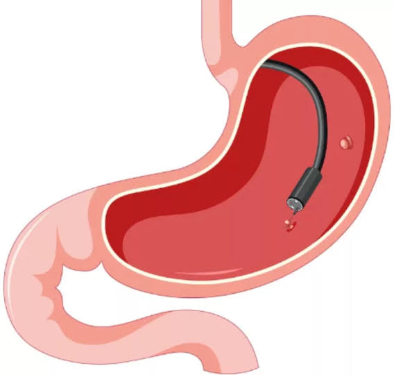
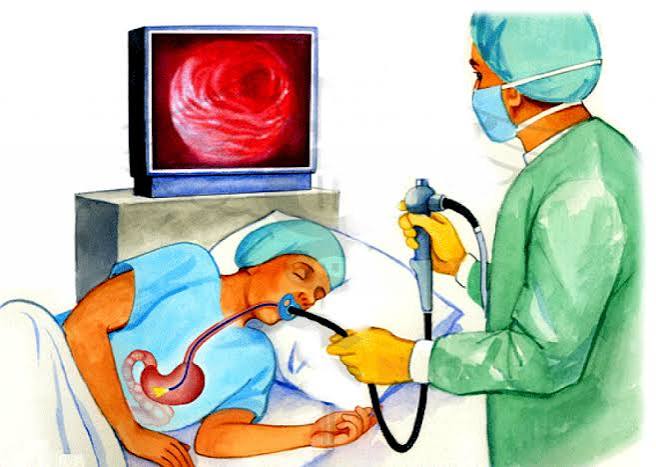
Endoscopy: OGD (upper GI endoscopy) is the definitive test for epigastric pathology — it identifies peptic ulcers, gastritis, oesophagitis, Barrett’s esophagus, and upper GI malignancy; mandatory when alarm features are present. Colonoscopy evaluates lower GI causes including colorectal malignancy and IBD. ERCP is both diagnostic and therapeutic for biliary and pancreatic pathology. According to NICE guideline NG12, urgent OGD referral within 2 weeks is recommended for patients aged 55 or over with new-onset dyspepsia, or at any age with alarm features.
When to Go to A&E, Urgent GP, or Routine GP
A practical decision framework for abdominal pain presentations:
Go to A&E or call 999 immediately: sudden severe “worst-ever” pain; vomiting blood or passing black tarry stools; feeling faint or near-collapse alongside pain; a rigid, board-like abdomen; fever above 39°C with jaundice and RUQ pain; pain after significant abdominal trauma.
Seek same-day urgent GP or emergency GP appointment: persistent severe pain not resolving over 4–6 hours; new RIF pain with fever and loss of appetite; new jaundice; first-episode significant rectal bleeding or melaena that has now settled; lower abdominal pain in pregnancy (possible ectopic).
Book a routine GP appointment: episodic dyspepsia or upper abdominal discomfort without alarm features in patients under 55; chronic recurrent abdominal discomfort without deterioration or change in character; bloating, constipation, or change in bowel habit without blood or weight loss in patients under 45; known functional condition (IBS, functional dyspepsia) with symptoms unchanged in character. For general NHS guidance on abdominal pain and when to get help, see the NHS stomach ache guidance.
Abdominal Pain in Special Populations
The presentation, differential diagnosis, and appropriate investigation threshold for abdominal pain differ significantly between population groups. Recognising these differences prevents dangerous misattribution of serious conditions to common benign causes.
Older adults (aged 65+): Older patients frequently present atypically — reduced pain perception (due to altered nociception and comorbid neuropathies), blunted fever responses, and reduced inflammatory markers mean that serious pathology can be present with minimal clinical signs. Appendicitis in the elderly may lack the classic periumbilical-to-RIF migration; mesenteric ischaemia, bowel obstruction from volvulus, and aortic pathology are all more common with advancing age. Any significant abdominal symptom in an older patient warrants a lower threshold for urgent investigation than the same symptom in a younger adult. Peritonism may be absent even with significant intra-abdominal pathology.
Women of childbearing age: Serum or urine HCG must be performed in any woman of childbearing age presenting with lower abdominal or pelvic pain — ectopic pregnancy is a leading cause of maternal mortality and can rupture with minimal preceding symptoms. Ovarian cyst torsion presents with sudden severe unilateral lower abdominal pain and requires urgent ultrasound and surgical assessment. Pelvic inflammatory disease (PID) causes lower abdominal pain, cervical motion tenderness, and fever, and is managed with broad-spectrum antibiotics.
Immunocompromised patients: Patients on corticosteroids, immunosuppressants, or with HIV/AIDS may present with atypical or blunted inflammatory responses. Peritoneal signs may be minimal even with perforation. Opportunistic infections (CMV colitis, Cryptosporidium, Microsporidium) cause abdominal pain in HIV patients and require specific testing. Any abdominal pain in an immunocompromised patient requires a lower threshold for cross-sectional imaging and specialist review.
Patients with diabetes: As noted above, inferior MI can present as epigastric pain with nausea in diabetic patients — the autonomic neuropathy of diabetes alters pain pathways and reduces the classic retrosternal component of cardiac ischaemia. Diabetic ketoacidosis (DKA) itself causes diffuse abdominal pain and vomiting, which can lead to confusion with an abdominal cause — a blood glucose and ketone test should be included in the initial workup of any diabetic patient with unexplained abdominal pain. Diabetic gastroparesis is a specific upper GI functional disorder unique to this population.
The Role of Self-Monitoring Between Episodes
For patients with recurrent or chronic abdominal pain — whether from a diagnosed condition like IBS or functional dyspepsia, or from an as-yet undiagnosed cause — systematic self-monitoring provides information that transforms the value of subsequent medical consultations.
A symptom diary recording pain location, severity (1–10 scale), timing (time of day, relation to meals and bowel movements), associated symptoms (nausea, bloating, change in stool), and potential triggers (food, stress, activity, menstrual cycle) over 4–6 weeks gives a clinician far more actionable information than a description of “stomach pain, on and off.” This level of documentation can make the difference between an endoscopy being requested (if the diary reveals progressive worsening, weight loss, or meal-correlated severe pain) or a functional diagnosis being confidently confirmed (if the diary shows intermittent, stress-correlated, fully resolving episodes without systemic features).
Patients should also note any changes from their established baseline — a new quality of pain, radiation to a new location, or pain that wakes them from sleep is a signal to seek earlier medical review rather than waiting for a pre-scheduled appointment. The principle that applies to monitoring is simple: the pattern of abdominal pain over time, not a single episode, is what guides the most appropriate next step in evaluation.
Frequently Asked Questions
The most dangerous causes of abdominal pain — ruptured aortic aneurysm, bowel perforation, mesenteric ischaemia, cholangitis, and ruptured ectopic pregnancy — can be rapidly fatal without emergency care. Any abdominal pain with haemodynamic instability (fainting, rapid pulse, pallor), a rigid abdomen, haematemesis, sudden “worst-ever” severity, or fever plus jaundice should be treated as an emergency. Appendicitis and acute pancreatitis require same-day assessment and can deteriorate rapidly if delayed.
Go to A&E immediately if you have sudden severe pain unlike anything before; vomiting blood or passing black tarry stools; feeling faint or nearly collapsing with pain; a rigid abdomen; severe pain with high fever and yellow skin; or severe pain after abdominal trauma. For ongoing but lower-grade pain with fever, loss of appetite, or right iliac fossa tenderness, seek same-day GP care rather than waiting. When in doubt, it is always safer to seek assessment — the consequences of missing a time-critical condition are far greater than the cost of a precautionary visit.
Yes. Inferior myocardial infarction (involving the inferior wall of the heart) can present as epigastric pain or nausea without chest pain — particularly in older patients, people with diabetes, and women. Any patient over 40–45 years with new unexplained epigastric or upper abdominal pain should have an ECG as part of the initial assessment to exclude a cardiac cause, especially if pain radiates to the jaw or left arm, or if cardiovascular risk factors are present. Never assume an upper abdominal pain is GI without considering cardiac causes in at-risk patients.
Acute pancreatitis typically presents as severe, persistent epigastric pain radiating directly to the back. The pain is constant (not colicky), worse with eating and lying flat, and partially relieved by sitting forward. Nausea and vomiting are almost universal. It is frequently described as the worst pain the patient has ever experienced. The key distinguishing feature from other epigastric causes is the direct back radiation and the constant, unrelenting character — unlike the episodic waves of biliary colic or the meal-related pattern of peptic ulcer pain.
Appendicitis classically begins with pain around the navel, which migrates to the right lower abdomen over 6–12 hours. The pain is constant and worse on movement, accompanied by loss of appetite, nausea, and a low-grade fever. Pressing on the left lower abdomen causing pain in the right lower abdomen (Rovsing’s sign) is a clinical pointer. Releasing pressure from the right lower abdomen and feeling a sudden sharp increase in pain (rebound tenderness) also suggests peritoneal involvement. Not all appendicitis follows this classic pattern — so any persistent right lower abdominal pain with systemic symptoms should be assessed promptly. CT confirms the diagnosis with 95% accuracy.
Daily abdominal pain is not normal and warrants medical evaluation. In many cases, systematic assessment identifies a manageable cause such as IBS or functional dyspepsia. However, daily pain can also reflect peptic ulcer disease, inflammatory bowel disease, gallstone disease, or occasionally early malignancy. The key threshold: if abdominal pain is present most days for more than 4–6 weeks, book a GP appointment. If accompanied by any alarm feature — weight loss, blood in vomit or stool, dysphagia, or anaemia — seek earlier evaluation without waiting.
The tests depend on the clinical picture. Initial blood tests typically include FBC, CRP, liver function, amylase, and HCG in women. Urine dipstick screens for UTI and kidney stones. Imaging starts with abdominal ultrasound for suspected gallbladder or upper abdominal pathology, or CT abdomen/pelvis for the acute abdomen. An erect chest X-ray checks for free air if perforation is suspected. Upper GI endoscopy (OGD) is the definitive test for epigastric causes when alarm features are present. According to the NICE dyspepsia guideline CG184, patients with persistent dyspepsia not responding to empirical treatment should be referred for OGD.
Medical Disclaimer: This article is for educational purposes only and does not constitute medical advice. If you experience severe abdominal pain, haematemesis, melaena, haemodynamic instability, or any alarm feature described above, seek emergency medical care immediately.
References
- NICE guideline NG12. Suspected cancer: recognition and referral. 2015 (updated 2023). Available at: nice.org.uk.
- NICE guideline CG141. Acute upper gastrointestinal bleeding in over 16s. 2012 (updated 2016). Available at: nice.org.uk.
- NICE guideline CG184. Dyspepsia and gastro-oesophageal reflux disease. 2014 (updated 2019). Available at: nice.org.uk.
- Silen W. Cope’s Early Diagnosis of the Acute Abdomen. 22nd ed. Oxford University Press; 2010.
- NHS. Stomach ache and abdominal pain. Available at: nhs.uk.
I went to A&E twice with severe abdominal pain and both times was told it was ‘just anxiety.’ It turned out to be acute cholecystitis. I wish I’d had this article earlier — the section on Murphy’s sign and persistent pain over 6 hours being different from biliary colic is exactly what I was experiencing. Really clear breakdown.
Thank you for sharing that, Rachel — and we’re glad you got the correct diagnosis eventually. Cholecystitis is one of those conditions where the distinction from simple biliary colic is crucial: colic resolves in 1–3 hours; cholecystitis persists, is accompanied by fever, and produces Murphy’s sign. The difficulty is that many people (and sometimes clinicians) default to reassurance when the initial presentation looks similar to previous benign episodes. If pain persists beyond 4–6 hours with fever, that transition point is when escalation is warranted. We hope the article helps you advocate for yourself if you ever need to in the future.
The section on mesenteric ischaemia is critical and underappreciated. Pain out of proportion to examination is exactly the right teaching point. I’ve seen that presentation mismanaged multiple times. Good to see it explained clearly in patient-facing content so people know to push for imaging when they’re in severe pain but look okay to the examiner.